Download presentation
Presentation is loading. Please wait.

1
PERITONEUM AND PERITONEAL CAVITY
Prepared by: Dr. Azmi hussien PhD. Md. Head of human anatomy department

2
peritoneum The peritoneum is a continuous, glistening, transparent serous membrane. It lines the abdominopelvic cavity and invests the viscera. The peritoneum consists of two continuous layers: the parietal peritoneum, which lines the internal surface of the abdominopelvic wall. the visceral peritoneum, which invests viscera such as the stomach and intestines. Both layers of peritoneum consist of mesothelium, a layer of simple squamous epithelial cells.

3
Intraperitoneal and Retroperitoneal Relationships:
intraperitoneal organ when it is almost totally covered with visceral peritoneum. The stomach, jejunum, ileum, and spleen are intraperitoneal organs. Retroperitoneal organs lie behind the peritoneum and are only partially covered with visceral peritoneum. The pancreas and the ascending and descending parts of the colon are retroperitoneal organs. No organ, however, is actually within the peritoneal cavity. Intraperitoneal in this case does not mean inside the peritoneal cavity

4
The peritoneal cavity The peritoneal cavity is within the abdominal cavity and continues inferiorly into the pelvic cavity. The peritoneal cavity is a potential space of capillary thinness between the parietal and visceral layers of peritoneum. It contains no organs but contains a thin film of peritoneal fluid, which is composed of water, electrolytes. Peritoneal fluid lubricates the peritoneal surfaces, enabling the viscera to move over each other without friction, and allowing the movements of digestion. the peritoneal fluid contains leukocytes and antibodies that resist infection.

5
Peritoneal Ligaments A peritoneal ligament consists of a double layer of peritoneum that connects an organ with another organ or to the abdominal wall. 1-The liver is connected to the: Anterior abdominal wall by the falciform ligament.

7
Peritoneal Ligaments B. Stomach by the hepatogastric ligament, the membranous portion of the lesser omentum.

8
Peritoneal Ligaments C. Duodenum by the hepatoduodenal ligament: the thickened free edge of the lesser omentum, which conducts the portal triad: portal vein, hepatic artery, and bile duct. The hepatogastric and hepatoduodenal ligaments are continuous parts of the lesser omentum and are separated only for descriptive convenience.

9
Peritoneal Ligaments 2-The stomach is connected to the:
Inferior surface of the diaphragm by the gastrophrenic ligament. Spleen by the gastrosplenic ligament. Transverse colon by the gastrocolic ligament. the apron-like part of the greater omentum, which descends from the greater curvature, turns under, and then ascends to the transverse colon.

10
Peritoneal Ligaments The greater omentum:
is a prominent, four-layered peritoneal fold that hangs down like an apron from the greater curvature of the stomach and the proximal part of the duodenum. After descending, it folds back and attaches to the anterior surface of the transverse colon and its mesentery.

11
Peritoneal Ligaments

13
Peritoneal Ligaments The lesser omentum:
is a much smaller, double-layered peritoneal fold that connects the lesser curvature of the stomach and the proximal part of the duodenum to the liver. It also connects the stomach to a triad of structures that run between the duodenum and liver in the free edge of the lesser omentum.

14
Mesenteries Mesenteries are two-layered folds of peritoneum connecting parts of the intestines to the posterior abdominal wall: the mesentery of the small intestine. the transverse mesocolon, and the sigmoid mesocolon. The peritoneal ligaments, omenta, and mesenteries permit blood, lymph vessels, and nerves to reach the viscera.

15
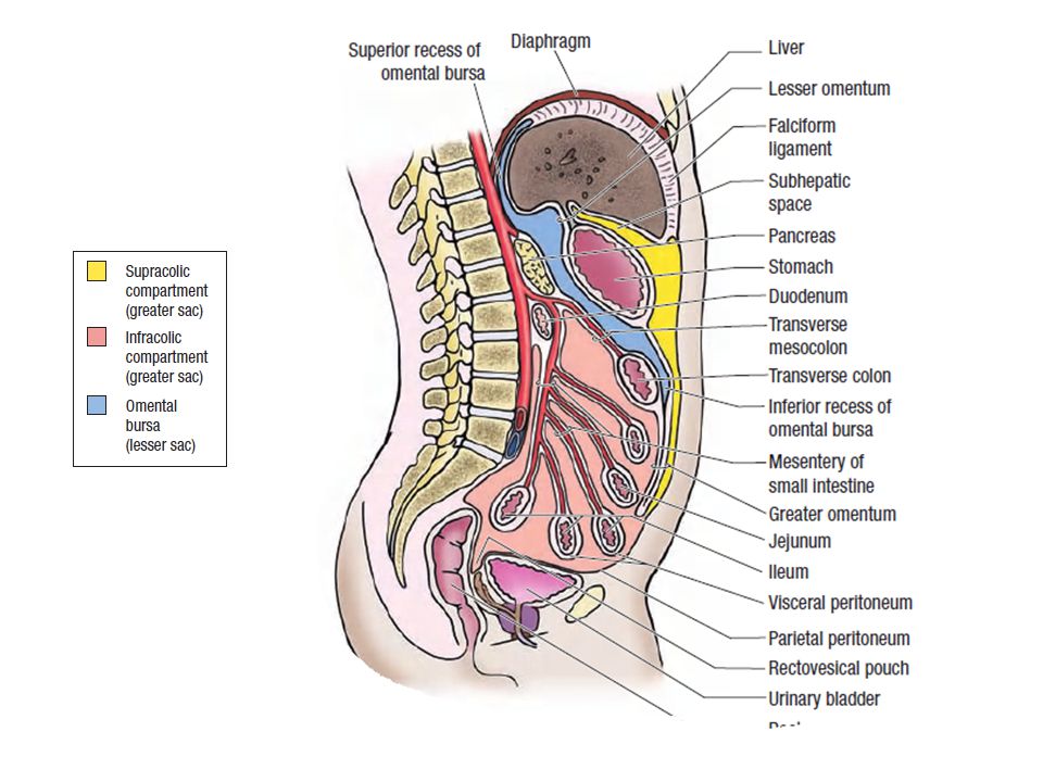
Subdivisions of Peritoneal Cavity
the peritoneal cavity is divided into: the greater peritoneal sac. lesser peritoneal sac (The omental bursa).
.")
17
lesser sac (The omental bursa).:
sac-like cavity that lies posterior to the stomach, lesser omentum, and adjacent structures . The omental bursa has a superior recess, limited superiorly by the diaphragm and the posterior layers of the coronary ligament of the liver, and an inferior recess between the superior parts of the layers of the greater omentum. Most of the inferior recess of the bursa becomes sealed off from the main part posterior to the stomach after adhesion of the anterior and posterior layers of the greater omentum

18
Omental foramen (epiploic foramen)
The omental bursa communicates with the greater sac through the omental foramen (epiploic foramen). an opening situated posterior to the free edge of the lesser omentum (hepatoduodenal ligament). The omental foramen can be located by running a finger along the gallbladder to the free edge of the lesser omentum.
. an opening situated posterior to the free edge of the lesser omentum (hepatoduodenal ligament). The omental foramen can be located by running a finger along the gallbladder to the free edge of the lesser omentum.")
19
The boundaries of the omental foramen are
Anteriorly: the hepatoduodenal ligament containing the hepatic portal vein, hepatic artery, and bile duct. Posteriorly: the IVC and a muscular band. Superiorly: the liver, covered with visceral peritoneum. Inferiorly: the superior or first part of the duodenum.

20
Supracolic, infracolic compartments
The transverse mesocolon (mesentery of the transverse colon) divides the abdominal cavity into: a supracolic compartment, containing the stomach, liver, and spleen. infracolic compartment, containing the small intestine and ascending and descending colon. The infracolic compartment lies posterior to the greater omentum and is divided into right and left infracolic spaces by the mesentery of the small intestine. Free communication occurs between the supracolic and the infracolic compartments through the paracolic gutters, the grooves between the lateral aspect of the ascending or descending colon and the posterolateral abdominal wall.
 divides the abdominal cavity into: a supracolic compartment, containing the stomach, liver, and spleen. infracolic compartment, containing the small intestine and ascending and descending colon. The infracolic compartment lies posterior to the greater omentum and is divided into right and left infracolic spaces by the mesentery of the small intestine. Free communication occurs between the supracolic and the infracolic compartments through the paracolic gutters, the grooves between the lateral aspect of the ascending or descending colon and the posterolateral abdominal wall.")
Similar presentations