Download presentation
Presentation is loading. Please wait.

1
Receptors & Signaling

2
Assumed Knowledge Structure of membrane proteins Ion concentrations across membranes Second messengers in signal transduction Regulation of protein activity through phosphorylation

3
Membrane Proteins Mainly transmembrane Act as receptors Ligand binding causes a conformational change – a change in the shape of the receptor

4
G Protein coupled receptors (GPCR) – these have a transmembrane bit with 7 helices spanning the membrane The extracellular part binds to the ligand The intracellular part binds to the G protein
 – these have a transmembrane bit with 7 helices spanning the membrane The extracellular part binds to the ligand The intracellular part binds to the G protein")
5
G Protein – these are proteins that bind to the guanine nucleotide (GTP – guanosine triphosphate, GDP – guanosine diphosphate) Hydrolysis of GTP releases a phosphate group which can act on other molecules – transmits the signal GTP > GDP + P
 Hydrolysis of GTP releases a phosphate group which can act on other molecules – transmits the signal GTP > GDP + P")
6
G proteins have three subunits – α (alpha), β (beta), and γ (gamma). β and γ subunits are tightly bound together. α binds to GDP

7
Ligand binding to the transmembrane protein causes a conformational change and release of the α subunit The α subunit exchanges GDP > GTP and becomes active The α subunit meets a target and phosphorylates it (adds a phosphate group from GTP converting it to GDP – this is hydrolysis of GTP) Hydrolysis = cleavage Phosphorylation = addition of a phosphate group
 Hydrolysis = cleavage Phosphorylation = addition of a phosphate group")
8
Now the α subunit is bound to GDP, it becomes inactive again and re-associates with the transmembrane protein and the β and γ subunits

10
Ion Concetrations Ions – these are molecules with a charge + or – Ion concentration – ions move down their concentrations gradient from [high] to [low]
![Ion Concetrations Ions – these are molecules with a charge + or – Ion concentration – ions move down their concentrations gradient from [high] to [low]](http://images.slideplayer.com/14/4335793/slides/slide_10.jpg "Ion Concetrations Ions – these are molecules with a charge + or – Ion concentration – ions move down their concentrations gradient from [high] to [low]")
11
[K + ] 160 mM [K + ] 5 mM [Na + ] 10 mM [Na + ] 150 mM [Cl - ] 5 mM [Cl - ] 115 mM [Ca 2+ ] 0.2 M [Ca 2+ ] 2 mM - - - - - - - - + + + + + + + + Membrane potential = -70 mV Anion –ve charge Cation +ve charge [A - ] 40 mM [A - ] 165 mM
![[K + ] 160 mM [K + ] 5 mM [Na + ] 10 mM [Na + ] 150 mM [Cl - ] 5 mM [Cl - ] 115 mM [Ca 2+ ] 0.2 M [Ca 2+ ] 2 mM Membrane potential = -70 mV Anion –ve charge Cation +ve charge [A - ] 40 mM [A - ] 165 mM](http://images.slideplayer.com/14/4335793/slides/slide_11.jpg "[K + ] 160 mM [K + ] 5 mM [Na + ] 10 mM [Na + ] 150 mM [Cl - ] 5 mM [Cl - ] 115 mM [Ca 2+ ] 0.2 M [Ca 2+ ] 2 mM Membrane potential = -70 mV Anion –ve charge Cation +ve charge [A - ] 40 mM [A - ] 165 mM")
12
Membrane Potential – this is the difference in charge between the interior and exterior of a cell Typically the interior is more negative

13
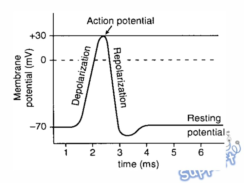
Action potentials – these are rapid rises and falls in the membrane potential. In neurons these act as nerve signals The interior becomes rapidly positive or less negative – depolarization. This is followed by a rapid return to a negative membrane potential – repolarization There is a transient hyperpolarization where the membrane potential becomes more negative than normal

15
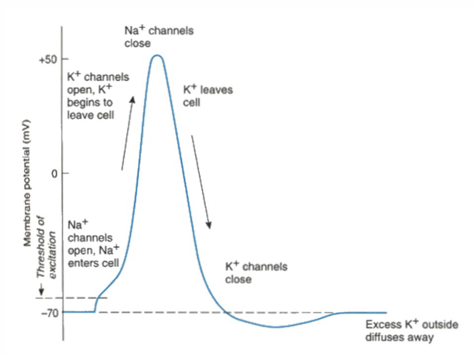
1.An stimulus is received by a nerve causing Na + channels to open – Na + moves into the cell. The membrane potential begins to become more positive

16
2.When the membrane potential reaches the threshold level (-55mV) there is an opening of more Na + channels allowing more Na + to enter the cell This is an ‘all-or-nothing’ moment meaning if the membrane potential doesn’t reach the threshold there will be no action potential but if it reaches the threshold there no turning back
 there is an opening of more Na + channels allowing more Na + to enter the cell This is an ‘all-or-nothing’ moment meaning if the membrane potential doesn’t reach the threshold there will be no action potential but if it reaches the threshold there no turning back")
17
3. As Na + enters the cell there is delayed opening of K + channels The membrane potential reaches +30mV where the Na + channels close

18
4. When the K + open, K + leaves the cell causing the membrane to start to become more negative again

19
5.When the K + channels finally close there is slightly more K + on the outside than Na + meaning the membrane potential dips below the normal resting potential (-70mV) Essentially more positive charges leave the cell than entered
 Essentially more positive charges leave the cell than entered")
20
5. There is a refractory period where the [K + ] and [Na + ] are returned to their original state Carried out by the Na + /K + -ATPase (a pump that uses ATP for energy)
.")
22
Second Messengers Second messengers – these are molecules that relay signals from receptors on the cell surface to target molecules inside the cell Examples include IP 3, Ca 2+, cAMP Allows for amplification of the signal

Similar presentations


. Types of signaling Autocrine Signaling Can Coordinate Decisions by Groups of Identical Cells Cells send signals to other.>")


 = difference in charge across.>")

