Download presentation
Presentation is loading. Please wait.

1
The Simple Acid/Base Disorders Dr. Dave Johnson Associate Professor Dept. Physiology UNECOM

2
Acid / Base Tutorial An acid base tutorial that I have used for years now is available at UCONN medical center: An acid base tutorial that I have used for years now is available at UCONN medical center: http://fitsweb.uchc.edu/student/selectives/TimurGraham/ Anion_Gap.html http://fitsweb.uchc.edu/student/selectives/TimurGraham/ Anion_Gap.html

3
Simple Acid Base Disorders 1) metabolic acidosis 2) respiratory acidosis 3) metabolic alkalosis 4) respiratory alkalosis A respiratory disorder is by definition an acid/base disorder that is due to a primary disturbance (increase or decrease) in pCO 2. A metabolic disorder is by definition an acid/base disorder that is due to a primary disturbance (increase or decrease) in HCO 3 -.
 in HCO")
4
Compensation A respiratory acid/base disorder is always compensated by an appropriate change in NaHCO 3. A metabolic acid/base disorder is always compensated by an appropriate change in pCO 2. The ‘appropriate change’ will always go in the same direction as the primary disturbance!

5
Acute vs Chronic We refer to respiratory acid/base disorders as “acute” (less than one day in duration) or “chronic” (more than three or four days in duration). That’s because it can take the kidneys three to four days before they are fully compensating a respiratory acid/base disorder These terms are generally not applied to metabolic disorders. That’s because the lungs can begin to compensate metabolic disorders within minutes or hours, by alterations in respiration rate.

6
Simple Metabolic Acidemia [CO 2 ] dissolved + H 2 0 H + + HCO 3 - - Usually caused by the appearance of excess fixed acids in the blood. - HCO 3 - is consumed, and equation above is shifted LEFT. - PCO 2 goes up transiently, but increased compensatory respirations eventually results in a LOW PCO 2, so usual findings in this disorder (if compensated) are low PCO 2 and low HCO 3 -.
![Simple Metabolic Acidemia [CO 2 ] dissolved + H 2 0 H + + HCO Usually caused by the appearance of excess fixed acids in the blood.](http://images.slideplayer.com/14/4335547/slides/slide_6.jpg "- HCO 3 - is consumed, and equation above is shifted LEFT. - PCO 2 goes up transiently, but increased compensatory respirations eventually results in a LOW PCO 2, so usual findings in this disorder (if compensated) are low PCO 2 and low HCO")
7
Anion Gap Once you have identified a metabolic acidosis, then look at the Anion Gap. The anion gap is estimated by subtracting the sum of Cl - and HCO 3 - concentrations from the plasma Na + concentration. Once you have identified a metabolic acidosis, then look at the Anion Gap. The anion gap is estimated by subtracting the sum of Cl - and HCO 3 - concentrations from the plasma Na + concentration. Anion gap = [Na + ] – ([Cl - ] + [HCO 3 - ]) The major UNMEASURED cations are calcium, magnesium, gamma globulins and potassium. The major UNMEASURED anions are negatively charged plasma proteins (albumin), sulphate, phosphates, lactate and other organic anions. The major UNMEASURED cations are calcium, magnesium, gamma globulins and potassium. The major UNMEASURED anions are negatively charged plasma proteins (albumin), sulphate, phosphates, lactate and other organic anions. The anion gap is defined as the quantity of anions not balanced by cations. This is usually equal to 12 ± 4 meq/L and is usually due to the negatively charged plasma proteins as the charges of the other unmeasured cations and anions tend to balance out. The anion gap is defined as the quantity of anions not balanced by cations. This is usually equal to 12 ± 4 meq/L and is usually due to the negatively charged plasma proteins as the charges of the other unmeasured cations and anions tend to balance out.
 The major UNMEASURED cations are calcium, magnesium, gamma globulins and potassium. The major UNMEASURED anions are negatively charged plasma proteins (albumin), sulphate, phosphates, lactate and other organic anions. The major UNMEASURED cations are calcium, magnesium, gamma globulins and potassium. The major UNMEASURED anions are negatively charged plasma proteins (albumin), sulphate, phosphates, lactate and other organic anions. The anion gap is defined as the quantity of anions not balanced by cations. This is usually equal to 12 ± 4 meq/L and is usually due to the negatively charged plasma proteins as the charges of the other unmeasured cations and anions tend to balance out. The anion gap is defined as the quantity of anions not balanced by cations. This is usually equal to 12 ± 4 meq/L and is usually due to the negatively charged plasma proteins as the charges of the other unmeasured cations and anions tend to balance out..")
8
Anion Gap If the anion of the acid added to plasma is Cl -, the anion gap will be normal (i.e., the decrease in [HCO 3 - ] is matched by an increase in [Cl - ]). For example: If the anion of the acid added to plasma is Cl -, the anion gap will be normal (i.e., the decrease in [HCO 3 - ] is matched by an increase in [Cl - ]). For example: HCl + NaHCO3 → NaCl + H 2 CO 3 → CO 2 + H 2 O HCl + NaHCO3 → NaCl + H 2 CO 3 → CO 2 + H 2 O -In this setting, there is a milliequivalent for milliequivalent replacement of extracellular HCO 3 - by Cl - ; thus, there is no change in the anion gap, since the sum of [Cl - ] + [HCO 3 - ] remains constant. -This disorder is called a HYPERCHLOREMIC acidosis, because of the associated increase in the Cl - concentration. -GI or renal loss of HCO 3 - produces the same effect as adding HCl as the kidney in its effort to preserve the ECV will retain NaCl leading to a net exchange of lost HCO 3 - for Cl -.
![Anion Gap If the anion of the acid added to plasma is Cl -, the anion gap will be normal (i.e., the decrease in [HCO 3 - ] is matched by an increase in [Cl - ]).](http://images.slideplayer.com/14/4335547/slides/slide_8.jpg "For example: If the anion of the acid added to plasma is Cl -, the anion gap will be normal (i.e., the decrease in [HCO 3 - ] is matched by an increase in [Cl - ]). For example: HCl + NaHCO3 → NaCl + H 2 CO 3 → CO 2 + H 2 O HCl + NaHCO3 → NaCl + H 2 CO 3 → CO 2 + H 2 O -In this setting, there is a milliequivalent for milliequivalent replacement of extracellular HCO 3 - by Cl - ; thus, there is no change in the anion gap, since the sum of [Cl - ] + [HCO 3 - ] remains constant. -This disorder is called a HYPERCHLOREMIC acidosis, because of the associated increase in the Cl - concentration. -GI or renal loss of HCO 3 - produces the same effect as adding HCl as the kidney in its effort to preserve the ECV will retain NaCl leading to a net exchange of lost HCO 3 - for Cl -..")
9
Anion Gap In contrast, if the anion of the acid is not Cl - (e.g. lactate, or β- hydroxybutyrate), the anion gap will increase (i.e. the decrease in [HCO 3 - ] will not be matched by an increase in the [Cl - ] but rather by an increase in the [unmeasured anion]: In contrast, if the anion of the acid is not Cl - (e.g. lactate, or β- hydroxybutyrate), the anion gap will increase (i.e. the decrease in [HCO 3 - ] will not be matched by an increase in the [Cl - ] but rather by an increase in the [unmeasured anion]: HA + NaHCO3 → NaA + H 2 CO 3 → CO 2 + H 2 O, where A - is the unmeasured anion of the acid that was added to the ECF. HA + NaHCO3 → NaA + H 2 CO 3 → CO 2 + H 2 O, where A - is the unmeasured anion of the acid that was added to the ECF. Causes of elevated Anion gap acidosis is best remembered by the mnemonic KULT or the popular MUDPILES Causes of elevated Anion gap acidosis is best remembered by the mnemonic KULT or the popular MUDPILES
, the anion gap will increase (i.e. the decrease in [HCO 3 - ] will not be matched by an increase in the [Cl - ] but rather by an increase in the [unmeasured anion]: In contrast, if the anion of the acid is not Cl - (e.g. lactate, or β- hydroxybutyrate), the anion gap will increase (i.e. the decrease in [HCO 3 - ] will not be matched by an increase in the [Cl - ] but rather by an increase in the [unmeasured anion]: HA + NaHCO3 → NaA + H 2 CO 3 → CO 2 + H 2 O, where A - is the unmeasured anion of the acid that was added to the ECF. HA + NaHCO3 → NaA + H 2 CO 3 → CO 2 + H 2 O, where A - is the unmeasured anion of the acid that was added to the ECF. Causes of elevated Anion gap acidosis is best remembered by the mnemonic KULT or the popular MUDPILES Causes of elevated Anion gap acidosis is best remembered by the mnemonic KULT or the popular MUDPILES.")
10
Causes of elevated Anion gap acidosis K = Ketoacidosis (DKA,alcoholic ketoacidosis, starvation) K = Ketoacidosis (DKA,alcoholic ketoacidosis, starvation) U = Uremia (Renal Failure) U = Uremia (Renal Failure) L =Lactic acidosis L =Lactic acidosis T = Toxins (Ethylene glycol, methanol, paraldehyde, salicylate) T = Toxins (Ethylene glycol, methanol, paraldehyde, salicylate) M = Methanol M = Methanol U = Uremia U = Uremia D = DKA (also AKA and starvation) D = DKA (also AKA and starvation) P = Paraldehyde P = Paraldehyde I = INH I = INH L = Lactic acidosis L = Lactic acidosis E = Ethylene Glycol E = Ethylene Glycol S = Salycilate S = Salycilate
 K = Ketoacidosis (DKA,alcoholic ketoacidosis, starvation) U = Uremia (Renal Failure) U = Uremia (Renal Failure) L =Lactic acidosis L =Lactic acidosis T = Toxins (Ethylene glycol, methanol, paraldehyde, salicylate) T = Toxins (Ethylene glycol, methanol, paraldehyde, salicylate) M = Methanol M = Methanol U = Uremia U = Uremia D = DKA (also AKA and starvation) D = DKA (also AKA and starvation) P = Paraldehyde P = Paraldehyde I = INH I = INH L = Lactic acidosis L = Lactic acidosis E = Ethylene Glycol E = Ethylene Glycol S = Salycilate S = Salycilate")
11
Simple Respiratory Acidemia [CO 2 ] dissolved + H 2 0 H + + HCO 3 - - Caused by a build-up in blood PCO 2, usually due to decreased respirations, or inability of CO 2 to diffuse into alveoli to be expired. This occurs secondarily CNS respiratory center defects, and intrinsic pulmonary diseases (COPD’s, alveolar filling, atelectasis, and loss of lung parenchyma). - The increase in pCO 2 shifts the equation above to the RIGHT, increasing plasma H + + HCO 3 - concentrations.
![Simple Respiratory Acidemia [CO 2 ] dissolved + H 2 0 H + + HCO Caused by a build-up in blood PCO 2, usually due to decreased respirations, or inability of CO 2 to diffuse into alveoli to be expired.](http://images.slideplayer.com/14/4335547/slides/slide_11.jpg "This occurs secondarily CNS respiratory center defects, and intrinsic pulmonary diseases (COPD’s, alveolar filling, atelectasis, and loss of lung parenchyma). - The increase in pCO 2 shifts the equation above to the RIGHT, increasing plasma H + + HCO 3 - concentrations..")
12
Simple Respiratory Acidemia [CO 2 ] dissolved + H 2 0 H + + HCO 3 - - The excess H + ion produced here can NOT be buffered by the HCO 3 - produced, because that would drive the equation BACK to the left again, which is kinetically impossible (there are higher levels of reactants on the left). - So in respiratory acidemia, the excess H + ions diffuse into cells, and are buffered by phosphate and protein buffers, including hemoglobin.
![Simple Respiratory Acidemia [CO 2 ] dissolved + H 2 0 H + + HCO The excess H + ion produced here can NOT be buffered by the HCO 3 - produced, because that would drive the equation BACK to the left again, which is kinetically impossible (there are higher levels of reactants on the left).](http://images.slideplayer.com/14/4335547/slides/slide_12.jpg "- So in respiratory acidemia, the excess H + ions diffuse into cells, and are buffered by phosphate and protein buffers, including hemoglobin..")
13
Simple Respiratory Acidemia [CO 2 ] dissolved + H 2 0 H + + HCO 3 - -Intracellular buffering of H + ions increases plasma HCO 3 - only slightly (remember, removal of H + ions from plasma is equivalent to addition of HCO 3 - to plasma). - Renal compensation occurs over 3-5 days resulting in an in urinary H + excretion, and therefore in a in new HCO 3 - synthesis. This drives plasma HCO 3 - levels up, in the same direction as the primary disturbance (which in this case was an in in PCO 2 ).
![Simple Respiratory Acidemia [CO 2 ] dissolved + H 2 0 H + + HCO 3 - -Intracellular buffering of H + ions increases plasma HCO 3 - only slightly (remember, removal of H + ions from plasma is equivalent to addition of HCO 3 - to plasma).](http://images.slideplayer.com/14/4335547/slides/slide_13.jpg "- Renal compensation occurs over 3-5 days resulting in an in urinary H + excretion, and therefore in a in new HCO 3 - synthesis. This drives plasma HCO 3 - levels up, in the same direction as the primary disturbance (which in this case was an in in PCO 2 )..")
14
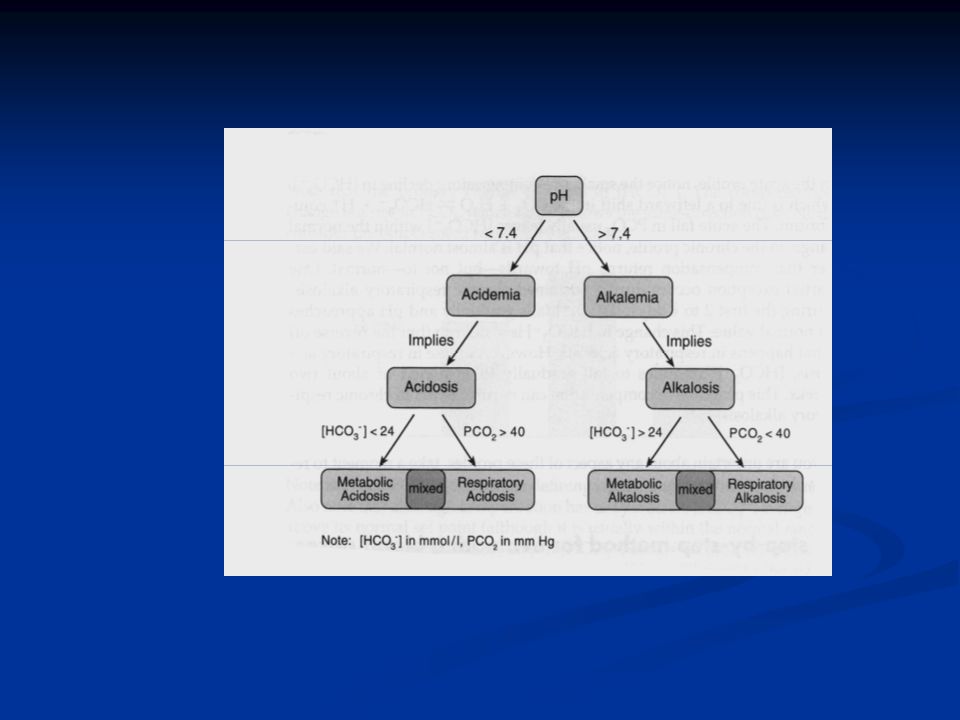
Method for Evaluating Blood Gasses Step 1: Assess the pH. Low pH ( 7.45) is alkalemia. Step 2: Determine the cause of the alkalemia or acidemia. Acidemia can only be caused by acidosis, and therefore implies the presence of an acidosis. Similarly, alkalemia can only be caused by alkalosis, and therefore implies the presence of an alkalosis. Step 3: Look at the change from normal values of the conjugate acid and conjugate base in your buffer (pCO 2 and HCO 3 - in the case of the bicarbonate buffering system) and determine which change can explain the change in pH.
 is alkalemia. Step 2: Determine the cause of the alkalemia or acidemia. Acidemia can only be caused by acidosis, and therefore implies the presence of an acidosis. Similarly, alkalemia can only be caused by alkalosis, and therefore implies the presence of an alkalosis. Step 3: Look at the change from normal values of the conjugate acid and conjugate base in your buffer (pCO 2 and HCO 3 - in the case of the bicarbonate buffering system) and determine which change can explain the change in pH..")
15
Method for Evaluating Blood Gasses Lets say pH is low, so you have an acidemia: An acidemia may either be respiratory or metabolic (remember, HCO 3 - is the conjugate base, and pCO 2 is the conjugate acid in this buffering pair). Therefore, either a DECLINE IN HCO 3 - or AN INCREASE IN PCO 2 can explain the presence of an acidemia. 1. 1. If a high PCO 2 is the primary disturbance, it’s a respiratory acidemia 2. 2. If a low HCO 3 - is the primary disturbance, it’s a metabolic acidemia WHAT IF BOTH A LOW HCO 3 - AND A HIGH PCO 2 - CAN ACCOUNT FOR THE ACIDEMIA THAT IS PRESENT? = mixed acid/base disorder

16
Method for Evaluating Blood Gasses Lets say pH is high, and you have an alkalemia: An alkalemia may either be respiratory or metabolic. 1. 1. Therefore, either a DECLINE IN PCO 2 - or AN INCREASE IN HCO 3 - can produce an alkalemia. 2. 2. If a low PCO 2 can explain the alkalemia, it’s a respiratory alkalemia. 3. 3. If high HCO 3 - can explain the alkalemia, it’s a metabolic alkalemia WHAT IF BOTH A HIGH HCO 3 - AND A LOW PCO 2 - CAN ACCOUNT FOR THE ALKALEMIA?

18
Compensation For Simple Acid Base Disorders If the primary disturbance is in plasma HCO 3 - (i.e., a metabolic disorder), then the compensator will be PCO 2. If the primary disturbance is in PCO 2 (i.e., a respiratory disorder), then the compensator will be HCO 3 -. The compensation always occurs in the same direction as the primary disturbance.
, then the compensator will be HCO 3 -. The compensation always occurs in the same direction as the primary disturbance..")
19
Compensation Rules of Thumb

20
A Few Examples…..

Similar presentations


 Blood Gas Analysis Dr. Prakash Mohanasundaram Department of Emergency & Critical Care medicine Vinayaka Missions University.>")


![Renal Acid-Base Balance. Acid An acid is when hydrogen ions accumulate in a solution. It becomes more acidic [H+] increases = more acidity CO 2 is an.](/20/5962628/big_thumb.jpg "Renal Acid-Base Balance. Acid An acid is when hydrogen ions accumulate in a solution. It becomes more acidic [H+] increases = more acidity CO 2 is an.>")
 concentration. More H + = more acidic = lower.>")
![Acid-Base balance Prof. Jan Hanacek. pH and Hydrogen ion concentration pH [H+] nanomol/l 6.0 1000 7.0 100 8.0 10 9.0 1.](/21/6241563/big_thumb.jpg "Acid-Base balance Prof. Jan Hanacek. pH and Hydrogen ion concentration pH [H+] nanomol/l 6.0 1000 7.0 100 8.0 10 9.0 1.>")

