Download presentation
Presentation is loading. Please wait.

1
جوینده دانش در کنف عنایت خداوند است. پيامبر اكرم (ص) می فرمایند : بسم الله الرحمن الرحیم
 می فرمایند : بسم الله الرحمن الرحیم")
2
Computer Tomography Technique September, 2012 Prepared by: Behzad Ommani Master of Medical Engineering Instructor Radiology Group

3
In routine abdominal scanning patients are given oral contrast to opacify the bowel and allow differentiation from adjacent pathological. lesions. In most instances patients are given 800 ml of positive oral contrast, either a dilute barium suspension, or a 3% solution of gastrografin (sodium/meglumine diatrizoate. Schering) or similar water-soluble contrast which may be flavoured with fruit squash. The contrast is given 30-45 minutes before the scan to opacify the small bowel and a further 200 ml of the same contrast is given immediately before the scan to opacify the stomach and proximal small bowel. Oral Contrast
 or similar water-soluble contrast which may be flavoured with fruit squash. The contrast is given minutes before the scan to opacify the small bowel and a further 200 ml of the same contrast is given immediately before the scan to opacify the stomach and proximal small bowel. Oral Contrast.")
4
To opacify the colon two doses of contrast are given: 800 ml of oral contrast the night before the scan to fill the colon and a second 800 ml to opacify the small bowel, given as usual before the scan. Alternatively, rectal contrast (using 200 ml of 6% water- soluble contrast) can be given immediately before the scan to opacify the rectum and sigmoid. Oral Contrast
 can be given immediately before the scan to opacify the rectum and sigmoid. Oral Contrast.")
5
Intravenous contrast may be used to opacify the vessels and aid their differentiation from retroperitoneal nodes. The timing of the administration of the contrast may be critical particularly with the modern spiral or helical CT scanners. Intra venous Contrast

6
Patient position : Supine, Both arms should be raised above the head. This is the planning scan and is usually an anteroposterior (AP) view. Start position : Dome of Diaphragm End position : Symphysis pubis Abdomen & Pelvic Protocl
 view. Start position : Dome of Diaphragm End position : Symphysis pubis Abdomen & Pelvic Protocl.")
7
Slice thickness 10 mm Table increment 10 mm Kilovoltage 120 kV mAs per slice 200 mAs Algorithm standard Scan field of view 35 - 48 cm Display field of view 35 - 48 cm Window width 450 / 500 Window level 50 Abdomen & Pelvic Protocl

9
The normal gastric wall is 3-5 mm thick. In order to assess gastric wall thickening, which occurs with gastric cancer and lymphoma, the stomach must be fully distended. Stomach Stomach wall well visualised when water is used as negative contrast a, water filled gastric lumen; b, gastric wall

10
1. To adequately distend the stomach, 20 mg of Buscopan (scopolamine butylbromide) is given intramuscularly 5 minutes before the scan, assuming there are no contraindications. The Buscopan decreases the artefacts from peristalsis and delays gastric emptying. 2. Immediately prior the scan, 400 ml of water is drunk to distend the stomach. 3. Intravenous contrast, 100 ml is given at a rate of 3 ml/sec. 4. Scanning is commenced 40 sec after the administration of contrast has begun Stomach
 is given intramuscularly 5 minutes before the scan, assuming there are no contraindications. The Buscopan decreases the artefacts from peristalsis and delays gastric emptying. 2. Immediately prior the scan, 400 ml of water is drunk to distend the stomach. 3. Intravenous contrast, 100 ml is given at a rate of 3 ml/sec. 4. Scanning is commenced 40 sec after the administration of contrast has begun Stomach.")
11
Normal attenuation values: Pancreases Noncontrast: 30 - 40 HU Postcontrast: 100 - 150 HU To optimise the visualisation of pancreatic calcification, negative bowel contrast (water) may be advantageous. in up to 75% of patients if thin section 3-5 mm collimation is used, and especially if intravenous contrast is given as the normal pancreas enhances, accentuating the difference between the gland and low attenuation duct. Pancreases

12
To optimise this enhancement, the delay between the onset of the injection and the scanning depends whether a conventional scanner (30-40 second delay) or a spiral scanner (45-60 second delay) is used.
 or a spiral scanner (45-60 second delay) is used.")
13
The splenic vessels are seen well without contrast, but for optimal visualisation of the splenic parenchyma intravenous contrast should be administered, particularly if focal masses, such as lymphomatous deposits are suspected. However, with bolus injections and early scanning, the spleen is non homogeneous due to blood flow in different compartments. Care must be taken not to misinterpret this as focal abnormality and rescanning at 60 seconds will resolve the issue. Spleen

14
Early scan through the spleen showing non-homogeneous enhancement (a) b, head of pancreas; (Note that the pancreas here is smooth in outline.) c, left adrenal; d, right adrenal Spleen
 b, head of pancreas; (Note that the pancreas here is smooth in outline.) c, left adrenal; d, right adrenal Spleen")
15
CT and, increasingly, magnetic resonance imaging (MRI) are the investigations of choice for adrenal pathology. Start position : Above 12 Thoracic vertebral End position : mid kidney or lower edge of 2 lumbar vertebral Adernal

16
If the patient is biochemically normal, with no known malignancy, a lesion less than 3 cm is likely to be an adenoma. Even in the presence of known malignancy, if the mass is small with a CT number of less than 18 HU on an unenhanced scan it will be an adenoma. Phaeochromocytomas are relatively rare tumours which originate from chromaffin cells in the adrenal medulla and which secrete the catecholamines adrenaline and noradrenaline. Adernal

17
Patient with myelolipoma containing fat (a). b, right adrenal; c, lobulated pancreas; d, Unopacified blood in the IVC, simulating thrombus in this early phase scan Adernal

18
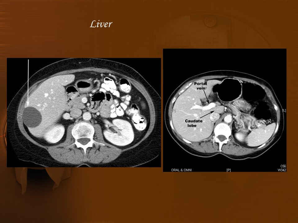
Normal attenuation values: Liver Noncontrast: 38 - 80 HU Start position : Dome Diaphragm End position : lower edge of 3 lumbar vertebral Liver

20
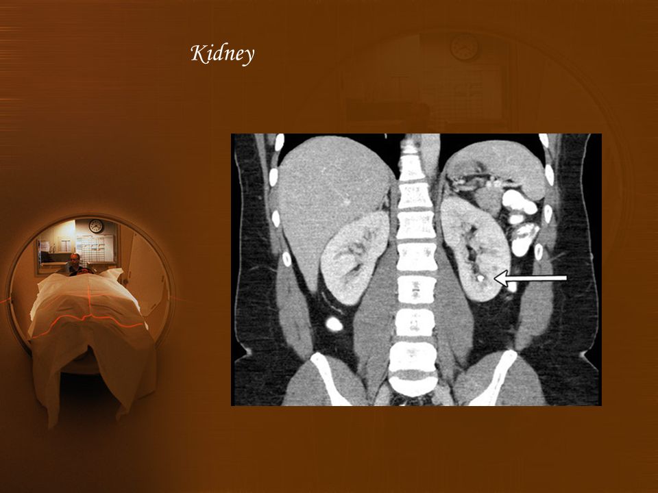
The kidneys and adrenal glands are surrounded by an envelope of fat and lie within the renal fascia. The perirenal space contains the kidneys, adrenals, renal vessels and proximal ureters and surrounding fat. Indications : 1. Assessment of renal masses and differentiation of solid and cystic lesions. 2. Tumour staging and surgical planning. 3. Renal trauma. 4. Non-invasive diagnosis of renal artery stenosis. 5. Renal colic. Kidney

21
Normal attenuation values: Cortex Kidney Noncontrast: 30 - 60 HU With contrast: 80 - 120 HU Start position : Dome Diaphragm End position : Symphysis pubis Kidney

22
1.Precontrast scans for the position of the kidneys are made, to identify calcification and to obtain accurate precontrast attenuation values of masses. 2. Contrast: 90-120 ml of 300 mg/ml injected at 3 ml/s. 3. Post contrast scans if using incremental dynamic scanning, 5 mm collimation with a 40 second delay. 4. Spiral scanner If this is used, the slice collimation depends on the patient's ability to hold their breath. Kidney

24
Patient position : Supine, Both arms should be raised above the head. This is the planning scan and is usually an anteroposterior (AP) view. Start position :Crest iliac End position : symphysis pubis or Ischial Tuberosity Pelvic
 view. Start position :Crest iliac End position : symphysis pubis or Ischial Tuberosity Pelvic.")
26
Slice thickness 10 mm Table increment 10 mm Kilovoltage 120 kV mAs per slice 300 - 480 mAs Algorithm standard Scan field of view 30 - 48 cm Display field of view 30 - 48 cm Window width 450 / 500 Window level 50 Window width 1500 / 2500 Window level 500 Pelvic Protocol

28
Femor Ante version

30
Tibia Torsion

Similar presentations


 می فرمایند : بسم الله الرحمن الرحیم.>")


 Dynamic scanning implies 15 or more scans in rapid sequence within one.>")

