Download presentation
Presentation is loading. Please wait.

2
In the name of GOD

3
Abdominal Trauma & hollow viscous injury
EVALUATION AND INDICATIONS FOR CELIOTOMY

5
Abdominal injuries Solid organ injuries Hollow viscous injuries

6
Clinical findings Abdominal pain Guarding

7
Hollow viscous injuries
Delay diagnosis (8-12 h) Hemorrhage Peritonitis Abdominal sepsis
 Hemorrhage. Peritonitis. Abdominal sepsis.")
8
Basic mechanisms of bowel and mesenteric injuries
Shearing injuries caused by deceleration Crush injuries from direct impact Burst injuries from sudden increases in intraluminal pressure

9
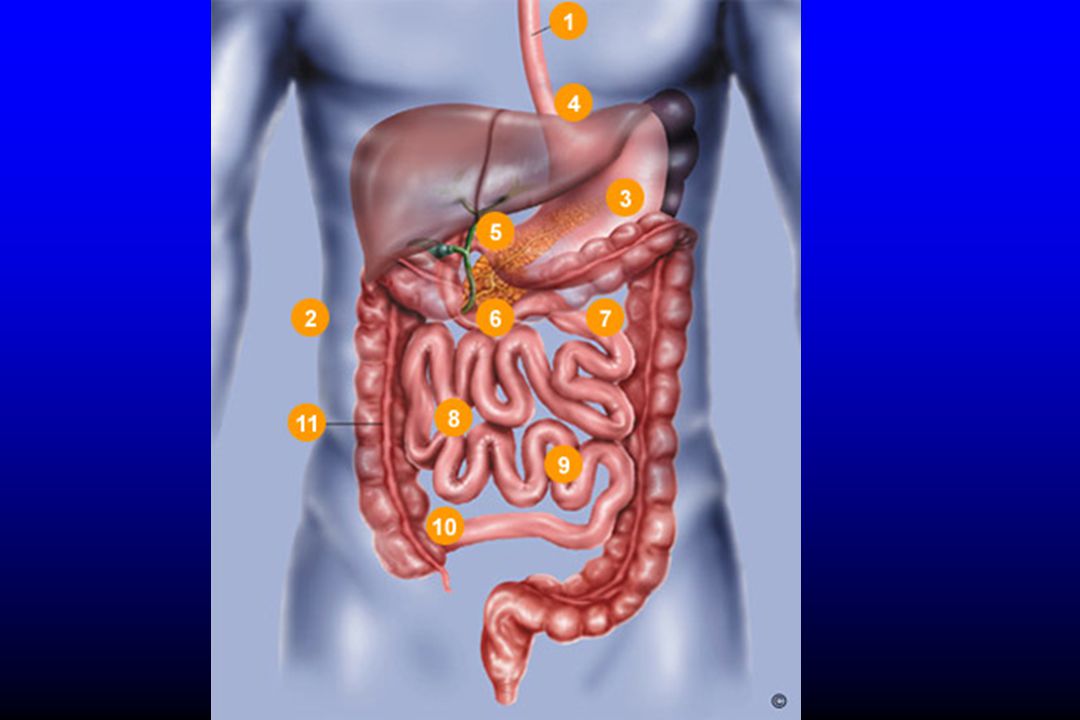
Site of injuries Small bowel Colon Duodenum Stomach

10
Imaging modalities Plain radiography Sonography CT scan

11
Plain radiography Chest X ray Abdominal radiography (supine & upright)
")
12
Pneumoperitoan

13
Pneumoperitoan

14
Pneumoperitoan

15
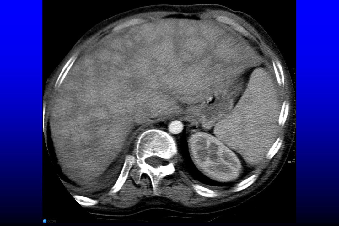
Peritoneal fluid

16
Ultrasound examination
Free fluid Pneumoperitoneum Air in bowel wall Wall thickening of bowel loops

17
Pneumoperitoan Sagital sonographic section of the right hypochondrium using a curvilinear probe showing enhanced peritoneal stripe (empty arrow) and reverberation artefacts (small arrows) which partially obscure the right lobe of the liver (L) and right kidney (K). Laparotomy confirmed that the patient had perforated diverticulitis
 and reverberation artefacts (small arrows) which partially obscure the right lobe of the liver (L) and right kidney (K). Laparotomy confirmed that the patient had perforated diverticulitis.")
18
Pneumoperitoan Transverse sonographic section of the right hypochondrium using a linear probe showing a hyperdence echogenic small area (arrow head) moving within a fluid collection. Laparotomy confirmed that the patient had a perforated duodenal ulcer
 moving within a fluid collection. Laparotomy confirmed that the patient had a perforated duodenal ulcer.")
19
Air in Morrison’s pouch
Sagital sonographic section of the right hypochondrium using a curvilinear probe showing a hyperdence interrupted echogenic lines under the liver in Morrison's pouch (arrow head), fluid collection (white arrow), and a hyperdense echogenic line in the anterior wall of the duodenum representing the scar of a duodenal ulcer (black arrow). Laparotomy confirmed that the patient had a perforated duodenal ulcer
, fluid collection (white arrow), and a hyperdense echogenic line in the anterior wall of the duodenum representing the scar of a duodenal ulcer (black arrow). Laparotomy confirmed that the patient had a perforated duodenal ulcer.")
20
Bowel loop hematoma

21
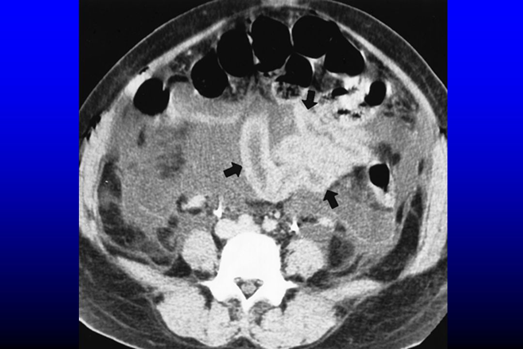
Bowel wall thickening, air bubble in its wall

22
CT Technique IV contrast (100-120 ml)
Portal venous phase (70 second delay) Delay film (7 minute) Oral contrast +/_ Rectal contrast +/_
 Delay film (7 minute) Oral contrast +/_. Rectal contrast +/_.")
23
CT signs of bowel loops injury
Wall transection with focal discontinuity (spe:100%& sen:7%) Extraluminal oral contrast Pneumoperitoneum (20-75%) Pneumoretroperitoneum Focal wall thickening Abnormal wall enhancement Ill defined increased attenuation of mesentry Intra peritoneal fluid
 Extraluminal oral contrast. Pneumoperitoneum (20-75%) Pneumoretroperitoneum. Focal wall thickening. Abnormal wall enhancement. Ill defined increased attenuation of mesentry. Intra peritoneal fluid.")
24
Hemoperitoan

25
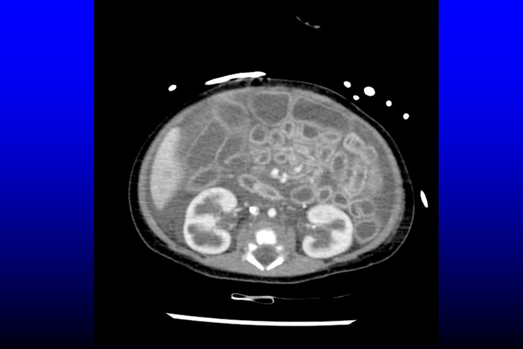
focal segment of thickened jejunum associated stranding of the small bowel mesentery

26
segment of thickened jejunum and hemoperitoneum

27
wall thickening in a segment of jejunum

28
segment of thickened and poorly enhancing small bowel

29
Mesenteric fat stranding

30
Other causes of pneumoperitoneum
Bladder rupture with an indwelling Foley catheter Massive pneumothorax Barotrauma Benign pneumoperitoneum Peritoneal lavage Pseudopneumoperitoneum (air between abdominal wall and parietal peritoneum)
")
31
Pneumoperitoneum and pseudopneumoperitoneum

32
Diffuse bowel wall thickening
Fluid over load Liver inhomogeneous enhancement(nutmeg appearance) Periportal edema Hypoperfusion complex(shock bowel) Flat IVC Increased enhancement of adrenal gland Retroperitoneal edema
 Periportal edema. Hypoperfusion complex(shock bowel) Flat IVC. Increased enhancement of adrenal gland. Retroperitoneal edema.")
35
Diffuse thickening and hyperenhancement of the loops due to aggressive resuscitation with intravenous fluids

39
Duodenal injuries More secondary to penetrating injuries and less likely due to blunt trauma CT findings: wall thickening, discontinuity, contrast extravasation, fluid adjacent to the duodenum and pancreatic head and retroperitoneum air or fluid

40
Duodenal wall thickening and extensive hemoperitoneum

41
Colonic injuries Wall transection with focal discontinuity
Contrast extravasation Pneumoperitoneum Pneumoretroperitoneum Focal wall thickening Abnormal wall enhancement Ill defined increased attenuation of mesentry Intra peritoneal fluid

42
Transverse colonic wall thickening

43
focal segment of ascending colonic wall thickening,worsened pericolonic fat stranding in delayfilm

44
Sign mesenteric trauma
Mesenteric hematoma Intraperitoneal extravasation of intravenous contrast Abrupt termination of mesenteric vessels Unequivocal irregularity of the wall of mesenteric vessels Increased attenuation of the mesentery

45
small focal hematoma in the root of the mesentery

46
Mesenteric hematoma

47
Mesenteric hematoma

48
Mesenteric bleeding

49
Focal collection of high attenuation fluid is seen in the root of the mesentery

50
abnormally positioned in the right hemiabdomen , with subtle stranding of the corresponding mesentery due to traumatic internal hernia

51
Anorectal injury Mortality rate three times more than colonic injury
Associated with pelvic fracture concomitant with bladder, urethral and vascular injuries Divided into intraperitoneal and extraperitoneal

52
Extraluminal air is seen in the presacral space

53
rectal wall tear with retroperitoneum and pseudopneumoperitoneum

54
Injury of mesentery and mesenteric vessels
Extravasation of IV contrast Mesentric hematoma Mesentric infiltration Beading or abrupt termination of mesentric vessels Mesentric rent with internal hernia

55
Mesenteric hematoma

56
Mesenteric tear

57
Causes of retroperitoneal air
Colonic perforation (ascending and descending) Duodenal injuries Pneumothorax Pneumomediastinum
 Duodenal injuries. Pneumothorax. Pneumomediastinum.")
58
Pneumoperitoneum and pseudopneumoperitoneum

59
Free peritoneal fluid Most common finding (most sensitive)
Absence of free fluid excludes surgical important injury The attenuation is highest in the vicinity of the injured organ (sentinel clot) Localized fluid (triangle sign) Attenuation of hemoperitoan is high (>30-40) , simple fluid H.U is about 13
 Localized fluid (triangle sign) Attenuation of hemoperitoan is high (>30-40) , simple fluid H.U is about 13.")
60
Lesser sac hematoma

61
Sentinel clot sign

62
Sentinel clot sign

63
hemoperitoneum (mean attenuation, 39 HU) due to small hepatic laceration
 due to small hepatic laceration")
64
A small amount free pelvic fluid with mean attenuation of the 8 HU

65
Thank you

Similar presentations