Download presentation
Presentation is loading. Please wait.

1
Pelvis, Hip, and Thigh Conditions
Chapter 17

2
Skeletal Features of Pelvis, Hip, and Thigh

3
Pelvis Function Protects organs
Transmits loads between trunk and lower extremity Provides site for muscle attachments 4 fused bones Sacrum Coccyx Innominate bones Ilium, ischium, and pubis

4
Pelvis (cont.) SI joint Critical link between the two pelvic bones
Strong ligamentous support Sacrococcygeal joint Fused line symphysis united by a fibrocartilaginous disc Pubic symphysis Interpubic disc located between the two joint surfaces

5
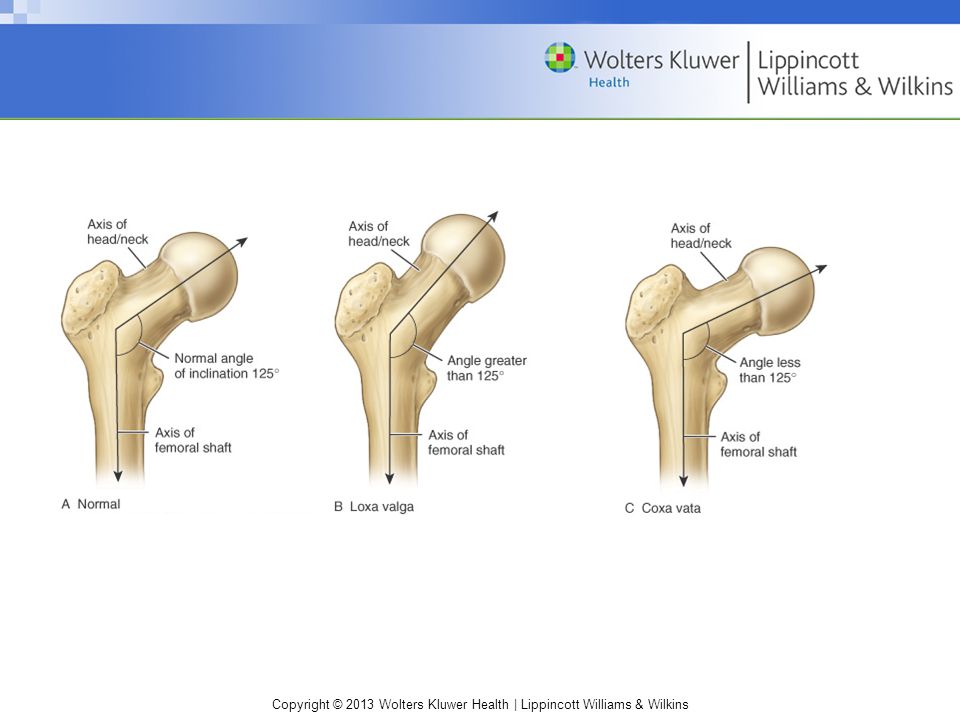
Bony Structure of Thigh
Femur Weakest at femoral neck Angle of inclination Angle of depression formed by a line drawn through the shaft of femur and a line passing through the long axis of femoral neck Approximately 125 in the frontal plane 125 coxa valga 125 coxa vara

7
Bony Structure of Thigh (cont.)
Femur Angle of torsion Relationship between femoral head and femoral shaft in transverse plane Approximately 12 12 anteversion 12 retroversion

9
Hip Joint Head of femur and acetabulum of pelvis Ball and socket joint
Very stable

10
Hip Joint Capsule Completely surrounds joint, attaching to the labrum of the acetabular socket Passes over a fat pad internally to join to the distal aspect of femoral neck Zona orbicularis

11
Ligaments of Hip Joint Iliofemoral ligament Limits hyperextension
Pubofemoral ligament Limits abduction and hyperextension Ischiofemoral ligament Limits extension

12
Femoral Triangle Borders Inguinal ligament— superior Sartorius—lateral
Adductor longus—medial Contents Femoral nerves Femoral artery Femoral vein

13
Bursae Iliopsoas Reduces friction between iliopsoas and articular capsule Deep trochanteric bursa Provides cushion between greater trochanter and gluteus maximus at its attachment to iliotibial tract Gluteofemoral bursa Separates gluteus maximus from origin of vastus lateralis Ischial bursa Weight-bearing structure during sitting Cushions ischial tuberosity where it passes over gluteus maximus

14
Q-Angle Angle between line of resultant force produced by quadriceps and line of patellar tendon Males 13°; females 18°

15
Muscles

16
Muscles (cont.)
")
17
Muscles (cont.)
")
18
Nerves Lumbar plexus Femoral nerve Obturator nerve Sacral plexus
Sciatic nerve

19
Blood Vessels External iliac Femoral Deep femoral Femoral circumflex

20
Kinematics Pelvis positioning
Pelvis “tilts” to facilitate movement in hip Posterior tilt—assists hip flexion Anterior tilt—assists hip extension Lateral tilt—assists hip abduction

21
Kinematics (cont.) Hip flexors
Iliopsoas, pectineus, rectus femoris, sartorius, and tensor fascia latae Two-joint muscles Rectus femoris—active during hip flexion and knee extension Sartorius—active during hip flexion and knee extension Hip extensors Gluteus maximus and hamstrings (biceps femoris, semitendinosus, and semimembranosus) Hamstrings—two-joint; hip extension and knee flexion
 Hamstrings—two-joint; hip extension and knee flexion.")
22
Kinematics (cont.) Hip abductors Gluteus medius, gluteus minimus
Active in stabilizing pelvis during single-leg support and during support phase of walking and running Hip adductors Adductor longus, adductor brevis, and adductor magnus

23
Kinematics (cont.) Lateral rotators
Piriformis, gemellus superior, gemellus inferior, obturator internus, obturator externus, and quadratus femoris Lateral rotation of femur of swinging leg accommodates lateral rotation of pelvis during stride Medial rotators Gluteus minimus Tensor fascia latae, semitendinosus, semimembranosus, gluteus medius, and adductors

24
Kinetics Body weight places compression on hip, as does tension in hip muscles Forces are less during standing than with running and walking Forces translated through the lower extremity; result ↑ compression on hip

25
Prevention Protective equipment
Hip joint well protected but iliac and pelvis need protection Thigh Physical conditioning Shoes Cushion forces

26
Contusions Hip pointer Mechanism: direct blow to iliac crest
Common—anterior or lateral portion of crest Often from improperly fitting (or absent) hip pads
 hip pads.")
27
Contusions (cont.) S&S Point tenderness; swelling; ecchymosis
Individual prefers slightly forward flexed position to relieve tension of abdominals and iliopsoas Antalgic gait with shortened swing phase ↑ pain with active trunk flexion and active hip flexion Pain with coughing, laughing, breathing Abdominal muscle spasm Management: standard acute; rest; protect with hard-shell pad for return to activity

28
Contusions (cont.) Quadriceps contusion Mechanism: direct blow
Common – anterolateral thigh S&S Transitory loss of function With continued play, progressively stiffer and unresponsive ↑ pain with active knee extension and hip flexion Limited AROM due to pain; knee flexion limited actively and passively

29
Contusions (cont.) Management:
Standard acute; with knee in maximum flexion Hard-shell pad for return to activity Physician referral if myositis ossificans or compartment syndrome is suspected

31
Contusions (cont.) Myositis ossificans
Develops secondary to single significant blow or repetitive blows to same area Evident on radiograph 3–4 weeks after injury

32
Contusions (cont.) S&S Warm, firm, swollen thigh; 2–4 cm larger
Palpable, painful mass may limit passive knee flexion to 20–30° Active quadriceps contractions and straight leg raises— difficult Management: standard acute; physician referral Self-limiting injury Maturation—6–12 months

33
Contusions (cont.) Compartment syndrome Neurovascular compression
Due to uncontrolled internal bleeding and swelling S&S Progressive, severe pain with passive motion and isometric contraction of quadriceps pressure → ↓ femoral sensation and motor weakness; distal pulse and capillary refill may be normal Management: ice (no compression); immediate physician referral
; immediate physician referral.")
34
Bursitis Mechanism Excessive friction or shear forces due to overuse
Posttraumatic bursitis from direct blows that cause bleeding in the bursa Greater trochanteric bursitis Influence of Q-angle

35
Bursitis (cont.) S&S Burning or aching over or posterior to greater trochanter Aggravated with: Hip abduction against resistance Hip flexion and extension on weight bearing Referred pain—lateral aspect of the thigh

36
Bursitis (cont.) Iliopsoas bursitis
Pain medial and anterior to joint; cannot be easily palpated pain with passive hip rotation; resisted hip flexion, abduction, and external rotation Ischial bursitis Pain aggravated by prolonged sitting and uphill running, Point tenderness directly over ischial tuberosity pain with passive and resisted hip extension

37
Bursitis (cont.) Bursitis management
Standard acute; deep friction massage; NSAIDs; stretching program for involved muscle On-going prevention: biomechanical analysis; technique analysis

38
Bursitis (cont.) Snapping hip syndrome
Causes: intra- and extra-articular (refer to Box 15.2) Types External—IT band or gluteus maximus snapping over greater trochanter during hip flexion → trochanteric bursitis Internal—iliopsoas snaps over structures deep to musculotendinous unit (e.g., iliopsoas bursa) Intra-articular—lesions of the joint (e.g., labral tear)
 Types. External—IT band or gluteus maximus snapping over greater trochanter during hip flexion → trochanteric bursitis. Internal—iliopsoas snaps over structures deep to musculotendinous unit (e.g., iliopsoas bursa) Intra-articular—lesions of the joint (e.g., labral tear)")
39
Bursitis (cont.) S&S Snapping sensation heard or felt during hip motion, especially with lateral rotation and flexion while balancing on one leg Iliopsoas bursa affected—snapping in medial groin Management: NSAIDs; rehabilitation program to address specific deficits

40
Hip Sprains and Dislocations
Mechanism Violent twisting actions With hip and knee flexed to 90°, force through shaft of femur

41
Hip Sprains and Dislocations (cont.)
S&S Mild/moderate: pain with internal rotation Severe: intense pain; inability to move hip Position of flexion and internal rotation Management Mild/moderate—standard acute Severe—activate EMS; immobilize in position found; assess distal vascular integrity; monitor and treat for shock; NPO

42
Hip Dislocation

43
Strains Mechanism Explosive movements
Tensile stress from overstretching Muscles Quadriceps Typically rectus femoris

44
Strains (cont.) Hamstrings
Initial swing—flex knee; late swing—eccentrically contract to decelerate knee extension and re-extend hip in prep for stance phase Overemphasis on stretching without strengthening Strength imbalance Adductors Common with quick change of direction and explosive propulsion and acceleration

45
Strains (cont.) S&S Point tender with palpable spasm
Possible palpable defect/divot Ecchymosis may or may not be present Pain with AROM; pain with PROM (muscles placed on stretch)
")
46
Strains (cont.) Piriformis strain
In some individuals, sciatic nerve passes through or above piriformis, subjecting nerve to compression from trauma, hemorrhage, or spasm

47
Strains (cont.) S&S History of prolonged sitting, overuse, recent ↑ in activity, or buttock trauma Dull ache in midbuttock—worse at night Numbness or weakness may extend down posterior leg ↑ pain or weakness during: Passive hip flexion, adduction, and internal rotation Active hip external rotation Resisted hip external rotation

48
Strains (cont.) Predisposing factors
Beginning of season – too much too soon Fatigue History of strains; reinjury common Restricted flexibility of involved muscle group Management: standard acute; restrict weight bearing if unable to assume normal gait

49
Vascular and Neural Disorders
Legg-Calvé-Perthes disease Avascular necrosis of proximal femoral epiphysis Seen esp in males ages 3–8 Osteochondrosis - femoral head S&S Gradual onset of limp and mild hip or knee pain of several months in duration Pain -activity related ROM in hip abduction, extension, and external rotation due to spasm in hip flexors and adductors

50
Vascular and Neural Disorders (cont.)
Venous disorders Direct blow may damage a vein causing Thrombophlebitis Superficial thrombophlebitis (ST) Deep venous thrombosis (DVT) Phlebothrombosis
 Deep venous thrombosis (DVT) Phlebothrombosis.")
51
Vascular and Neural Disorders (cont.)
S&S ST—acute, red, hot, palpable, tender cord in course of a superficial vein Extension of ST to deep veins—via proximal long and short saphenous veins to common femoral and popliteal veins, respectively Management: anticoagulant therapy; external support (e.g., compression stockings); therapeutic exercise
; therapeutic exercise.")
52
Vascular and Neural Disorders (cont.)
Toxic synovitis of hip Transient inflammatory condition Painful hip joint with an antalgic gait Management: physician referral Obturator nerve entrapment Possible causes: pelvic tumors, obturator hernias, or pelvic and proximal femoral fractures S&S: exercise-induced medial thigh pain; described as vague groin or medial knee pain

53
Hip Fractures Avulsion fractures Apophyseal sites
ASIS with displacement of sartorius AIIS with rectus femoris displacement Ischial tuberosity with hamstrings displacement Lesser trochanter with iliopsoas displacement Due to rapid, sudden acceleration and deceleration

54
Hip Fractures (cont.) S&S
Sudden, acute, localized pain—may radiate down muscle Swelling and discoloration Palpable gap between tendon attachment and bone pain with AROM, PROM, RROM of involved muscle Management: immobilize with elastic bandage; fit with crutches; immediate physician referral

55
Hip Fractures (cont.) Slipped capital femoral epiphysis
Boys ages 12–15 Femoral head slips at epiphyseal plate— displaces inferiorly and posteriorly relative to femoral neck

56
Hip Fractures (cont.) S&S Early stages—diffuse knee pain Later stages
More comfortable holding leg in slight flexion Unable to touch abdomen with thigh because hip externally rotates with flexion Unable to rotate femur internally or stand on one leg Management: fit with crutches; physician referral

57
Hip Fractures (cont.) Stress fractures
Pubis, femoral neck, and proximal one-third of femur Risk factors

58
Hip Fractures (cont.) S&S
Diffuse or localized aching pain in anterior groin or thigh during weight-bearing activity, relieved with rest Night pain Antalgic gait may be present Pain with deep palpation in inguinal ↑ pain on extremes of hip rotation + Trendelenburg sign Management: physician referral

59
Hip Fractures (cont.) Osteitis pubis
Continued stress on pubic symphysis From repeated overload of the adductor muscles From repetitive running activities S&S Gradual onset of pain in the adductor musculature, aggravated by kicking, running, and pivoting on one leg pain with sit-ups and abdominal strengthening exercises Pain may radiate distally into groin or medial thigh Management: standard acute—treat symptoms

60
Sacral and Coccygeal Fractures
Rare in sports Direct blow to area due to fall on buttock S&S: extremely painful; unable to sit Management: immediate referral to a physician

61
Femoral Fractures Mechanism Tremendous impact forces
Direct compressive forces Potential for neurovascular damage

62
Femoral Fractures (cont.)
S&S Previous history of femoral stress fracture ↑ risk of complete fracture Extreme pain and inability/unwillingness to move involved side Shock Neck Individual supine, lower extremity in external rotation and abduction; appears shortened compared with other side Shaft Limb appears shortened; thigh appears externally rotated

63
Femoral Fractures (cont.)
Management Activate EMS Assess distal vascular integrity Monitor and treat for shock Defer immobilization until emergency medical personnel arrive (traction splint will typically be applied) NPO—possible surgical intervention
 NPO—possible surgical intervention.")
64
Assessment History Observation/inspection Contranutation and nutation
Palpation Physical examination tests

65
Observation Contranutation at the SI joint
Indicates anterior torsion of joint, or posterior rotation of sacrum on ilium on one side Nutation Backward rotation of ilium on sacrum

66
Range of Motion (ROM) Active range of motion (AROM) Hip Flexion
Extension Abduction Adduction Lateral rotation Medial rotation

67
ROM (cont.) Knee Flexion Extension
 Knee Flexion Extension")
68
ROM (cont.) Normal ranges Hip flexion (110–120°) with knee flexed
Hip extension (10–15°) Abduction (30–50°) Adduction (30°) Lateral rotation (40–60°) Medial rotation (30–40°) Knee flexion (0–135°) Knee extension (0–15°)
 Abduction (30–50°) Adduction (30°) Lateral rotation (40–60°) Medial rotation (30–40°) Knee flexion (0–135°) Knee extension (0–15°)")
69
ROM (cont.)
")
70
ROM (cont.) Passive range of motion (PROM) Normal end feel
Hip flexion and adduction—tissue approximation Hip extension, abduction, and medial and lateral rotation—tissue stretch Passive movements at pelvic joint also stress the ligamentous structures Sacroiliac compression and distraction test

71
ROM (cont.) RROM
 RROM")
72
ROM (cont.)
")
73
ROM (cont.)
")
74
Stress Tests Sacroiliac compression and distraction test “Squish” test
Sacroiliac rocking test

75
Stress Tests Approximation test Patrick’s (FABER) test
 test")
76
Special Tests Leg length measurement Anatomic Apparent

77
Special Tests (cont.) Thomas Test for flexion contractures
 Thomas Test for flexion contractures")
78
Special Tests (cont.) Gaenslen’s test
 Gaenslen’s test")
79
Special Tests (cont.) Kendall test for rectus femoris contracture
Hamstring contracture test 90° – 90° straight leg raising test

80
Special Tests (cont.) Straight leg raising (Lasegue's) test
Trendelenburg test

81
Special Tests (cont.) Piriformis test Long sitting test Ober’s test
 Piriformis test Long sitting test Ober’s test")
82
Special Tests (cont.) Sign of the buttock test
 Sign of the buttock test")
83
Neurologic Tests Myotomes Hip flexion—L1, L2 Knee extension—L3
Ankle dorsiflexion—L4 Toe extension—L5 Ankle plantarflexion, foot eversion, or hip extension—S1 Knee flexion—S2 Reflexes No specific reflexes to test the pelvic or hip area Lower extremity reflexes Patella—L3, L4 Achilles tendon—S1

84
Neurologic Tests (cont.)
Dermatomes F16.35

85
Neurologic Tests (cont.)
Cutaneous patterns

86
Rehabilitation Restoration of motion
Refer to Field Strategies 16.1 and 17.1 Restoration of proprioception and balance Closed-chain exercises Muscular strength, endurance, and power Open-chain exercises PNF-resisted exercises Cardiovascular fitness

Similar presentations