Download presentation
Presentation is loading. Please wait.

1
Anatomy II THE THORAX BY: DR.Yahya Al-Farra

2
Respiratory system Nose Pharynx larynx trachea bronchi the lungs.

3
Thorax Thorax wall: Structure of thorax wall 1- sternum
2- ribs & intercotal spaces 3- vertebral column. Thorax cavity : Middle/ mediastenium :heart ,trachea,esophagus,big vesseles Laterally/ pleura , lungs

4
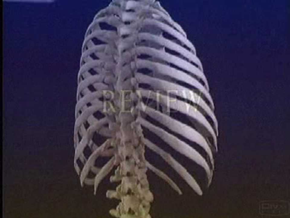
THE THORACIC WALL The thorax (or chest) is the region of the body between the neck and the abdomen . The thoracic cage : is formed by : the vertebral column behind, the ribs and intercostal spaces on either side, the sternum and costal cartilages in front

5
Superiorly: the thorax communicates with the neck
Inferiorly: it is separated from the abdomen by the diaphragm Function of thoracic cage: Protect lungs & heart and give attachment for Mm.

6
Structure of the Thoracic Wall
1- sternum. 2- ribs & intercotal spaces 3- vertebral column

8
:Sternum The sternum lies in the midline of the anterior chest wall.
It is a flat bone that can be divided into three parts: 1- manubrium sterni, 2- body of the sternum, 3- xiphoid process

9
1- The manubrium : is the upper part of the sternum. It articulates with the body of the sternum at the manubriosternal joint.

11
2- The body of the sternum:
articulates above with the manubrium at the manubriosternal joint and below with the xiphoid process at the xiphisternal joint Note: On each side it articulates with the second (lower part of 2nd rib )to the seventh costal cartilages .
to the seventh costal cartilages .")
13
No ribs or costal cartilages are attached to it .
3- The xiphoid process No ribs or costal cartilages are attached to it . angle of Louis) :) The sternal angle formed by the articulation of the manubrium with the body of the sternum. * The sternal angle is facing 2nd rib (ant.) * The sternal angle lies opposite the intervertebral disc between the fourth and fifth thoracic vertebrae .( Post.) * The xiphisternal joint lies opposite the body of the ninth(9th) thoracic vertebra .
 :) The sternal angle. formed by the articulation of the manubrium with the body of the sternum. * The sternal angle is facing 2nd rib (ant.) * The sternal angle lies opposite the intervertebral disc between the fourth and fifth thoracic vertebrae .( Post.) * The xiphisternal joint lies opposite the body of the ninth(9th) thoracic vertebra .")
15
The Ribs : There are 12 pairs of ribs, all of which are attached posteriorly to the thoracic vertebrae . The ribs are divided into three types : True ribs : The upper seven pairs are attached anteriorly to the sternum by their costal cartilages . False ribs : The 8th, 9th, and 10th pairs of ribs are attached anteriorly to each other and to the 7th rib . Floating ribs : The 11th and 12th pairs have no anterior attachment

18
The Intercostal Spaces :
The spaces between the ribs(rib+rib) contain three muscles of respiration: the external intercostal. the internal intercostal. the innermost intercostal muscle.__lies internally by parietal pleura The intercostal nerves and blood vessels VAN run betweenthe internal intercostal.(middle)& the innermost intercostal muscle(internal)
 contain three muscles of respiration: the external intercostal. the internal intercostal. the innermost intercostal muscle.__lies internally by parietal pleura. The intercostal nerves and blood vessels VAN run betweenthe internal intercostal.(middle)& the innermost intercostal muscle(internal)")
19
Increase in anteroposterior dimension by true ribs

20
Increase in lateral dimension by false ribs

21
Increase in superoinferior dimension by diapphragm

23
The Openings of the Thorax Chest cavity
Upper part (open with thoracic outlet) The chest cavity communicates with the root of the neck through an opening called the thoracic outlet.(important for B.V.) Opening is bounded by: Posteriorly: by the first thoracic vertebra,T1 Laterally:1st rib anteriorly :by the superior border of the manubrium sterni
 The chest cavity communicates with the root of the neck through an opening called the thoracic outlet.(important for B.V.) Opening is bounded by: Posteriorly: by the first thoracic vertebra,T1. Laterally:1st rib. anteriorly :by the superior border of the manubrium sterni.")
24
Lower part (closed by diaphragm)
The thoracic cavity communicates with the abdomen through a large opening which is closed by diaphragm. Opening is bounded by: posteriorly :by the 12th thoracic vertebre T12 laterally :by the curving costal margin anteriorly :by the xiphisternal joint.

26
The Diaphragm : The diaphragm is a thin muscular and tendinous septum LIKE DOME SHAPED(peripheral muscular part & and a centrally placed tendon ) that separates the chest cavity above from the abdominal cavity below . function of The Diaphragm : respiration
 that separates the chest cavity above from the abdominal cavity below . function of The Diaphragm : respiration.")
28
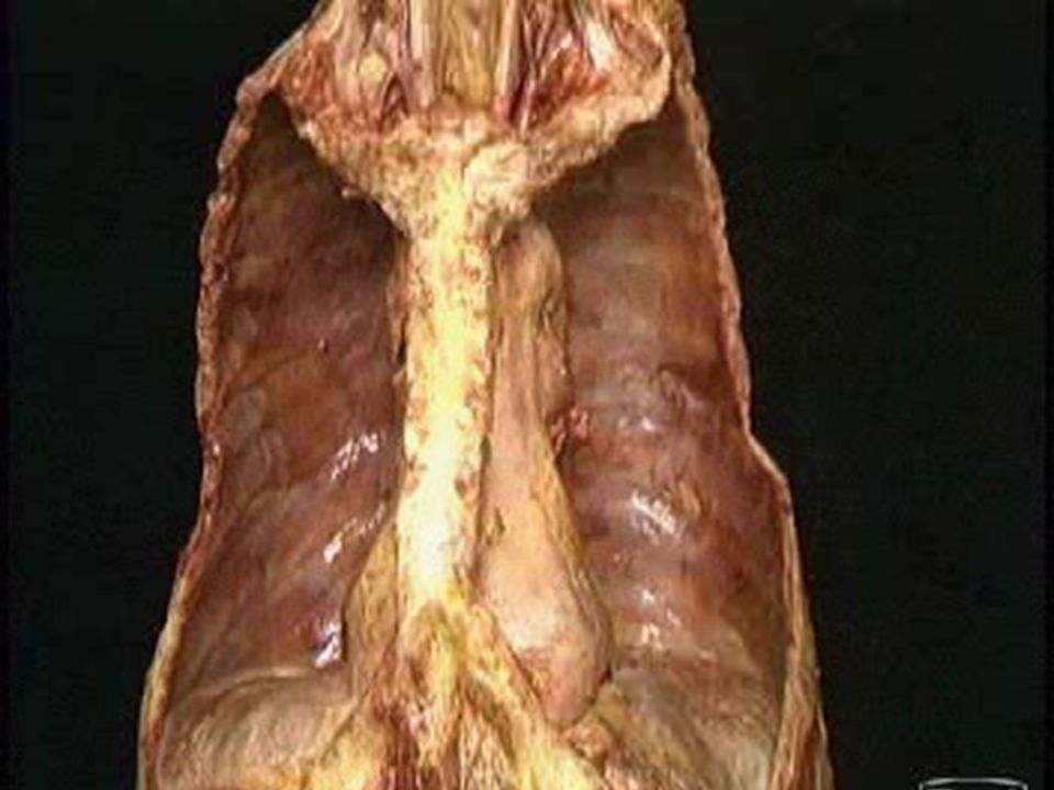
The Thoracic Cavity The thoracic cavity is bounded by the chest wall and below by the diaphragm The chest cavity can be divided into a median partition, called the mediastinum, and the laterally placed pleurae and lungs. Middle-mediastenium :(heart ,trachea,esophagus,big vesseles) laterally( pleura , lungs(
 laterally( pleura , lungs(")
29
Mediastinum The mediastinum is divided into:
superior and inferior mediastina by an imaginary plane passing from the sternal angle anteriorly to the lower border of the body of the fourth thoracic vertebra posteriorly The inferior mediastinum is subdivided into the middle mediastinum, the anterior mediastinum, the posterior mediastinum

30
Subdivision of mediastinum as seen on cross section.
anterior mediastinum (1) middle mediastinum (2) posterior mediastinum (3)
 middle mediastinum (2) posterior mediastinum (3)")
31
Subdivision of mediastinum as seen on sagittal section
superior mediastinum (1) anterior mediastinum (2) medial mediastinum (3) posterior mediastinum (4)
 anterior mediastinum (2) medial mediastinum (3) posterior mediastinum (4)")
34
Nose Nasal cavity is divided by nasal septum.
The lateral wall has three bony projections the chonchae. The nose is separated from the mouth by the palate, the anterior part is bony, posterior part is fleshy.

35
Larynx Is the voice box, below pharynx?
It is formed of nine cartilages, the largest are thyroid, cricoid, and the leaf like which cover the laryngeal opening, the epiglottis The vocal cords are ligaments covered by mucous membrane. The lower part of larynx and upper part of trachea.

36
Pharynx Is muscular passage 13 cm in length? It is divided into :
Nasopharynx , Oropharynx Laryngopharynx Auditory tube opens into naso pharynx. Tonsil is a cluster of lymphoid tissue. Pharyngeal tonsil is located high in nasopharynx called adenoids The palatine tonsils located in oropharynx, lingual tonsil at base of the tongue.

37
The Trachea The trachea is a mobile cartilaginous and membranous tube.
In adults the trachea is about 4 in. (11.25 cm) long and 1 in. (2.5 cm) in diameter It is continuation of the larynx at the lower border of the cricoid cartilage. The end of trachea is called the carina by dividing into right and left principal (main) bronchi at the level of the sternal angle.
 long and 1 in. (2.5 cm) in diameter. It is continuation of the larynx at the lower border of the cricoid cartilage. The end of trachea is called the carina by dividing into right and left principal (main) bronchi at the level of the sternal angle.")
39
- Wider , shorter - narrower,longer 1 in.(2.5cm) long - 2in.(5cm)long
Rt. Principle bronchus: Lt. bronchus : - Wider , shorter narrower,longer 1 in.(2.5cm) long in.(5cm)long 3 lobes lobes
 long - 2in.(5cm)long. 3 lobes - 2 lobes.")
42
Esophagus Run from the pharynx to stomach Is 25cm in length?
Enter abdomen through esophageal opening of the diaphragm at level of T10

45
The Pleurae It placed lateral to mediastenium
Each pleura has two parts: parietal pleura (inner surface of chest) Visceral pleura (inner surface of lung). In between 2 pleura – pleural space which contain pleural fluid Nerve Supply of the Pleura : The parietal pleura is sensitive to pain, temperature, touch, and pressure and is supplied by the intercostal Nn. & Phrenic N. The visceral pleura covering the lungs is sensitive to stretch but is insensitive to common sensations such as pain and touch. It receives an autonomic nerve supply from the pulmonary plexus.
 Visceral pleura (inner surface of lung). In between 2 pleura – pleural space which contain pleural fluid. Nerve Supply of the Pleura : The parietal pleura is sensitive to pain, temperature, touch, and pressure and is supplied by the intercostal Nn. & Phrenic N. The visceral pleura covering the lungs is sensitive to stretch but is insensitive to common sensations such as pain and touch. It receives an autonomic nerve supply from the pulmonary plexus.")
46
The Lungs it’s apex: toward thoracic outlet Base: diaphragm
The right lung is slightly larger than the left and is divided by the oblique and horizontal fissures into three lobes: the upper, middle, and lower lobes ; The left lung is divided by a similar oblique fissure into two lobes: the upper and lower lobes . There is no horizontal fissure in the left lung.

47
The lungs fill the pleural cavities and are divided into lobes.
The left lung has 2 lobes and the right lung has 3 lobes. The bulk of the lung surface is against the ribs

50
The Heart Surfaces of the Heart: The heart has three surfaces:
* sternocostal (anterior) * diaphragmatic (inferior) * base (posterior). Chambers of the Heart: The heart is divided by vertical septa into four chambers: the right and left atria and the right and left ventricles
 * diaphragmatic (inferior) * base (posterior). Chambers of the Heart: The heart is divided by vertical septa into four chambers: the right and left atria and the right and left ventricles.")
52
The wall of the heart Outer layer epicardium Middle layer myocardium
Deep layer endocardium

53
Major arteries of systemic circulation
Aorta It is the largest artery in the body. It start as the ascending aorta, then continue as aortic arch, curve downward as descending aorta, ends in the diaphragmatic opening by becoming abdominal aorta. Brachicephalic, lt common carotid, lt subclavian arterie

54
Brachiocephalic artery divides in to RT common carotid, RT subclavian arteries behind rt sternoclavicular joint. Common carotid artery divides into: External, and internal carotid arteries at level of thyroid cartilage C4 External carotid artery is the artery to the neck, supply face tongue, thyroid. Internal carotid artery supplies the brain. Subclavian artery gives vertebral artery to the brain. Continue as axillary artery in axilla, then brachial artery in the arm, divide into radial and ulnar arteries at elbow joint.

56
Major veins of systemic circulation
Superior vena cava for upper part of the body, head neck, and upper limb Inferior vena cava for lower part of the body, abdomen pelvis and lower limb.

Similar presentations


 THE THORACIC WALL Posteriorly by the thoracic part of the vertebral column Posteriorly by the thoracic.>")