Download presentation
Presentation is loading. Please wait.

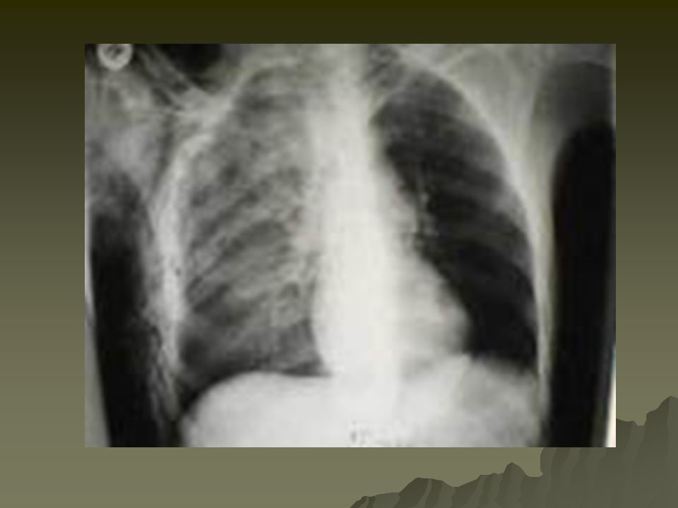
1
Thoracic Trauma Dave Lloyd, MD

2
Introduction to Thoracic Injury
Vital Structures Heart, Great Vessels, Esophagus, Tracheobronchial Tree, & Lungs 25% of MVC deaths are due to thoracic trauma 12,000 annually in US Abdominal injuries are common with chest trauma. Prevention Focus Gun Control Legislation Improved motor vehicle restraint systems Passive Restraint Systems Airbags

3
Anatomy and Physiology of the Thorax
Thoracic Skeleton 12 Pair of C-shaped ribs Ribs 1-7: Join at sternum with cartilage end-points Ribs 8-10: Join sternum with combined cartilage at 7th rib Ribs 11-12: No anterior attachment Sternum Manubrium Joins to clavicle and 1st rib Jugular Notch Body Sternal angle (Angle of Louis) Junction of the manubrium with the sternal body Attachment of 2nd rib Xiphoid process Distal portion of sternum
 Junction of the manubrium with the sternal body. Attachment of 2nd rib. Xiphoid process. Distal portion of sternum.")
5
Anatomy and Physiology of the Thorax
Thoracic Skeleton Topographical Thoracic Reference Lines Midclavicular line Anterior axillary line Mid-axillary line Posterior axillary line Intercostal space Artery, Vein and Nerve on inferior margin of each rib Thoracic Inlet Superior opening of the thorax Curvature of 1st rib with associated structures Thoracic Outlet Inferior opening of the thorax 12th rib and associated structures & Xiphisternal joint

6
Anatomy and Physiology of the Thorax
Diaphragm Muscular, dome-like structure Separates abdomen from the thoracic cavity Affixed to the lower border of the rib cage Central and superior margin extends to the level of the 4th rib anteriorly and 6th rib posteriorly Major muscle of respiration Draws downward during inspiration Moves upward during exhalation

7
Anatomy and Physiology of the Thorax
Associated Musculature Shoulder girdle Muscles of respiration Diaphragm Intercostal muscles Contract to elevate the ribs and increase thoracic diameter Increase depth of respiration Sternocleidomastoid Raise upper rib and sternum

9
Anatomy and Physiology of the Thorax
Physiology of Respiration Changing pressure assists: Venous return to heart Pumping blood to systemic circulation Inhalation Diaphragm contracts and flattens Intercostals contract expanding ribcage Thorax volume increases Less internal pressure than atmospheric Air enters lungs Exhalation Musculature relaxes Diaphragm & intercostals return to normal Greater internal pressure than atmospheric Air exits lungs

10
Anatomy and Physiology of the Thorax
Trachea, Bronchi & Lungs Trachea Hollow & cartilage supported structure Bronchi Right & left extend for 3 centimeters Enters lungs at Pulmonary Hilum Also where pulmonary arteries & veins enter Further subdivide and terminate as alveoli Basic unit of structure & function in the lungs Single cell membrane External versus Internal Respiration Lungs Right = 3 lobes Left = 2 lobes

12
Anatomy and Physiology of the Thorax
Trachea, Bronchi & Lungs Pleura Visceral Pleura Cover lungs Parietal Pleura Lines inside of thoracic cavity Pleural Space POTENTIAL SPACE Air in Space = PNEUMOTHORAX Blood in Space = HEMOTHORAX Serous (pleural) fluid within Lubricates & permits ease of expansion
 fluid within. Lubricates & permits ease of expansion.")
13
Anatomy and Physiology of the Thorax
Mediastinum Central space within thoracic cavity Boundaries Lateral: Lungs Inferior: Diaphragm Superior: Thoracic outlet Structures Heart Great Vessels Esophagus Trachea Nerves Vagus Phrenic Thoracic Duct

15
Anatomy and Physiology of the Thorax
Heart Chambers Valves Vessels External Vessels Coronary Arteries Contraction Cycle Systole Diastole Filling of the coronary arteries occur

16
Anatomy and Physiology of the Thorax
Heart General Structure Pericardium Surrounds heart Visceral Parietal Serous 35-50 ml fluid Epicardium Outer Layer Myocardium Muscular layer Endocardium Innermost layer Nervous Structure SA Node Right Atrium Intra-atrial Pathways AV Node AV Junction Bundle of His Left & Right Bundle Branches Purkinje Fibers

18
Anatomy and Physiology of the Thorax
Great Vessels Aorta Fixed at three sites Annulus Attaches to heart Ligamentum Arteriosum Near bifurcation of pulmonary artery Aortic hiatus Passes through diaphragm Superior Vena Cava Inferior Vena Cava Pulmonary Arteries Pulmonary Veins Esophagus Enters at thoracic inlet Posterior to trachea Exits at esophageal hiatus

19
Pathophysiology of Thoracic Trauma
Blunt Trauma Results from kinetic energy forces Subdivision Mechanisms Blast Pressure wave causes tissue disruption Tear blood vessels & disrupt alveolar tissue Disruption of tracheobronchial tree Traumatic diaphragm rupture Crush (Compression) Body is compressed between an object and a hard surface Direct injury of chest wall and internal structures Deceleration Body in motion strikes a fixed object Blunt trauma to chest wall Internal structures continue in motion Ligamentum Arteriosum shears aorta Age Factors Pediatric Thorax: More cartilage = Absorbs forces Geriatric Thorax: Calcification & osteoporosis = More fractures
 Body is compressed between an object and a hard surface. Direct injury of chest wall and internal structures. Deceleration. Body in motion strikes a fixed object. Blunt trauma to chest wall. Internal structures continue in motion. Ligamentum Arteriosum shears aorta. Age Factors. Pediatric Thorax: More cartilage = Absorbs forces. Geriatric Thorax: Calcification & osteoporosis = More fractures.")
20
Pathophysiology of Thoracic Trauma
Penetrating Trauma Low Energy Arrows, knives, handguns Injury caused by direct contact and cavitation High Energy Military, hunting rifles & high powered hand guns Extensive injury due to high pressure cavitation Trauma.org

21
Pathophysiology of Thoracic Trauma
Penetrating Injuries (cont.) Shotgun Injury severity based upon the distance between the victim and shotgun & caliber of shot Type I: >7 meters from the weapon Soft tissue injury Type II: 3-7 meters from weapon Penetration into deep fascia and some internal organs Type III: <3 meters from weapon Massive tissue destruction
 Shotgun. Injury severity based upon the distance between the victim and shotgun & caliber of shot. Type I: >7 meters from the weapon. Soft tissue injury. Type II: 3-7 meters from weapon. Penetration into deep fascia and some internal organs. Type III: <3 meters from weapon. Massive tissue destruction.")
22
Trauma.org

23
Injuries Associated with Penetrating Thoracic Trauma
Closed pneumothorax Open pneumothorax (including sucking chest wound) Tension pneumothorax Pneumomediastinum Hemothorax Hemopneumothorax Laceration of vascular structures Tracheobronchial tree lacerations Esophageal lacerations Penetrating cardiac injuries Pericardial tamponade Spinal cord injuries Diaphragm trauma Intra-abdominal penetration with associated organ injury
 Tension pneumothorax. Pneumomediastinum. Hemothorax. Hemopneumothorax. Laceration of vascular structures. Tracheobronchial tree lacerations. Esophageal lacerations. Penetrating cardiac injuries. Pericardial tamponade. Spinal cord injuries. Diaphragm trauma. Intra-abdominal penetration with associated organ injury.")
24
Pathophysiology of Thoracic Trauma Chest Wall Injuries
Contusion Most Common result of blunt injury Signs & Symptoms Erythema Ecchymosis DYSPNEA PAIN on breathing Limited breath sounds HYPOVENTILATION BIGGEST CONCERN = “HURTS TO BREATHE” Crepitus Paradoxical chest wall motion

25
Pathophysiology of Thoracic Trauma Chest Wall Injuries
Rib Fractures >50% of significant chest trauma cases due to blunt trauma Compressional forces flex and fracture ribs at weakest points Ribs 1-3 requires great force to fracture Possible underlying lung injury Ribs 4-9 are most commonly fractured Ribs 9-12 less likely to be fractured Transmit energy of trauma to internal organs If fractured, suspect liver and spleen injury Hypoventilation is COMMON due to PAIN

28
Pathophysiology of Thoracic Trauma Chest Wall Injuries
Sternal Fracture & Dislocation Associated with severe blunt anterior trauma Typical MOI Direct Blow (i.e. Steering wheel) Incidence: 5-8% Mortality: % Myocardial contusion Pericardial tamponade Cardiac rupture Pulmonary contusion Dislocation uncommon but same MOI as fracture Tracheal depression if posterior
 Incidence: 5-8% Mortality: 25-45% Myocardial contusion. Pericardial tamponade. Cardiac rupture. Pulmonary contusion. Dislocation uncommon but same MOI as fracture. Tracheal depression if posterior.")
29
Pathophysiology of Thoracic Trauma Chest Wall Injuries
Flail Chest Segment of the chest that becomes free to move with the pressure changes of respiration Three or more adjacent rib fracture in two or more places Serious chest wall injury with underlying pulmonary injury Reduces volume of respiration Adds to increased mortality Paradoxical flail segment movement Positive pressure ventilation can restore tidal volume

30
Pathophysiology of Thoracic Trauma Pulmonary Injuries
Simple Pneumothorax AKA: Closed Pneumothorax Progresses into Tension Pneumothorax Occurs when lung tissue is disrupted and air leaks into the pleural space Progressive Pathology Air accumulates in pleural space Lung collapses Alveoli collapse (atelectasis) Reduced oxygen and carbon dioxide exchange Ventilation/Perfusion Mismatch Increased ventilation but no alveolar perfusion Reduced respiratory efficiency results in HYPOXIA Typical MOI: “Paper Bag Syndrome”
 Reduced oxygen and carbon dioxide exchange. Ventilation/Perfusion Mismatch. Increased ventilation but no alveolar perfusion. Reduced respiratory efficiency results in HYPOXIA. Typical MOI: Paper Bag Syndrome")
31
Pathophysiology of Thoracic Trauma Pulmonary Injuries
Open Pneumothorax Free passage of air between atmosphere and pleural space Air replaces lung tissue Mediastinum shifts to uninjured side Air will be drawn through wound if wound is 2/3 diameter of the trachea or larger Signs & Symptoms Penetrating chest trauma Sucking chest wound Frothy blood at wound site Severe Dyspnea Hypovolemia

32
Pathophysiology of Thoracic Trauma Pulmonary Injuries
Tension Pneumothorax Buildup of air under pressure in the thorax. Excessive pressure reduces effectiveness of respiration Air is unable to escape from inside the pleural space Progression of Simple or Open Pneumothorax

34
Pathophysiology of Thoracic Trauma Pulmonary Injuries Tension Pneumothorax Signs & Symptoms
Dyspnea Tachypnea at first Progressive ventilation/perfusion mismatch Atelectasis on uninjured side Hypoxemia Hyperinflation of injured side of chest Hyperresonance of injured side of chest Diminished then absent breath sounds on injured side Cyanosis Diaphoresis AMS JVD Hypotension Hypovolemia Tracheal Shifting LATE SIGN

35
Pathophysiology of Thoracic Trauma Pulmonary Injuries
Hemothorax Accumulation of blood in the pleural space Serious hemorrhage may accumulate 1,500 mL of blood Mortality rate of 75% Each side of thorax may hold up to 3,000 mL Blood loss in thorax causes a decrease in tidal volume Ventilation/Perfusion Mismatch & Shock Typically accompanies pneumothorax Hemopneumothorax

36
Trauma.org

37
Pathophysiology of Thoracic Trauma Pulmonary Injuries Hemothorax Signs & Symptoms
Blunt or penetrating chest trauma Shock Dyspnea Tachycardia Tachypnea Diaphoresis Hypotension Dull to percussion over injured side

38
Pathophysiology of Thoracic Trauma Pulmonary Injuries
Pulmonary Contusion Soft tissue contusion of the lung 30-75% of patients with significant blunt chest trauma Frequently associated with rib fracture Typical MOI Deceleration Chest impact on steering wheel Bullet Cavitation High velocity ammunition Microhemorrhage may account for 1- 1 ½ L of blood loss in alveolar tissue Progressive deterioration of ventilatory status Hemoptysis typically present

39
Pathophysiology of Thoracic Trauma Cardiovascular Injuries
Myocardial Contusion Occurs in 76% of patients with severe blunt chest trauma Right Atrium and Ventricle is commonly injured Injury may reduce strength of cardiac contractions Reduced cardiac output Electrical Disturbances due to irritability of damaged myocardial cells Progressive Problems Hematoma Hemoperitoneum Myocardial necrosis Dysrhythmias CHF & or Cardiogenic shock

40
Pathophysiology of Thoracic Trauma Cardiovascular Injuries Myocardial Contusion Signs & Symptoms
Bruising of chest wall Tachycardia and/or irregular rhythm Retrosternal pain similar to MI Associated injuries Rib/Sternal fractures Chest pain unrelieved by oxygen May be relieved with rest THIS IS TRAUMA-RELATED PAIN Similar signs and symptoms of medical chest pain

41
Pathophysiology of Thoracic Trauma Cardiovascular Injuries
Pericardial Tamponade Restriction to cardiac filling caused by blood or other fluid within the pericardium Occurs in <2% of all serious chest trauma However, very high mortality Results from tear in the coronary artery or penetration of myocardium Blood seeps into pericardium and is unable to escape ml of blood can restrict effectiveness of cardiac contractions Removing as little as 20 ml can provide relief

42
Pathophysiology of Thoracic Trauma Cardiovascular Injuries Pericardial Tamponade Signs & Symptoms
Dyspnea Possible cyanosis Beck’s Triad JVD Distant heart tones Hypotension or narrowing pulse pressure Weak, thready pulse Shock Kussmaul’s sign Decrease or absence of JVD during inspiration Pulsus Paradoxus Drop in SBP >10 during inspiration Due to increase in CO2 during inspiration Electrical Alterans P, QRS, & T amplitude changes in every other cardiac cycle PEA

43
Pathophysiology of Thoracic Trauma Cardiovascular Injuries
Myocardial Aneurysm or Rupture Occurs almost exclusively with extreme blunt thoracic trauma Secondary due to necrosis resulting from MI Signs & Symptoms Severe rib or sternal fracture Possible signs and symptoms of cardiac tamponade If affects valves only Signs & symptoms of right or left heart failure Absence of vital signs

44
Pathophysiology of Thoracic Trauma Cardiovascular Injuries
Traumatic Aneurysm or Aortic Rupture Aorta most commonly injured in severe blunt or penetrating trauma 85-95% mortality Typically patients will survive the initial injury insult 30% mortality in 6 hrs 50% mortality in 24 hrs 70% mortality in 1 week Injury may be confined to areas of aorta attachment Signs & Symptoms Rapid and deterioration of vitals Pulse deficit between right and left upper or lower extremities

45
Pathophysiology of Thoracic Trauma Cardiovascular Injuries
Other Vascular Injuries Rupture or laceration Superior Vena Cava Inferior Vena Cava General Thoracic Vasculature Blood Localizing in Mediastinum Compression of: Great vessels Myocardium Esophagus General Signs & Symptoms Penetrating Trauma Hypovolemia & Shock Hemothorax or hemomediastinum

46
Pathophysiology of Thoracic Trauma Other Thoracic Injuries
Traumatic Esophageal Rupture Rare complication of blunt thoracic trauma 30% mortality Contents in esophagus/stomach may move into mediastinum Serious Infection occurs Chemical irritation Damage to mediastinal structures Air enters mediastinum Subcutaneous emphysema and penetrating trauma present

47
Pathophysiology of Thoracic Trauma Other Thoracic Injuries
Tracheobronchial Injury MOI Blunt trauma Penetrating trauma 50% of patients with injury die within 1 hr of injury Disruption can occur anywhere in tracheobronchial tree Signs & Symptoms Dyspnea Cyanosis Hemoptysis Massive subcutaneous emphysema Suspect/Evaluate for other closed chest trauma

48
Pathophysiology of Thoracic Trauma Other Thoracic Injuries
Traumatic Asphyxia Results from severe compressive forces applied to the thorax Causes backwards flow of blood from right side of heart into superior vena cava and the upper extremities Signs & Symptoms Head & Neck become engorged with blood Skin becomes deep red, purple, or blue NOT RESPIRATORY RELATED JVD Hypotension, Hypoxemia, Shock Face and tongue swollen Bulging eyes with conjunctival hemorrhage

49
Assessment of the Thoracic Trauma Patient
Scene Size-up Initial Assessment Rapid Trauma Assessment Observe JVD, SQ Emphysema, Expansion of chest Question Palpate Auscultate Percuss Blunt Trauma Assessment Penetrating Trauma Assessment Ongoing Assessment

50
Management of the Chest Injury Patient General Management
Ensure ABC’s High flow O2 via NRB Intubate if indicated Consider RSI Consider overdrive ventilation If tidal volume less than 6,000 mL BVM at a rate of 12-16 May be beneficial for chest contusion and rib fractures Promotes oxygen perfusion of alveoli and prevents atelectasis Anticipate Myocardial Compromise Shock Management Consider PASG Only in blunt chest trauma with SP <60 mm Hg Fluid Bolus: 20 mL/kg AUSCULTATE! AUSCULATE! AUSCULATE!

51
Management of the Chest Injury Patient
Rib Fractures Consider analgesics for pain and to improve chest excursion Versed Morphine Sulfate CONTRAINDICATION Nitrous Oxide May migrate into pleural or mediastinal space and worsen condition

52
Management of the Chest Injury Patient
Sternoclavicular Dislocation Supportive O2 therapy Evaluate for concomitant injury Flail Chest Place patient on side of injury ONLY if spinal injury is NOT suspected Expose injury site Dress with bulky bandage against flail segment Stabilizes fracture site High flow O2 Consider PPV or ET if decreasing respiratory status DO NOT USE SANDBAGS TO STABILIZE FX

53
Trauma.org

54
Management of the Chest Injury Patient
Open Pneumothorax High flow O2 Cover site with sterile occlusive dressing taped on three sides Progressive airway management if indicated

55
Management of the Chest Injury Patient
Tension Pneumothorax Confirmation Auscultaton & Percussion Pleural Decompression 2nd intercostal space in mid-clavicular line TOP OF RIB Consider multiple decompression sites if patient remains symptomatic Large over the needle catheter: 14ga Create a one-way-valve: Glove tip or Heimlich valve

56
Management of the Chest Injury Patient
Hemothorax High flow O2 2 large bore IV’s Maintain SBP of EVALUATE BREATH SOUNDS for fluid overload Myocardial Contusion Monitor ECG Alert for dysrhythmias IV if antidysrhythmics are needed

57
Management of the Chest Injury Patient
Pericardial Tamponade High flow O2 IV therapy Consider pericardiocentesis; rapidly deteriorating patient Aortic Aneurysm AVOID jarring or rough handling Initiate IV therapy enroute Mild hypotension may be protective Rapid fluid bolus if aneurysm ruptures Keep patient calm

58
Management of the Chest Injury Patient
Tracheobronchial Injury Support therapy Keep airway clear Administer high flow O2 Consider intubation if unable to maintain patient airway Observe for development of tension pneumothorax and SQ emphysema Traumatic Asphyxia Support airway Provide O2 PPV with BVM to assure adequate ventilation 2 large bore IV’s Evaluate and treat for concomitant injuries If entrapment > 20 min with chest compression Consider 1mEq/kg of Sodium Bicarbonate

Similar presentations