Download presentation
Presentation is loading. Please wait.

1
CHEST TRAUMA RIFLES LIFESAVERS

2
CHEST ANATOMY Heart Lungs Major vessels Thoracic Cage – –Ribs, thoracic vertebrae and sternum

3
CAUSES OF CHEST INJURIES BLUNT TRAUMA – –Motor vehicle accidents – –Auto vs. pedestrian – –Falls – –Blast injuries PENETRATING TRAUMA –Gunshot wounds –Stab wounds –Shrapnel wounds

4
TYPES OF CHEST WOUNDS Tension pneumothorax Sucking chest wounds Hemothorax Flail chest Rib fractures

5
CATEGORIES OF CHEST WOUNDS OPEN –Tension pneumothorax –Sucking chest wound –Hemothorax –Impaled object CLOSED –Tension pneumothorax –Hemothorax –Flail chest –Rib fractures

6
OPEN CHEST WOUND

7
TENSION PNEUMOTHORAX 33% of preventable combat deaths Injured chest or lung acts as one-way valve Air becomes trapped between the lung and chest wall causing the lung to collapse The heart is pushed to the other side causing blood vessels to kink Death will result if not quickly recognized and treated with needle decompression May occur in open and closed chest wounds

8
TENSION PNEUMOTHORAX

9
Tension Pneumothorax Air collapses lung and pushes heart to other side Blood return to heart restricted by kinked vessels, heart unable to pump Air between lung and chest wall

10
TACTICAL FIELD CARE: TENSION PNEUMOTHORAX Progressive severe respiratory distress in setting of unilateral penetrating chest trauma Do not rely on typical signs as breath sounds, tracheal shift, and hyperresonance on percussion Decompress immediately with 14-gauge catheter

11
OTHER SIGNS AND SYMPTOMS OF TENSION PNEUMOTHORAX Difficulty breathing Chest pain Unilateral decreased/absent breath sounds Anxiety or agitation Increased pulse Tracheal deviation Jugular venous distention (JVD) Cyanosis
 Cyanosis")
12
TRACHEAL DEVIATION AND JVD The trachea is shifted away from the collapsed lung The jugular veins become engorged from restricted blood return to heart LATE SIGNS! JVD Tracheal Deviation

13
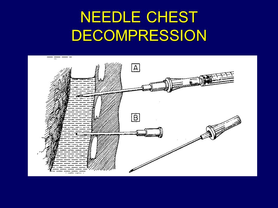
NEEDLE CHEST DECOMPRESSION Locate 2d intercostal space at midclavicular line Insert 14-gauge catheter-over-needle into chest cavity over superior edge of rib Listen for gush of air and observe for improvement of symptoms Tape catheter in place with cap or valve in place to prevent re-entry of air May also place Asherman chest seal over catheter Dress open chest wound if present

14
NEEDLE CHEST DECOMPRESSION

17
SUCKING CHEST WOUND (OPEN PNEUMOTHORAX) Open chest wound allows air entry into chest and escape Although lung is collapsed (pneumothorax), pressure is relieved by air escape and tension pneumothorax is avoided Tension pneumothorax may develop later Continually reassess the casualty for signs and symptoms of tension pneumothorax
 Open chest wound allows air entry into chest and escape Although lung is collapsed (pneumothorax), pressure is relieved by air escape and tension pneumothorax is avoided Tension pneumothorax may develop later Continually reassess the casualty for signs and symptoms of tension pneumothorax")
18
SUCKING CHEST WOUND

19
SIGNS AND SYMPTOMS OF SUCKING CHEST WOUND Penetrating chest wound A “sucking” or “hissing” sound with inhaling Difficulty breathing Impaled object in chest Froth or bubbles around injury Coughing up blood or blood-tinged sputum Pain in chest or shoulder

20
MANAGEMENT OF SUCKING CHEST WOUND Expose the wound Check for exit wound Seal the wounds with airtight material, covering the larger wound first Cover wound completely and tape down 3 sides to provide flutter-type valve for air escape May use Asherman chest seal NOTE: Treat ALL penetrating chest wounds in this manner Continually reassess for tension pneumothorax and shock

21
SUCKING CHEST WOUND Upon exhaling, air in the chest escapes through the flutter- type valve created by taping 3 sides only With inhaling, the patch should suck against the skin, preventing air entry

22
ASHERMAN CHEST SEAL

23
HEMOTHORAX Blood accumulation in chest cavity May occur slowly or rapidly depending on size of disrupted blood vessel May occur due to penetrating or blunt trauma In massive hemothorax, blood loss is complicated by low oxygen levels in blood (hypoxia)
")
24
SIGNS AND SYMPTOMS OF HEMOTHORAX Usually open chest wound Chest pain and tightness Shock Cyanosis Dullness to percussion Coughing up frothy red blood

25
TREATMENT OF HEMOTHORAX Cover and dress open chest wounds Tension pneumothorax may also be present, therefore treat with needle chest decompression if suspected If massive hemothorax, must be treated with IV fluids for shock Immediate evacuation to surgical assets

26
FLAIL CHEST Three or more ribs fractured in two or more places or a fractured sternum Severe pain at site Rapid shallow breathing Paradoxical respirations (may be difficult to detect initially) Pneumothorax may be present Possible underlying contusion to lung could lead to hypoxia
 Pneumothorax may be present Possible underlying contusion to lung could lead to hypoxia")
27
FLAIL CHEST

28
PARADOXICAL RESPIRATIONS

29
MANAGEMENT OF FLAIL CHEST AND FRACTURED RIBS Stabilize the flail segment – –Apply manual pressure – –Tape a field jacket or poncho in place – –Place casualty on injured side Pain control

30
MANAGEMENT OF IMPALED OBJECT IN THE CHEST Immobilize the impaled object Stabilize object with support dressings – –Use bulky dressings – –Construct protective structure using splint or sling Cover and dress open wounds

31
QUESTIONS?

Similar presentations