Download presentation
Presentation is loading. Please wait.

1
TOOTH dens, dentis odus, odonotos

2
Teeth (Dentes) arcus dentalis superior (maxillaris) – ellipse
arcus dentalis inferior (mandibularis) – parabola permanent teeth (dentes permanentes) – 32 deciduous teeth (dentes decidui) – 20 dens incisivus (= incisor tooth) 8/8 dens caninus (= canine tooth) 4/4 dens premolaris (= premolar tooth) 8/0 dens molaris (= molar tooth) 12/8
 – parabola. permanent teeth (dentes permanentes) – 32. deciduous teeth (dentes decidui) – 20. dens incisivus (= incisor tooth) 8/8. dens caninus (= canine tooth) 4/4. dens premolaris (= premolar tooth) 8/0. dens molaris (= molar tooth) 12/8.")
4
Teeth – parts crown (corona) neck (cervix) root (radix) pulp (pulpa)
 neck (cervix) root (radix) pulp (pulpa)")
5
Surfaces and directions
occlusalis vestibularis (buccalis/labialis) lingualis (lower teeth) palatinalis (upper teeth) mesialis distalis
 lingualis (lower teeth) palatinalis (upper teeth) mesialis. distalis.")
6
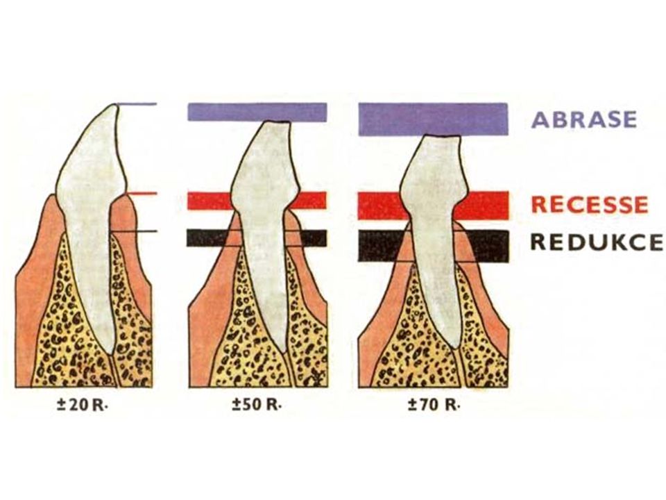
Teeth – fixation gomphosis (socket) = dentoalveolar joint
located in bony alveolus dentalis of jaw periodontium parodontium = all structures around tooth

8
Peridontium between tooth and dental alveolus (fixed to the bone of alveolus) collagen fibers (serve as alveolar periosteum) fixation apparatus of tooth = fibers of various directions penetrates into cement rapid change of connective tissue, plasticity – orthodontics atrophy in lack of proteins and vitamin C → scurvy (= skorbut) Vit. C - kofaktor prolin hydroxylázy (hydroxyprolin je specifický pro kolagen)
 Vit. C - kofaktor prolin hydroxylázy (hydroxyprolin je specifický pro kolagen)")
9
Periodontium

10
Scurvy (scorbut)
")
11
Macroscopy of tooth and its fixation

12
Denture as a whole mordex = denture
orthodental position – teeth vertically occlusion (occlusio) 80 % psalidodontia (scissors-like occlusion) = norm progenia = lower jaw longer (lower teeth in front of upper ones) stegodontia = roof-like occlusion prognathia = upper jaw longer (upper teeth in front of lower ones) opisthodontia = lower teeth too far behind upper ones hiatodontia (= mordex apertus)
 80 % psalidodontia (scissors-like occlusion) = norm. progenia = lower jaw longer (lower teeth in front of upper ones) stegodontia = roof-like occlusion. prognathia = upper jaw longer (upper teeth in front of lower ones) opisthodontia = lower teeth too far behind upper ones. hiatodontia (= mordex apertus)")
13
Dental chart / scheme crossed with letters crossed with numbers
tooth number designed with lower INDEX lowercase = decicuous UPPERCASE = PERMANENT crossed with numbers Roman numerals = decicuous Arabic numerals = PERMANENT

14
Dental chart / scheme of deciduous teeth
Dental chart / scheme of permanent teeth

15
Dental chart / scheme binumeral (Féderation Dentaire Internationale)
1-4 quadrants (from right side above clock-wise) = PERMANENT 5-8 (similar) = deciduous numeral (American Dental Association) numerals 1-32 (from right upper third mollar clock-wise) = PERMANENT letters A-T (similar from right upper second molar) = deciduous
 = PERMANENT. 5-8 (similar) = deciduous. numeral (American Dental Association) numerals 1-32 (from right upper third mollar clock-wise) = PERMANENT. letters A-T (similar from right upper second molar) = deciduous.")
16
Teeth – structure dentine – dentinum (substantia eburnea)
enamel – enamelum (substantia adamantina) cement – cementum (substantia ossea) pulp – pulpa loose connective tissue, vessels, nerves
 cement – cementum (substantia ossea) pulp – pulpa. loose connective tissue, vessels, nerves.")
17
Enamel hardest tissue of body organic part anorganic part 95%
secreted by ameloblasts (enameloblastus) glycoproteins (amelogenins, enamelins) anorganic part 95% hydroxyapatite arranged vertically in prisms (rods) in between interprismatic substance
 glycoproteins (amelogenins, enamelins) anorganic part 95% hydroxyapatite. arranged vertically in prisms (rods) in between interprismatic substance.")
18
Fluoridation fluorine is sired under the enamel surface, posteruptivelly from saliva and tooth-paste re-covers defects fluorapatite is more resistant to acids (Ph 4,5) and is produced more quickly than hydroxyapatite (pH 5,5) the more fluorapatite is in enamel, the more resistant to dental caries (tooth decay) supplement: tooth-paste, salt, at dentist
 and is produced more quickly than hydroxyapatite (pH 5,5) the more fluorapatite is in enamel, the more resistant to dental caries (tooth decay) supplement: tooth-paste, salt, at dentist.")
19
Dentine calcified connective tissue organic part anorganic part
collagen I, proteoglycans secreted by odontoblasts (dentinoblastus) located on internal surface of dentine Tomes fibers (fibrae dentinales) anorganic part hydroxyapatite non-calcified dentine predentine close to enamel and cement
 located on internal surface of dentine. Tomes fibers (fibrae dentinales) anorganic part. hydroxyapatite. non-calcified dentine. predentine. close to enamel and cement.")
20
Pulp loose connective tissue vessels nerve fibers (senstitive to pain)
fibroblasts immune cells soustavy vessels nerve fibers (senstitive to pain)
")
21
Cement thin layer at neck thick layer at root fibrilar type of bone
cellular part – cementocytes

22
Gum (Gingiva) mucosa attached to periosteum
stratified nonkeratinizing squamous epithelium papilla gingivalis no glands no submucosa gingivodental junction

23
Gum (Gingiva) linea mucogingivalis
gingiva alveolaris (pars affixa gingivae) pink, stippled, keratizing „free gingival groove“ gingiva marginalis (pars libera gingivae) shiny, red, nonkeratinizing sulcus gingivalis junctio dentogingivalis epithelium junctionale
 pink, stippled, keratizing. „free gingival groove gingiva marginalis (pars libera gingivae) shiny, red, nonkeratinizing. sulcus gingivalis. junctio dentogingivalis. epithelium junctionale.")
25
Teeth development (Odontogenesis)
oral ectoderm mesoderm cells of neural crest ectomesenchyme enamel is derived from ectoderm other tissues are derived from ectomesenchyme

26
Teeth development Week 6: lamina dentalis (dental lamina) appears
thickening of stomodeum epithelium in each lamina 10 proliferation centers dental buds Zubní lišta: a - retní val; b - žlábek předsíně; c - základ zubu (červeně) v zubní liště (růžově); d - Meckelova chrupavka; e - kost dolní čelisti;
 v zubní liště (růžově); d - Meckelova chrupavka; e - kost dolní čelisti;")
27
Stages of tooth development
dental bud (status gemmalis) local thickening of epithelium, 10 in each jaw dental cap (status galearis) ectodermal part → enamel organ (organum enameleum) invagination of mesenchyme → dental papilla a – začátek zubní lišty, b – pupen, c - čepička
 local thickening of epithelium, 10 in each jaw. dental cap (status galearis) ectodermal part → enamel organ (organum enameleum) invagination of mesenchyme → dental papilla. a – začátek zubní lišty, b – pupen, c - čepička.")
28
čepička

29
Stages of tooth development
dental cap → dental bell (status campanalis) outer dental epithelium enamel reticulum inner dental epithelium dental papilla → dental pulp dental sac → cement, periodontal ligaments
 outer dental epithelium. enamel reticulum. inner dental epithelium. dental papilla → dental pulp. dental sac → cement, periodontal ligaments.")
31
Stages of tooth development – bell
odontoblasts (dentinoblasti) derived from mesenchyme cells at inner enamel organ produce (pre)dentine Tomes fibers (fibrae dentinales) cytoplasmatic processes left within dentine Pulpa vyvíjejícího se zubu s pravidelným uložením odontoblastů. Proti dentinu je vrstva u produkovaná vnitřními ameloblasty: a - vnitřní ameloblasty; b - ; c - dentin; d - odontoblasty;
 derived from mesenchyme cells at inner enamel organ. produce (pre)dentine. Tomes fibers (fibrae dentinales) cytoplasmatic processes left within dentine. Pulpa vyvíjejícího se zubu s pravidelným uložením odontoblastů. Proti dentinu je vrstva u produkovaná vnitřními ameloblasty: a - vnitřní ameloblasty; b - ; c - dentin; d - odontoblasty;")
32
D – dentin, O – odontoblasty

33
Stages of tooth development – bell
ameloblasts (enameloblasti) from inner enamel epithelium basal surface becomes secretory production of enamel Pulpa vyvíjejícího se zubu s pravidelným uložením odontoblastů. Proti dentinu je vrstva u produkovaná vnitřními ameloblasty: a - vnitřní ameloblasty; b - ; c - dentin; d - odontoblasty; E – sklovina, A - ameloblasty
 from inner enamel epithelium. basal surface becomes secretory. production of enamel. Pulpa vyvíjejícího se zubu s pravidelným uložením odontoblastů. Proti dentinu je vrstva u produkovaná vnitřními ameloblasty: a - vnitřní ameloblasty; b - ; c - dentin; d - odontoblasty; E – sklovina, A - ameloblasty.")
34
Stages of tooth development – bell
epithelial root sheath (vagina epithelialis radicis) = Hertwig sheath transition between outer and inner enamel epithelium ingrowth into mesenchyme and induction of root formation
 = Hertwig sheath. transition between outer and inner enamel epithelium. ingrowth into mesenchyme and induction of root formation.")
36
Tooth eruption decidual teeth: 6th – 24th month
enamel organ disrupted during tooth eruption

37
Permanent teeth develop similarily to decidual teeth
secondary dental lamina located at lingual side of dental lamina prolonged distally (molars) eruption from 6th year (finished in 30th – 40th year)
 eruption from 6th year. (finished in 30th – 40th year)")
38
Clinical note tetracycline antibiotics
are contraindicated in children up to 8 years of age, pregnant and nursing women high affinity to newly produced enamel brown-yellow color enamel hypoplasia

Similar presentations


>")


 Lamina Propria- loose CT.>")
, major.>")


>")