Download presentation
Presentation is loading. Please wait.

1
THE DEVELOPMENT OF PALATE NASAL CAVITIES AND TOOTH
By Dr Samina Anjum

3
FORMATION OF INTERMAXILLARY SEGMENT

4
COMPONENTS OF INTERMAXILLARY SEGMENT
Labial Components (Philtrum) Upper Jaw Component (4 Incisor teeth) Palatal Component (Triangular Primary Palate)
 Upper Jaw Component (4 Incisor teeth) Palatal Component (Triangular Primary Palate)")
5
SECONDARY PALATE

11
Congenital malformations
1) cleft lip: a. unilateral cleft lip: results from failure of the maxillary prominence to merge with medial nasal prominence on the affected side
 cleft lip: a. unilateral cleft lip: results from failure of the maxillary prominence to merge with medial nasal prominence on the affected side.")
12
b. bilateral cleft lip : results from failure of the maxillary prominences to merge with the medial nasal prominence on both sides c. median cleft lip: results from failure of the medial nasal prominences to merge and form the intermaxillary segments

13
2) oblique facial cleft: results from failure of the maxillary prominence to fuse with the lateral nasal prominence
 oblique facial cleft: results from failure of the maxillary prominence to fuse with the lateral nasal prominence")
15
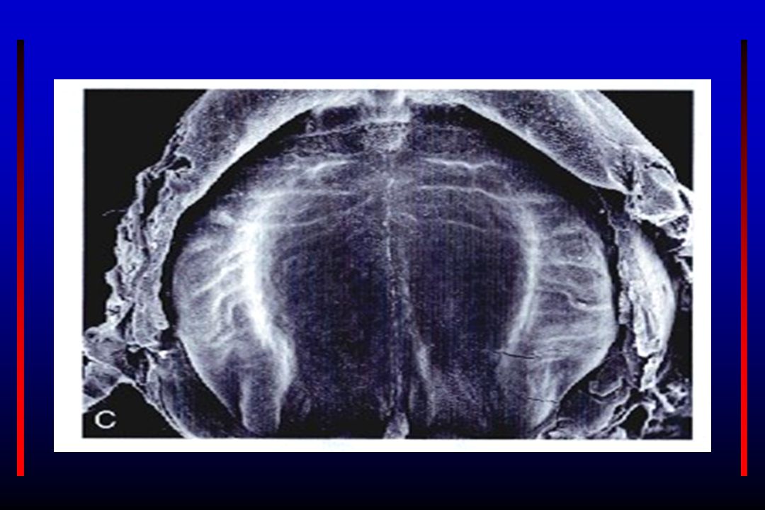
DEVELOPMENT OF NASAL CAVITIES

16
TEETH Teeth arise from an epithelial - mesenchymal interaction between overlying oral epithelium and underlying mesenchyme derived from neural crest cells.

17
DENTAL LAMINA The basal layer of oral epithelium forms a C shaped dental lamina along the upper and lower jaw by 6th week of development. Connects the developing tooth bud to the oral epithelium.

18
Fate – DL’s total activity is about 5 years
Fate – DL’s total activity is about 5 years. After it has disappeared everywhere, it still is present in the region of 3rd molar. As the teeth continue to develop, they lose the connection with the dental lamina. Remnants of dental lamina persist as epithelial pearls or islands within the jaw as well as in the gingiva.

19
Stages of Tooth Development
Bud Cap Bell ©Copyright 2007, Thomas G. Hollinger, Gainesville, Fl

20
THE BUD STAGE The dental lamina give rise to the dental buds/ enamel organs (10) in each jaw which form ectodermal components of teeth. At 8 wks

22
THE CAP STAGE

23
Cont… Stellate reticulum At 10 wks

24
CAP STAGE

25
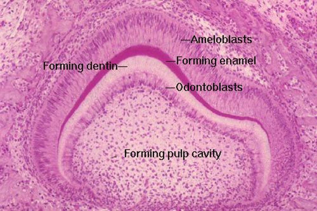
BELL STAGE Dental cap grows and indentation deepens, tooth takes the appearance of bell.

26
Cont… Enamel knot: Cluster of cells in inner dental epithelium that regulates early tooth development Dental cuticle: Once ameloblasts retreat into stellate reticulum, leave a thin membrane on the surface of enamel. After tooth eruption this membrane sloughs off. Dental process 3 months 6 months

27
BELL STAGE The dental lamina disintegrates, leaving the developing teeth completely separated from the epithelium of the oral cavity; the two will not join again until the final eruption of the tooth into the mouth.

30
Root of tooth --- begins when dental epithelial layers penetrate into underlying mesenchyme and form epithelial root sheath. Pulp chamber narrows, forms a canal containing blood vessels and nerves of the tooth.

31
Tooth just before birth and after eruption
Mesenchymal cells on the outside of tooth and in contact with dentin of root differentiates into cementoblasts. Lengthening of root pushes the crown up through the overlying tissue layers into the oral cavity.

32
Outside the cementum mesenchyme give rise to periodontal ligaments,
which holds the tooth firmly in position and function as shock absorber. Buds for permanent teeth lie on lingual aspect of milk teeth, are formed during 3rd month. These buds will remain dormant until 6th year of postnatal life. Then they begin to grow, push against the underside of milk teeth and aid in their shedding. As permanent tooth grows its root is resorbed by osteoclasts.

33
Photomicrograph of section of crown & neck of tooth

35
THANK YOU

Similar presentations


>")


>")


