Download presentation
Presentation is loading. Please wait.

1
X-rays: Pelvis, Hip & Shoulder
Feb. 22, 2006 J. Huffman, PGY-1 Thanks to Dr. J. Lord Also thanks to Moritz, Adam and Steve Lan for some borrowed slides and images

2
Goals: As per instructions, this is a radiology talk ONLY. The focus is on reading as many films as possible. Therefore, try your best to describe what you see as you would when on the phone with a consultant. No epidemiology No management No associated injuries (i.e. vascular injury with pelvic #)
")
3
Outline Pelvis Hip Shoulder Anatomy Views Classification of fractures
Practice Hip Fractures Dislocations Shoulder Anatomy Views Dislocations Fractures Practice

4
Pelvis: Anatomy Pelvis = sacrum, coccyx + 2 inominate bones
Inominate bones = ilium, ischium, pubis Strength from ligamentous + muscular supports

5
Pelvis: Anatomy Anterior Support: Posterior Support: ~40% of strength
Symphysis pubis Fibrocartilaginous joint covered by ant & post symphyseal ligaments Pubic rami Posterior Support: ~60% of strength Sacroiliac ligament complex Pelvic floor Sacrospinous ligament Sacrotuberous ligament Pelvic diaphragm Sacrospinous resists external rotation Sacrotuberous resists rotational and vertical shearing forces

6
Pelvis: Anatomy Very strong posterior ligaments
Disruption of these is the cause of mechanical instability Arteries and veins lie adjacent to posterior arch

7
Pelvis: Anatomy Divided into 3 columns: Anterior superior column
(= ilium) Anterior inferior column (= pubis) Posterior Column (= ischium) Ant superior column is the primary wt bearing structure The ant inferior column is thin and easily fractured The post column is thick and strong bust most commonly fractured
 Anterior inferior column. (= pubis) Posterior Column. (= ischium) Ant superior column is the primary wt bearing structure. The ant inferior column is thin and easily fractured. The post column is thick and strong bust most commonly fractured.")
8
Pelvis: Imaging Plain films CT scans AP Inlet view / Outlet view
Judet view (oblique – shows columns, acetabulum) AP alone ~90% sensitive; combined w/ inlet/outlet views ~94% Limited in ability to clearly delineate posterior injuries Pelvic films are NOT necessary in pts with normal physical exam, GCS >13, no distracting injury and not intoxicated At least one study shows clinical exam reliable in EtOH Gonzalez et al. J Am Coll Surg. 2002; 194: 121-5 CT scans Evaluates extent of posterior injury better Superior imaging of sacrum and acetabulum More detailed info about associated injuries EtOh levels up to 104 mmol/l, most were > 21.7 Prospective study of 2176 consecutive blunt trauma pts of which 4.5% had pelvic #’s AP plevis alone missed more injuries than clinical exam even in intoxicated pts On the other hand, plain films can help to predict bleeding complications and should be done if pelvis is suspected to be busted as the first step in the work up
 AP alone ~90% sensitive; combined w/ inlet/outlet views ~94% Limited in ability to clearly delineate posterior injuries. Pelvic films are NOT necessary in pts with normal physical exam, GCS >13, no distracting injury and not intoxicated. At least one study shows clinical exam reliable in EtOH. Gonzalez et al. J Am Coll Surg. 2002; 194: CT scans. Evaluates extent of posterior injury better. Superior imaging of sacrum and acetabulum. More detailed info about associated injuries. EtOh levels up to 104 mmol/l, most were > Prospective study of 2176 consecutive blunt trauma pts of which 4.5% had pelvic #’s. AP plevis alone missed more injuries than clinical exam even in intoxicated pts. On the other hand, plain films can help to predict bleeding complications and should be done if pelvis is suspected to be busted as the first step in the work up.")
9
6 lines of the pelvis: 1. Iliopubic (arcuate) line – disruption indicates ant column injury 2. Ilioischial line which defines the posterior column 3. Teardrop or Roentgenographic U formed by roof of acetabaulum and ilioischial spine defines quadrangular plate – disruption means intraplevic penetration 4. Roof of acetabulum 5. Post rim of acetabulum 6. Ant rim of acetabulum 7. Shenton’s line = medial femoral shaft obturator foramen: disruption in hip dislocation or femoral neck #’s

11
Pelvis: Imaging - Acetabulum
Arcuate line Ileoischial line Radiographic U (teardrop) Acetabular roof Anterior lip of acetabulum Posterior lip of acetabulum
 Acetabular roof. Anterior lip of acetabulum. Posterior lip of acetabulum.")
12
Pelvis: Imaging - Acetabulum

13
Pelvis: Imaging – Normal Inlet

14
Pelvis: Imaging – Normal Outlet

15
Pelvis: Imaging Radiographic clues to posterior arch fractures:
L5 transverse process avulsion* (iliolumbar ligament) Avulsion of the lower, lateral sacral lip* (sacrotuberous ligament) Ischial spine avulsion* (sacrospinous ligament) Assymmetry of sacral foramina Displacement at the site of a pubic ramus fracture Ist 2 always denote mechanical instability
 Avulsion of the lower, lateral sacral lip* (sacrotuberous ligament) Ischial spine avulsion* (sacrospinous ligament) Assymmetry of sacral foramina. Displacement at the site of a pubic ramus fracture. Ist 2 always denote mechanical instability.")
16
Pelvis: Fracture Classification Systems
2 most common are Tile and Young systems Tile Classification system: Advantages Comprehensive Predicts need for operative intervention Disadvantages Does NOT predict morbidity or mortality Young Classification System: Based on mechanism of injury predicts ass’d injury Estimates mortality Excludes more minor injuries

17
Tile Classification System
Type A: Stable: Posterior structures intact Type B: Partially stable: Posterior structures incompletely disrupted Type C: Unstable: Posterior structures completely disrupted *Each type further classified into 3 sub-types based on fracture.

18
Tile Classification System
Type A: Stable pelvis: post structures intact A1: avulsion injury A2: iliac wing or ant arch # A3: Transverse sacrococcygeal #

19
Tile Classification System
Type B: Partially stable pelvis: incomplete posterior structure disruption B1: open-book injury B2: lateral compression injury B3: contralateral / bucket handle injuries

20
Tile Classification System
Type C: Unstable pelvis: complete disruption of posterior structures C1: unilateral C2: bilateral w/ one side Type B, one side Type C C3: bilateral Type C

21
Young Classification System
Lateral Compression Anteroposterior Compression Vertical Shear Combination *LC and APC further classified into 3 sub-types based on fracture Pros and cons to each – Tile is comprehensive but Youngs predicts mortality, GU complications and risk of bleeding

22
Young Classification System:
Lateral Compression (50%) transverse # of pubic rami, ipsilateral or contralateral to posterior injury LC I – sacral compression on side of impact LC II – iliac wing # on side of impact LC III – LC-I or LC-II on side of impact w/ contralateral APC injury
 transverse # of pubic rami, ipsilateral or contralateral to posterior injury. LC I – sacral compression on side of impact. LC II – iliac wing # on side of impact. LC III – LC-I or LC-II on side of impact w/ contralateral APC injury.")
23
Young Classification System:
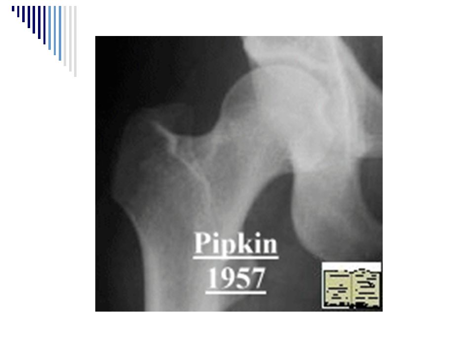
AP Compression (25%) Symphyseal and/or Longitudinal Rami Fractures APC I – slight widening of the pubic symphysis and/or anterior SI joint APC II – disrupted anterior SI joint, sacrotuberous, and sacrospinous ligaments APC III – complete SI joint disruption w/ lateral displacement and disruption of sacrotuberous and sacrospinous ligaments
 Symphyseal and/or Longitudinal Rami Fractures. APC I – slight widening of the pubic symphysis and/or anterior SI joint. APC II – disrupted anterior SI joint, sacrotuberous, and sacrospinous ligaments. APC III – complete SI joint disruption w/ lateral displacement and disruption of sacrotuberous and sacrospinous ligaments.")
24
Young Classification System:
Vertical Shear (5%) Symphyseal diastasis or vertical displacement andteriorly and posteriorly Combined Mechanism combination of injury patterns
 Symphyseal diastasis or vertical displacement andteriorly and posteriorly. Combined Mechanism. combination of injury patterns.")
25
Young Classification System: Morbidity and Mortality

26
Type A1 avulsion Tile A1

27
Tile B1 Tile B1 / Young APC II

28
Pubic ramus # = Tile A2 Tile C1/ Young VS

29
Tile A1

30
No Fracture, just an IUD

31
Tile B3 / Young APC

32
Right iliac wing # also called Duverney
Tile A2 / Young LC II

33
No #, just SC air from rib fractures

34
Pelvis: Acetabular Fractures
Four Categories: Posterior lip fracture Commonly assoc. w/ posterior hip dislocation Central or transverse fracture Fracture line crosses acetabulum horizontally Anterior column fracture Disrupts arcuate line, ileoischial line intact, U displaced medially Posterior column fracture Ileoischial line disrupted and separated from the U Judet (oblique views) or CT helpful if suspicious
 or CT helpful if suspicious.")
35
Pelvis: Imaging - Acetabulum

36
Focus on the acetabular fractures.
Posterior Column #

37
Posterior Column #

38
Anterior Column #

39
Bilateral Anterior Column #

40
Posterior Lip #

41
Central (Transverse) fracture
 fracture")
42
Proximal Femur & Hip

43
Proximal Femur & Hip: Injuries
Fractures: Femoral neck, intertrochanteric, femoral head, greater & lesser trochanter, subtrochanteric Dislocations: Anterior, posterior, central, (inferior) Any elderly pt c/o hip, thigh or knee pain has a proximal femur fracture until proven otherwise Femoral neck & intertrochanteric #’s account for 90% of hip #’s
 Any elderly pt c/o hip, thigh or knee pain has a proximal femur fracture until proven otherwise. Femoral neck & intertrochanteric #’s account for 90% of hip #’s.")
44
Proximal Femur: Anatomy
Ward’s Triangle

45
Proximal Femur: Images
AP Internal rotation! Lateral Cross-table Lateral Frog-leg Lateral

46
Proximal Femur: Images
Cross-table lateral view * = ischial tuberosity

47
Proximal Femur: Fracture Classification
Relationship to capsule Intracapsular, extracapsular Anatomic location Neck, trochanteric, intertrochanteric, subtrochanteric, shaft Degree of displacement

48
Proximal Femur: Approach to the film
Shenton’s Line Femoral neck # Dislocation ‘S’ and ‘Reverse S’ patterns Position of lesser trochanter Femoral head size Trace trabecular groups

49
Left posterior dislocation – note Shenton’s line

50
Proximal Femur: Approach to the film
Lowell’s ‘S’ patterns

51
Impacted femoral neck #

52
Hip: Dislocations Etiology Types: Orthopedic emergencies:
Adults: high energy mechanism (MVA) Elderly, prosthetic joints, kids < 6yo: minor mech Types: Posterior >> anterior > central (> inferior) Orthopedic emergencies: Urgent reduction after ABC’s / stabilization Significant neurovascular complications Often multiple associated injuries Mandate CT post-reduction CT post reduction for intra-articular #’s, acetabular #’s
 Elderly, prosthetic joints, kids < 6yo: minor mech. Types: Posterior >> anterior > central (> inferior) Orthopedic emergencies: Urgent reduction after ABC’s / stabilization. Significant neurovascular complications. Often multiple associated injuries. Mandate CT post-reduction. CT post reduction for intra-articular #’s, acetabular #’s.")
53
Hip: Dislocation imaging
Plain Films: ant vs. post dislocations Femoral head size Posterior dislocation femoral head smaller Lesser trochanter visibility Post dislocation adduction & internal rotation, lesser trochanter not seen Ant dislocation external rotation; lesser trochanter clearly visible CT Indicated for more detailed evaluation of femoral neck, intra-articular #’s, and acetabulm

54
Anterior dislocation

55
Posterior dislocation
Lesser trochanter

56
Proximal Femur: Fractures
Femoral head fracture: Usually 2° to dislocation Pipkin classification Femoral neck fracture: Can be subtle (check lines, ‘S’) Describe as nondisplaced (15-20%) vs displaced Intertrochanteric fracture: High energy or weak bone Classify according to number of bone fragments (e.g. two-part)
 Describe as nondisplaced (15-20%) vs displaced. Intertrochanteric fracture: High energy or weak bone. Classify according to number of bone fragments (e.g. two-part)")
58
Displaced femoral neck fracture

59
Nondisplaced femoral neck #

60
Two-part intertrochanteric fracture

61
Three-part intertrochanteric #

62
Proximal Femur: Fractures
Isolated trochanter fracture: Rare (women more than men) Direct fall or avulsion by iliopsoas Outpt management Subtrochanteric fracture: #’s b/w lesser trochanter & point 5 cm distal Common site for pathologic fractures Vague symptoms Occult fracture: ~%5 of hip fractures not seen radiographically
 Direct fall or avulsion by iliopsoas. Outpt management. Subtrochanteric fracture: #’s b/w lesser trochanter & point 5 cm distal. Common site for pathologic fractures. Vague symptoms. Occult fracture: ~%5 of hip fractures not seen radiographically.")
63
Isolated greater trochanter #

64
Isolated lesser trochanter #

65
Subtrochanteric fracture

66
Proximal Femur & Hip Practice

67
Intertrochanteric fracture 2° to mets from prostate CA

68
Pipkin III femoral head fracture and posterior dislocation

69
AC separation Clavicle fracture Scapula fracture Shoulder dislocation

70
Shoulder: Anatomy 3 bones: 3 joints: 1 articulation: Clavicle Humerus
Scapula 3 joints: Acromioclavicular Glenohumeral Sternoclavicular 1 articulation: Scapulothoracic

71
Shoulder: Anatomy

72
Shoulder: Anatomy

73
Shoulder: Images True AP Lateral (transcapular) Axillary AC view
Should see no overlap of humerus over the glenoid Lateral (transcapular) Scapula looks like a ‘Y’) Axillary Best “true lateral” view of the shoulder AC view 100° abduction
 Scapula looks like a ‘Y’) Axillary. Best true lateral view of the shoulder. AC view. 100° abduction.")
74
More useful for soft-tissue evaluation
Shoulder: Images Internal rotation External rotation More useful for soft-tissue evaluation

75
Normal True AP of the Shoulder

76
Normal lateral film of the shoulder

78
Normal axillary film of the shoulder

79
Trauma Axillary View: - does not require abduction of the arm (nor removal from sling); - the patient leans backward; - the x-ray plate is placed directly under the shoulder, and the x-ray tube is positioned directly above;
; - the patient leans backward; - the x-ray plate is placed directly under the shoulder, and the x-ray tube is positioned directly above;")
80
AC Separation: Classification
Type I Sprain of the AC joint CC distance maintained (N = 11-13mm) Type II AC ligaments disrupted Joint space widened CC distance maintained Clavicle rides upward (<50% its width)
 Type II. AC ligaments disrupted. Joint space widened. CC distance maintained. Clavicle rides upward (<50% its width)")
81
AC Separation: Classification
Type III (and IV, V, VI) Complete disruption of AC and coracoclavicular ligaments as well as muscle attachements Joint space widened CC space is increased (5mm difference from uninjured side) Clavicle is displaced
 Complete disruption of AC and coracoclavicular ligaments as well as muscle attachements. Joint space widened. CC space is increased. (5mm difference from uninjured side) Clavicle is displaced.")
82
Type III AC separation – AC view (100° Abduction)
")
83
Clavicle Fracture Classified anatomically:
Medial third (5%) – direct blow to the anterior chest Middle third (80%) – direct force to lateral aspect of shoulder Lateral third (15%) – direct blow to the top of shoulder Lateral to the coracoclavicular lig. (stable) Medial to the coracoclavicular lig. (tend to displace) Involves the articular surface
 – direct blow to the anterior chest. Middle third (80%) – direct force to lateral aspect of shoulder. Lateral third (15%) – direct blow to the top of shoulder. Lateral to the coracoclavicular lig. (stable) Medial to the coracoclavicular lig. (tend to displace) Involves the articular surface.")
84
Fracture of the middle third of the clavicle

85
Comminuted fracture of the middle third of the clavicle

86
Distal third clavicle fracture – type II

87
Scapula Fracture Classified Anatomically:
Acromion process, scapular spine or coracoid process Scapular neck involved Intra-articular fractures of the glenoid fossa Scapular body involved (most common)
")
88
Type I scapular fracture (coracoid fracture)
")
89
Type III scapular fracture

90
Comminuted, type III scapular fracture

91
Shoulder: Dislocation
Classification Anterior (95-97%) Subcoracoid (most common) Subglenoid (1/3 associated with # greater tuberosity, or # glenoid rim) Subclavicular Intrathoracic Also important to note primary vs. recurrent
 Subcoracoid (most common) Subglenoid. (1/3 associated with # greater tuberosity, or # glenoid rim) Subclavicular. Intrathoracic. Also important to note primary vs. recurrent.")
92
Anterior dislocation - subcoracoid

93
Shoulder: Dislocation
Classification – cont’d Posterior Subacromial (98% of posterior dislocations) Subglenoid Subspinous Inferior (Luxatio Erecta) - rare superior - rare
 Subglenoid. Subspinous. Inferior (Luxatio Erecta) - rare. superior - rare.")
94
Shoulder: Dislocation
Signs of posterior shoulder dislocation: ↑distance from anterior glenoid rim and humeral head “rim” sign Humeral head internally rotated “Light bulb” or “drum stick” sign True AP shows humeral/glenoid overlap Impaction # of the anteromedial humeral head “reverse Hill-Sachs deformity” “Trough sign”

95
Posterior dislocation
Arrow = impaction # of anteromedial humeral head

96
Posterior dislocation
Note the humeral head roatation

97
Posterior dislocation – lateral view

98
Posterior dislocation – axillary view

99
Shoulder: Dislocation
Associated fractures: Compression # of the posterolateral aspect of the humeral head “Hill-Sachs deformity” 11-50% of anterior dislocations Anterior glenoid rim fracture “Bankart’s fracture” ~5% of cases Avulsion fracture of the greater tuberosity ~10-15% of cases

100
Anterior dislocation Arrow = # of the posterolateral aspect of humerus

101
Post-reduction film Avulsion # of the greater tuberosity

103
Shoulder Practice

104
Clavicle fracture – distal third – type II

105
Scapula fracture – type III

106
AC separation - grade I

107
Anterior shoulder dislocation

108
Posterior dislocation (False AP – note overlap)
")
Similar presentations


 Pelvic girdle.>")


 fxs.- m.c. type; m.c. levels.>")


