Download presentation
Presentation is loading. Please wait.

1
VMB 976 A Ultrasound Laura Paasch Case report

2
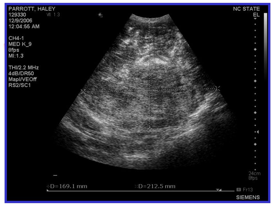
Haley Parrott 10 year old FS Labrador Presented on emergency for possible splenic mass Seen at rDVM 5 days previously for abnormal urination and flatulence Prescribed a gastroprotectant Seen by rDVM for vomiting PCV-29% Large abdominal mass on radiographs

4
Initial Work-Up PE Depressed, pale mm, P-140, RR-28, T-102, CRT 4 sec Large abdominal mass on palpation Bloodwork PCV-32%, TP-6.0, BUN-30-40, GLU-47, icteric plasma, thrombocytopenic (55,000), elevated APTT- 23.3 sec Imaging Chest radiographs- no evidence of metastasis Abdominal U/S
, elevated APTT sec Imaging Chest radiographs- no evidence of metastasis Abdominal U/S")
10
Post-Operative Recovery ICU, Fluids, Fentanyl CRI, KCl, Metoclopramide, Lidocaine (VPC’s), Famotidine ….. Still recovering in ICU following morning Alert, ambulatory, drinking, not eating, fewer VPC’s, almost moved to intermediate! Next morning Severe hemorrhagic diarrhea Hemorrhage from nares/mouth Euthanized Necropsy

11
Necropsy Report Multiple pulmonary and mesenteric thrombi Liver thrombus, multifocal necrosis Marked, diffuse hemorrhage of stomach and intestine Myocardial degeneration and necrosis Splenic hematoma with areas of infarction and marked diffuse hemorrhage DIC Large amount of blood loss, platelets, clotting factors hypercoaguability Hypoxia, acidosis, inflammation endothelial damage

12
Normal Spleen <25mm dogs, <8mm cats Upper left quadrant Tail and body along L body wall or ventral abdomen Echogenic capsule Parenchyma finely textured, homogenous More echogenic than liver L kidney cortex Splenic A & V at hilus, not abnormal to see fat surrounding

13
Abnormalities Splenomegaly Anesthetics Immune mediated Infectious, parasites Neoplasia Torsion Passive congestion

14
Abnormalities Hypoechoic Congestion Infections Infiltrative disease Amyloidisis EMH Hyperechoic Chronic inflammation Vascular compromise Peritonitis Infections Infiltrative disease Fibrosis Mineralization

15
Abnormalities Focal/Multifocal lesions Nodular hyperplasia EMH Abscess Neoplasia Necrosis Fibrosis Calcification Hematoma

16
Hematomas Trauma Clotting disorders Neoplasia (hemangiosarcoma, lymphosarcoma) Extreme variation in appearance Local hemorrhage hemoabdomen Unclotted blood (anechoic to hypoechoic) Clotted blood (isoechoic to hyperechoic) FNA Non-diagnostic False negatives
 Extreme variation in appearance Local hemorrhage hemoabdomen Unclotted blood (anechoic to hypoechoic) Clotted blood (isoechoic to hyperechoic) FNA Non-diagnostic False negatives")
Similar presentations


 measures the number of cells of different types circulating in the bloodstream three major types.>")


