Download presentation
Presentation is loading. Please wait.

1
Adrenals, Lymphnodes, Gall Bladder, and Pancreas
Jane MacLellan

2
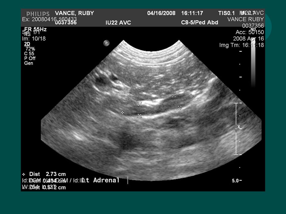
Adrenal Glands In the dog - peanut shaped In the cat - more oval
Located cranial and medial to kidneys Left - caudal to the branches of the aorta Right - adjacent to caudal vena cava Locate kidney, then fan medially with probe Hypoechoic - similar to blood vessels Can be hard to distinguish - use doppler Overlying bowel can obscure

4
Adrenal Gland Disease Measure length and width
Length with vary between animals Proportional to body weight Width does not Width may increase with disease Normal width = < 0.74 cm Note: New paper suggests dogs < 10Kg normal width < 0.6 cm

5
Adrenal Gland Disease Pituitary dependent hyperadrenocorticism
Bilaterally enlarged Normal shape - but ‘plump’ Thickened poles Uniformly hypoechoic Nodular hyperplasia Normal size does not r/o PDH

6
Adrenal Gland Disease Adrenal tumor Gland enlargement Abnormal shape
Change in echotexture Unilateral masses more common Can’t distinguish benign from malignant tumors May be able to tell if invading surrounding tissue

8
Lymph nodes More sensitive then radiographs
Medial iliac and jejunal lymph nodes Large More often seen when normal Normal - same echogenicity as surrounding mesentery Easier to see in young, thin animals When enlarged, more hypoechoic Can do ultrasound guided FNA

9
Lymph nodes Medial iliac Visceral
Near terminal portion of aorta and caudal vena cava Not normally seen unless enlarged Bladder, prostatic neoplasia Visceral Seen when doing routine scan

11
Gall Bladder Visualized just right of midline in liver
Size is variable - depending on last meal Fasting or anorexia In cats, can be bi-lobed Things you might see Thickened wall Stones Mucoceles Cholestasis Cholecystitis “Sludge” Icterus

12
Thickened Wall Wall normally thin, echogenic, poorly visualized
<1mm in cats, slightly thicker in dog Double layered - inside and outside surfaces – Halo sign Thickening is a non-specific sign Chronic hepatitis Cholecystitis Cholangiohepatitis Right CHF Hypoalbuminemia Sepsis Neoplasia

13
Sludge Commonly seen Especially if haven’t eaten recently Dependent

14
Mucocele Cystic mucinous hyperplasia Proliferation of GB epithelium
Increased mucin production Marked distension of the GB Kiwi appearance Cystic mucinous hyperplasia is characterised by papillary proliferation of the gall bladder epithelium with increased mucin production. Sometimes there is extreme mucin production with marked distension of the gall bladder (mucocele)
")
15
Choleliths Uncommon Incidental finding
Should be noted - cholecystitis or biliary obstruction Hyperechoic Acoustic shadowing Mobile

16
Bile Duct Seen as a continuation of the GB
Dogs - not consistently seen Should be < 3mm Cats - more often seen Should be < 4mm Ventral to portal vein Extrahepatic obstruction Dilation of GB and bile duct

17
GB Artifacts Mirror image duplicate Refraction Acoustic enhancement
Sound wave bounces off the diaphragm, echos off the gall bladder back towards diaphragm, reflected towards the transducer Refraction When sound waves go through tissues of different acoustic impedance Acoustic enhancement Less attenuation compared to liver

18
Pancreas Normal is routinely difficult to visualize
Echogenicity similar to surrounding fat No defined capsule Less echogenic then spleen, more echogenic then liver Right limb just dorsal to duodenum More likely to see in puppies, thin dogs, or with free abdominal fluid

19
Pancreatitis Acute - surrounded by a hyperechoic area
Due to peri-pancreatic fat necrosis Severe - mixed echogenicity Chronic - hyperechoic pancreas Due to pancreatic fat necrosis Pancreatic pseudocysts Mass effect In acute cases a hypoechoic pancreas is often surrounded by a hyperechoic area that is due to peripancreatic fat necrosis. In dogs with severe pancreatitis, the sensitivity for various degrees of peripancreatic fluid accumulation was 68%. 1 Chronic pancreatitis can be associated with a hyperechoic pancreas indicating the presence of pancreatic fibrosis.

22
Pancreas Neoplasia Difficult to identify
Looks similar to pancreatitis (mass effect) Fluid accumulation Invasion of surrounding tissues Evidence of metastasis in other organs
 Fluid accumulation. Invasion of surrounding tissues. Evidence of metastasis in other organs.")
Similar presentations


 Glands Anatomy & Embryology>")


