Download presentation
Presentation is loading. Please wait.

1
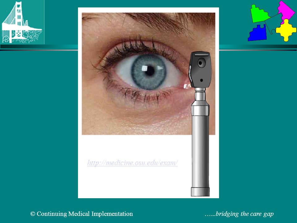
Fundoscopic Examination
Window to the blood vessels

3
Normal Ocular Fundus Arterioles Optic cup Fovea Optic disc Vein

4
Fundoscopic Examination
Hypertensive retinopathy Diabetic retinopathy Bacterial endocarditis Athero-emboli Cholesterol emboli

5
Hypertensive Retinopathy
Modified Scheie Classification Grade 0: No changes Grade 1: Minimal arteriolar narrowing Grade 2: Obvious arteriolar narrowing with focal irregularities Grade 3: Grade 2 + retinal hemorrhages and/or exudate Grade 4: Grade 3 + swollen optic nerve (Malignant hypertension)
")
6
Hypertensive Retinopathy Grade 2
Arteriovenous nicking in association with hypertension Grade 2 (yellow arrow)
")
7
Hypertensive Retinopathy Grade 3
Flame-shaped hemmorhage in association with severe hypertension Grade 3 (yellow arrow)
")
8
Hypertensive Retinopathy Grade 4
Papilledema from malignant hypertension. There is blurring of the borders of the optic disk with hemorrhages (yellow arrows) and exudates (white arrow)
 and exudates (white arrow)")
9
Current Perspectives of Diabetic Retinopathy A Photo-Essay for Health Professionals- John G. O'Shea MD, Robert B. Harvey FRCSE

10
A Classification of Diabetic Retinopathy
Non-proliferative diabetic retinopathy (NPDR) Mild non-proliferative diabetic retinopathy Microaneurysms Dot and blot haemorrhages Hard ( intra-retinal ) exudates Moderate-to-severe non-proliferative diabetic retinopathy The above lesions, usually with exacerbation, plus: Cotton-wool spots Venous beading and loops Intra-retinal microvascular abnormalities ( IRMA ) Proliferative diabetic retinopathy Neovascularization of the retina, optic disc or iris Fibrous tissue adherent to vitreous face of retina Retinal detachment Vitreous haemorrhage Pre retinal haemorrhage Maculopathy Clinically significant macular oedema (CSME ) Ischaemic Maculopathy
 Mild non-proliferative diabetic retinopathy. Microaneurysms. Dot and blot haemorrhages. Hard ( intra-retinal ) exudates. Moderate-to-severe non-proliferative diabetic retinopathy The above lesions, usually with exacerbation, plus: Cotton-wool spots. Venous beading and loops. Intra-retinal microvascular abnormalities ( IRMA ) Proliferative diabetic retinopathy Neovascularization of the retina, optic disc or iris. Fibrous tissue adherent to vitreous face of retina. Retinal detachment. Vitreous haemorrhage. Pre retinal haemorrhage Maculopathy. Clinically significant macular oedema (CSME ) Ischaemic Maculopathy")
11
Perifoveal microaneuryisms and haemorrhages
Retinal microaneurysms are focal dilatations of retinal capillaries, 10 to 100 microns in diameter, and appear as red dots. They are usually seen at the posterior pole, especially temporal to the fovea. They may apparently disappear whilst new lesions appear at the edge of areas of widening capillary non-perfusion. Microaneurysms are the first ophthalmoscopically detectable change in diabetic retinopathy. Beginning as dilatations in areas in the capillary wall where pericytes are absent, microaneurysms are initially thin-walled. Later, endothelial cells proliferate and lay down layers of basement membrane material around themselves. Fibrin and erythrocytes may accumulate within the aneurysm. Despite multiple layers of basement membrane, they are permeable to water and large molecules, allowing the accumulation of water and lipid in the retina. Since fluorescein passes easily through them, many more microaneurysms are usually seen on fluorescein angiography than are apparent on ophthalmoscopy

12
Cotton Wool Spots Cotton wool spots result from occlusion of retinal pre-capillary arterioles supplying the nerve fibre layer with concomitant swelling of local nerve fibre axons. Also called "soft exudates" or "nerve fibre layer infarctions" they are white, fluffy lesions in the nerve fibre layer.

13
Hard exudates (Intra-retinal lipid exudates)
Hard exudates ( Intra-retinal lipid exudates ) are yellow deposits of lipid and protein within the sensory retina. Accumulations of lipids leak from surrounding capillaries and microaneuryisms, they may form a circinate pattern. Hyperlipidaemia may correlate with the development of hard exudates.
 are yellow deposits of lipid and protein within the sensory retina. Accumulations of lipids leak from surrounding capillaries and microaneuryisms, they may form a circinate pattern. Hyperlipidaemia may correlate with the development of hard exudates.")
14
Bacterial Endocarditis: Roth Spots
Roth spot. The yellow arrow indicates a hemmorhage with a white central spot typical of subacute bacterial endocarditis

15
Pseudoxanthoma elasticum
Blue sclera Angioid streaks Coronary artery calcification Systemic hypertension Intermittent claudication Arrhythmias Angioid retinal streaks in association with pseudoxanthoma elasticum. The yellow arrows demarcate a reddish-brown line which radiates from the optic disk. This represents an interruption in the elastic lamella of the choroid.

Similar presentations


 Waxman MD PhD>")


 Giovanni Caboto Club October 3, 2012>")
 pigment.>")

![Prevalence of Diabetic Retinopathy In Diabetic patients cared for at the Family Care Center at RCRMC Kam Chan, DO [ role of BP & glycemic control ]](/13/3842378/big_thumb.jpg "Prevalence of Diabetic Retinopathy In Diabetic patients cared for at the Family Care Center at RCRMC Kam Chan, DO [ role of BP & glycemic control ]>")


 occludes a branch of the central retinal vein Blockage causes bleeding from that branch Concerned about neovascularization.>")

