Download presentation
Presentation is loading. Please wait.

1
Ch. 9 Joints

2
Articulation- A point of contact between two bones or between a bone and cartilage
Can produce a wide variety of motions Arthrology: The study of the structure of joints Arthrologist- help design joint replacements Kinesiology: The study of the movement of joints Learn how a joint goes through a range of motions. The field of study for physical/occupational therapists.

3
Functional Classifications of Joints
This is the easy way to classify joints. Synarthroses: The group of joints that produce no movement. Amphiarthroses: Slightly moveable joints- little separation between bones Diarthroses: Fully moveable joints

4
Structural Classifications of Joints
This is a more difficult way to classify joints. Fibrous joints: lack a synovial cavity and the articulating bones are held together by a thin layer of dense irregular connective tissue. Bones are in direct contact with one another. Bones are joined in a way that no movement is produced. This is a type of synarthrosis Be careful on the on-line quiz- If the question asks for structural classification, then don’t put the functional classification.

5
Types of Fibrous Joints
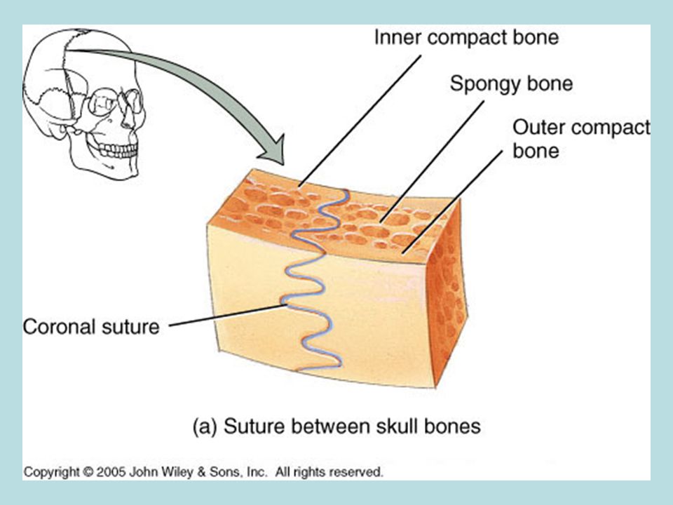
Suture joints: fibrous joints in which the bones forming the joint are in direct contact. Found between bones of the cranium. Neighboring bones have “saw-like” projections to help lock the bones together producing a very strong joint. Joints are actually stronger than the bone itself! Synostosis: A joint in which there is a complete fusion of the two separate bones into one bone. The saw-like projections completely ossify If this happens too quickly in the skull, it could affect brain development.

7
Types of Fibrous Joints
Syndesmoses: fibrous joints in which there is a greater distance between the articulating surfaces and more dense iregular connective tissue than in a suture. Found between the bones of the forearm (radius and ulna) and the bones of the lower leg (tibia and fibula). Bones are held in place by the interosseus membrane which prevents bones from separating when weight is applied.
 and the bones of the lower leg (tibia and fibula). Bones are held in place by the interosseus membrane which prevents bones from separating when weight is applied.")
9
Types of Fibrous Joints
Gomphoses: fibrous joints in which a cone-shaped peg fits into a socket. Hold the teeth in place A thin piece of connective tissue called the periodontal ligament helps secure the root of a tooth into the socket of the upper/lower jaw.

11
Cartilaginous Joints Lack a synovial cavity and the articulating bones are tightly connected by either hyaline cartilage or by fibrocartilage. Two Types: Synchondroses: cartilaginous joints in which the connecting material is hyaline cartilage. Often found between the ends (epiphyses) and shaft (diaphysis) of a long bone. Epiphyseal plate Once it gets turned into bone, it could be considered a fibrous joint. Allow the ends of bones to shift slightly to compensate for muscle development.
 and shaft (diaphysis) of a long bone. Epiphyseal plate. Once it gets turned into bone, it could be considered a fibrous joint. Allow the ends of bones to shift slightly to compensate for muscle development.")
13
Cartilaginous Joints Symphyses: cartilaginous joints in which the ends of the articulating bones are covered with hyaline cartilage but a broad, flat disc of fibrocartilage connects the bones. This is found between the bones of the spine and hip Allows bones to shift slightly during movement The hormone relaxin is secreted during childbirth to soften the connective tissue.

15
Synovial Joints- have a synovial cavity
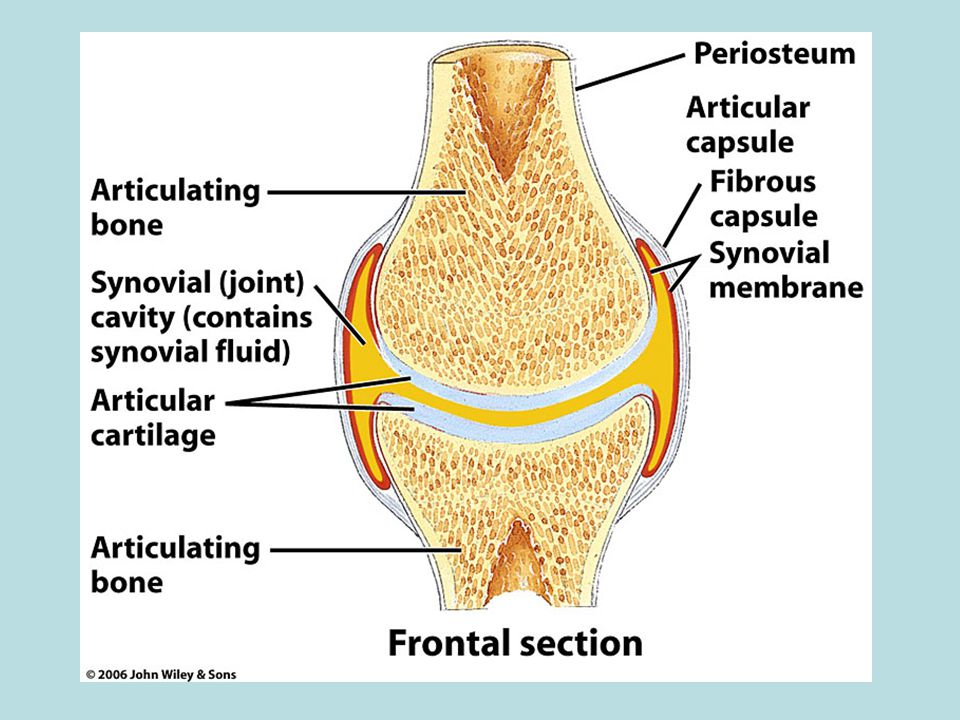
This is the most complex of the 3 structural joints. These are forms of diarthroses. Synovial cavity: a space between the articulating bones. Filled with synovial fluid This space allows joints to move freely. Synovial capsule: surround a synovial joint, encloses a synovial joint and unites the articulating bones. Combination of fibrous connective tissue and the synovial membrane.

17
Layers of the Synovial Capsule
Fibrous capsule: a layer of dense irregular connective tissue that attaches to the periosteum of the articulating bones. Functions to help hold the articulating bones together and limits the range of motion for that joint (to limit injury). Synovial membrane: a layer of areolar connective tissue that contains elastic fibers. Functions to produce and secrete synovial fluid that acts as an additional shock absorber. Synovial fluid is slippery and viscous.
. Synovial membrane: a layer of areolar connective tissue that contains elastic fibers. Functions to produce and secrete synovial fluid that acts as an additional shock absorber. Synovial fluid is slippery and viscous.")
18
6 Types of Synovial Joints
Planar: The articulating surfaces of the bones are flat or lightly curved. Also called a gliding joint. Found between carpal (wrist) and tarsal (ankle) bones. Sliding movement to even the distribution of forces to the arms and legs.
 and tarsal (ankle) bones. Sliding movement to even the distribution of forces to the arms and legs.")
20
6 Types of Synovial Joints
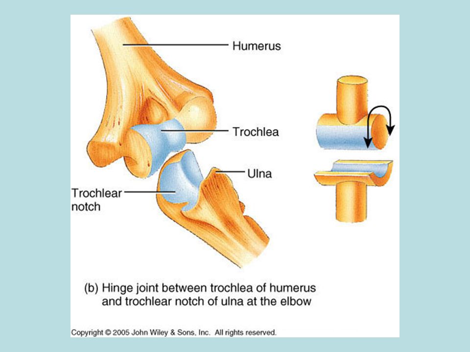
Hinge: The convex articulating surface of one bone fits into the concave articulating surface of another bone. Found at the elbow and knee. Opening and closing movements in 1 plane. Shape of the joint limits movement so this is the easiest synovial joint to damage.

22
6 Types of Synovial Joints
Pivot: The rounded or pointed surface of one bone articulates with a ring formed partly by another bone and partly by a ligament. Found between the 1st 2 vertebrae (atlas and axis) and the bones of the forearm (radius and ulna) Moves by the rotation of 1 bones around its own long axis. (Rotation of the radius to turn the palm over)
 and the bones of the forearm (radius and ulna) Moves by the rotation of 1 bones around its own long axis. (Rotation of the radius to turn the palm over)")
24
6 Types of Synovial Joints
Condyloid: The convex, oval-shaped projection of one bone fits into the oval-shaped depression of another bone. Found at the following: Between the head and 1st vertebra Between the skull and lower jaw Between the forearm and wrist Between the palm and fingers Between the lower leg and ankle of the radius to turn the palm over) Moves front to back and side to side like in chewing
 Moves front to back and side to side like in chewing.")
26
6 Types of Synovial Joints
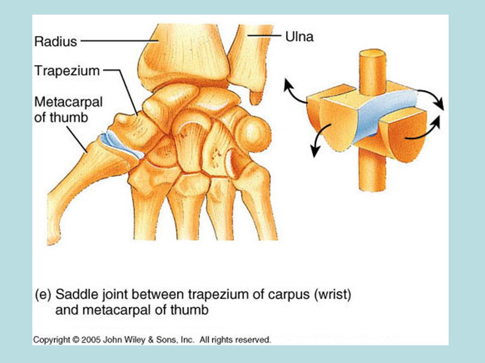
Saddle: the articular surface of one bone is saddle-shaped and the articular surface of the other bone fits into the saddle. Found at the base of each thumb Moves front to back, side to side, circular and opposing the pinky

28
6 Types of Synovial Joints
Ball and socket: the ball-like articulating surface of one bone fits into the cup-like depression of another bone. Found at the shoulder and hip Provides the most versatile movement- front to back, side to side, circular, and rotating

30
ACT-UP

31
ACT-UP 1) What part of the body is being shown in the slide?
2) What is the structural classification of the joint shown?
 What is the structural classification of the joint shown")
32
ACT-UP 3) What part of the body is being shown in the slide?.
4) What is the structural classification of the joint shown?
 What is the structural classification of the joint shown")
33
ACT-UP 5) Which of the joints pictured offers the greatest range of motion and why?
 Which of the joints pictured offers the greatest range of motion and why")
Similar presentations


![Articulations. Articulations- points where two or more bones come together to form a joint [ maybe rigid or movable] Classified by Structure or Function.](/13/3878517/big_thumb.jpg "Articulations. Articulations- points where two or more bones come together to form a joint [ maybe rigid or movable] Classified by Structure or Function.>")


 A point of contact between bones, between cartilage and bone or between teeth and bone.>")
: a point of contact between bones. Some allow movement, others are immovable (sutures). Most joints.>")


 and type of substance.>")


 Fibrous Joints 1) connections between adjacent bones 2) syndesmoses to gomphoses 3) ex.suture c) Cartilagenous.>")


