Download presentation
Presentation is loading. Please wait.

1
The Knee Complex

2
The Knee Complex General Structure & Function
Structure & Function of Specific Joints Muscular Considerations

3
General Structure

5
Joints of the Knee Complex

6
General Function Provides very mobile link in an otherwise stable lower extremity Transmits loads from tibia/fibula to femur

9
Knee Complex Movements

10
Transverse plane Medial and lateral rotation Sagittal plane Flexion, extension

11
Knee Complex Movements
Frontal plane Varus, valgus Anteroposterior translation Mediolateral translation

12
The Knee Complex General Structure & Function
Structure & Function of Specific Joints Muscular Considerations

13
Structure & Function of Specific Joints
Tibiofibular Joint Patellofemoral Joint Tibiofemoral Joint

14
Tibiofibular Joint: Bony Structure
Amphiarthrodial membranous syndesmosis joint

15
Structure & Function of Specific Joints
Tibiofibular Joint Patellofemoral Joint Tibiofemoral Joint

16
Purpose of Patella Increase leverage of QF
Protect joint during knee flexion ↓ pressure and distribute forces on femur Prevent Fcompression on PT in resisted knee flexion Disadvantage: ANT shear of QF Increase leverage of QF Protect joint during knee flexion ↓ pressure and distribute forces on femur Prevent Fcompression on PT with resisted knee flexion as in deep knee bends

17
Patella Structure Medial facet Lateral facet Odd facet (30%) M L
 M L")
18
PF Articular Surfaces Largest sesamoid bone Least congruent joint
Articular cartilage Vertical ridge Facets M L Largest sesamoid bone Least congruent joint in body Posterior surface covered with articular cartilage Vertical ridge on posterior, typically in center but can be situated more medially 30% of patellae have a second vertical ridge toward medial border, creating the odd facet on the extreme medial edge Angle of femoral sulcus (angle formed by medial & lateral facets of femur) averages 138° but varies widely (116° -151°)
 averages 138° but varies widely (116° -151°)")
19
PF Articular Surfaces Largest sesamoid bone Least congruent joint
Articular cartilage Vertical ridge Facets Angle of femoral sulcus Largest sesamoid bone Least congruent joint in body Posterior surface covered with articular cartilage Vertical ridge on posterior, typically in center but can be situated more medially 30% of patellae have a second vertical ridge toward medial border, creating the odd facet on the extreme medial edge Angle of femoral sulcus (angle formed by medial & lateral facets of femur) averages 138° but varies widely (116° -151°)
 averages 138° but varies widely (116° -151°)")
20
Patellar Motion INF & SUP Sliding Patellar tilt 11 MT as KN FL Med
Lat Sliding during knee flexion – enters sulcus at 20° of knee flexion Patellar tilt rotation about vertical axis to medial side of 11° occurs as knee flexion occurs

21
Patellar Motion Lateral rotation Medial rotation ACC MR of femur
6 through KN FL Medial rotation ACC LR of femur Lateral rotation of patella (around AP axis) with medial rotation of femur on tibia (top of patella goes with femur (7° - most by 60 ° of knee flexion) Medial rotation of patella (around AP axis) with lateral rotation of femur on tibia Failure to slide, tilt, or rotate will lead to restriction of knee joint ROM, instability of PF joint, or pain due to erosion of PF surfaces
 with medial rotation of femur on tibia (top of patella goes with femur (7° - most by 60 ° of knee flexion) Medial rotation of patella (around AP axis) with lateral rotation of femur on tibia. Failure to slide, tilt, or rotate will lead to restriction of knee joint ROM, instability of PF joint, or pain due to erosion of PF surfaces.")
22
Patellalectomy ↓ MA of QF (↓ strength 49%) Q tendon friction
compressive stress on groove by Q tendon Most evident in closed chain EXT ECC QF in CC Coupled w/ & assisted by hip & ankle movement QF not needed in erect posture of CC

23
Extension Little effect overall

24
Slight Flexion Noticeable weakness

25
Extreme Flexion Noticeable weakness

26
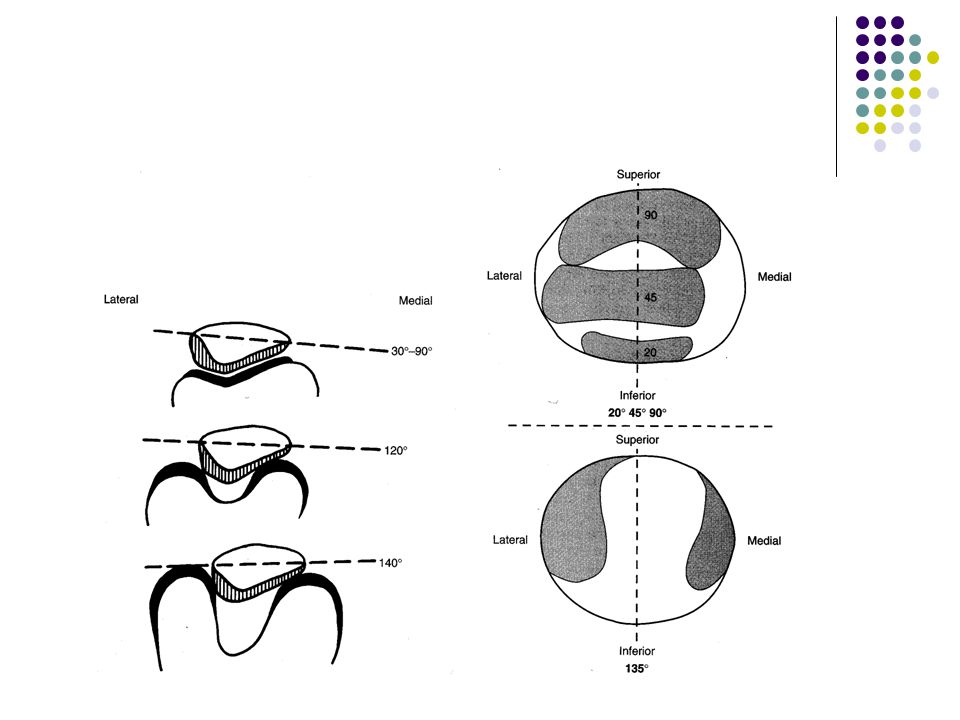
From 0° to 60° of Knee Flexion

27
0-60 Contact area MA of QF; 60 ANT shear of QF 0-60 Facet contact at 20

28
From 60° to 140° of Knee Flexion

29
60-140 contact area MA of QF No leverage in full FL

30
Overall Medial facet most contact Odd facet least contact

31
During Full Extension Full EXT MA of QF QF length
Patella very unstable

32
PF JRF Amount of knee FL Strength of QF contraction
Collinear pull of patella tendon (inferior) and quadriceps tendon (superior) results in little or no contact with femur in full extension Justification for use of straight-leg raises to improve quad strength without creating or exacerbating PF problems With knee flexion, the pull of these tendons create a compressive force on patella into femur (Fig ) (occurs with active or passive mechanism due to elasticity of tendon) FQ and FP are generally not equal – depend on knee joint angle (Fig. 6.2) Creates JRF (magnitude of JRF depends on magnitude of active and passive pulls, and degree of knee flexion) JRF during gait at foot contact (10° -15° of knee flexion) is 50% of body weight In running, JRF may increase to 3.3X (60° of knee flexion & more vigorous muscle contraction) 7.8X BW (130° of knee flexion with strong muscle contraction) in deep knee bends Lack of congruency at this joint results in this force distribution occurring over smaller area – more stress/pressure Medial facet bears the brunt of this, but there are mechanisms to minimize this Lowest at knee flexion < 30 degrees
 and quadriceps tendon (superior) results in little or no contact with femur in full extension. Justification for use of straight-leg raises to improve quad strength without creating or exacerbating PF problems. With knee flexion, the pull of these tendons create a compressive force on patella into femur (Fig ) (occurs with active or passive mechanism due to elasticity of tendon) FQ and FP are generally not equal – depend on knee joint angle (Fig. 6.2) Creates JRF (magnitude of JRF depends on magnitude of active and passive pulls, and degree of knee flexion) JRF during gait at foot contact (10° -15° of knee flexion) is 50% of body weight. In running, JRF may increase to 3.3X (60° of knee flexion & more vigorous muscle contraction) 7.8X BW (130° of knee flexion with strong muscle contraction) in deep knee bends. Lack of congruency at this joint results in this force distribution occurring over smaller area – more stress/pressure. Medial facet bears the brunt of this, but there are mechanisms to minimize this. Lowest at knee flexion < 30 degrees.")
34
PF Compressive Forces Descending stairs 4000 N Max isometric extension
Kicking 6800 N Parallel squat 14,900 N (7-8X BW) Isokinetic knee extension 8300 N Rising from chair 3800 N Running/jogging 5000 N (3-4X BW) Ascending stairs 1400 N Walking N ( X BW) Cycling 880 N
 Isokinetic knee extension N. Rising from chair N. Running/jogging N (3-4X BW) Ascending stairs N. Walking N ( X BW) Cycling. 880 N.")
35
Compensatory Mechanisms for Compressive Force Distribution
Contact area with knee flexion Medial facet contact from 30-70 Thickest hyaline cartilage in body

37
Compensatory Mechanisms for Compressive Force Distribution
Contact area with knee flexion Medial facet contact from 30-70 Thickest hyaline cartilage in body Largest QF MA 30-70 QF torque as MA decreases QF tendon contacts condyles 70-90

38
Normal Patella Tracking
Maintains maximum congruence Passive restraints Active restraints
![]()
39
Abnormal Patella Tracking
↓ congruence Stretches capsule & retinacula ↓ contact area Lateral Medial
![]()
40
Causes of Abnormal Tracking
Skeletal abnormalities Strength imbalance in QF Strength imbalance in fibrous tissues Compensatory movements in knee due to abnormal foot movement
![]()
41
Causes of Abnormal Tracking
Skeletal abnormalities Strength imbalance in QF Strength imbalance in fibrous tissues Compensatory movements in knee due to abnormal foot movement
![]()
42
Skeletal Abnormalities: Q-angle

43
Skeletal Abnormalities: Genu Varum & Genu Valgum
Q angle w/ age Varum common in very young children Valgum seen in growing children Menisectomy effects

44
Skeletal Abnormalities: Patella Alta & Patella Baja
Index of Insall & Salviti LT/LP Normal = 1.0 Patella alta = 0.8 Patella baja = 1.2 Women ratio

45
Skeletal Abnormalities: Patella Surface Lateral Border
Appositional forces ↓ in full extension Prominence of lateral border prevents lateral displacement Underdevelopment common in children as growing

46
Skeletal Abnormalities: Femoral & Tibial Torsion
Lateral tracking

47
Causes of Abnormal Tracking
Skeletal abnormalities Strength imbalance in QF Strength imbalance in fibrous tissues Compensatory movements in knee due to abnormal foot movement
![]()
48
QF Strength Imbalance

49
Causes of Abnormal Tracking
Skeletal abnormalities Strength imbalance in QF Strength imbalance in fibrous tissues Compensatory movements in knee due to abnormal foot movement
![]()
50
Fibrous Tissue Strength Imbalance
IT

51
Causes of Abnormal Tracking
Skeletal abnormalities Strength imbalance in QF Strength imbalance in fibrous tissues Compensatory movements in knee due to abnormal foot movement
![]()
52
Compensatory Movement
Pronation of foot accompanied by medial rotation of tibia medial rotation & medial translation of patella Pronation coupled w/ forceful quadriceps femoris leads to anterior tilt EX: jumping, landing, running

53
Summary

Similar presentations


 by Susan J. Hall, Ph.D.>")


 articulating with 2 concave surfaces (tibia) Poor bony stability Stability increased.>")


 practical section>")


 Medial condyle (8 left) Intercondylar fossa (7 left)>")
