Download presentation
Presentation is loading. Please wait.

1
Innate Host Resistance
33 Innate Host Resistance Copyright © McGraw-Hill Global Education Holdings, LLC. Permission required for reproduction or display.

2
Immunity Nonspecific immune response – Ch 33
Aka nonspecific resistance, innate, or natural immunity acts as a first line of defense offers resistance to any microbe or foreign material lacks immunological memory Specific immune response – Ch 34 Aka acquired, adaptive, or specific immunity resistance to a particular foreign agent has “memory” effectiveness increases on repeated exposure to agent

4
White Blood Cells of Innate and Adaptive Immunity
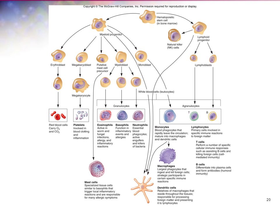
White blood cells (WBCs) play a major role in the innate and specific responses Hematopoesis development of white blood cells in bone marrow of mammals WBCs that mature prior to leaving bone marrow, e.g., macrophages and dendritic cells, become part of innate immune system and will respond to all antigens WBCs that are mature but not yet activated after leaving bone marrow become part of the adaptive immune response, e.g., B and T cells and could differentiate in response to specific antigens
 play a major role in the innate and specific responses. Hematopoesis. development of white blood cells in bone marrow of mammals. WBCs that mature prior to leaving bone marrow, e.g., macrophages and dendritic cells, become part of innate immune system and will respond to all antigens. WBCs that are mature but not yet activated after leaving bone marrow become part of the adaptive immune response, e.g., B and T cells and could differentiate in response to specific antigens.")
5
Physical Barriers in Nonspecific (Innate) Resistance
Effectiveness impacted by: direct factors nutrition, physiology, fever, age, and genetics indirect factors personal hygiene, socioeconomic status, and living conditions Along with host’s secretions (flushing), barriers = first line of defense against microbes
, barriers = first line of defense against microbes.")
6
Skin Strong mechanical barrier to microbial invasion
keratin produced by keratinocytes in outer layer Inhospitable environment for microbes attached organisms removed by shedding of outer skin cells pH is slightly acidic high NaCl concentration subject to periodic drying

7
Mucous Membranes Form protective covering that resists penetration and traps many microbes Are often bathed in antimicrobial secretions which contain a variety of antimicrobial substances (chemical mediators) lysozyme hydrolyzes bond connecting sugars in peptidoglycan lactoferrin secreted by activated macro- phages sequesters iron from plasma lactoperoxidase produces superoxide radicals
 lysozyme. hydrolyzes bond connecting sugars in peptidoglycan. lactoferrin. secreted by activated macro- phages. sequesters iron from plasma. lactoperoxidase. produces superoxide radicals.")
8
Respiratory System Turbulent air flow deposits microbes onto mucosal surfaces Mucociliary blanket mucous secretions trap microbes once trapped, microbes transported away from the lungs (mucociliary escalator) expelled by coughing or sneezing salivation washes microbes to stomach Alveolar macrophages phagocytic cells in alveoli of lungs
 expelled by coughing or sneezing. salivation washes microbes to stomach. Alveolar macrophages. phagocytic cells in alveoli of lungs.")
9
Gastrointestinal Tract
Intestines shedding of columnar epithelial cells secretory IgA normal microbiota Paneth cells produce lysozyme produce cryptins Stomach gastric acid Intestines pancreatic enzymes bile intestinal enzymes GALT peristalsis

10
Genitourinary Tract Unfavorable environment for foreign microbes
low pH of urine and vagina vagina has lactobacilli urea and other toxic metabolic end products in urine hypertonic nature of kidney medulla Flushing with urine and mucus Distance barrier of male urethra

11
The Eye Physical protection from the eye lid and eye lashes
Mucus secreting epithelial membrane Flushing action of tears Lysozyme, lactoferrin, and secretory IgA in tears

12
Chemical Mediators in Nonspecific (Innate) Resistance
Many already noted (e.g., gastric juices, lysozyme, lactoferrin, urea) A variety of defensive chemicals such as defensins and other polypeptides are also found in blood, lymph, and other body fluids
 A variety of defensive chemicals such as defensins and other polypeptides are also found in blood, lymph, and other body fluids.")
13
Antimicrobial Peptides
Cationic peptides - three classes whose biological activity is related to their ability to damage bacterial plasma membranes First class: linear, alpha-helical peptides that lack cysteine amino acid residues e.g., cathelicidin, produced by a variety of cells Second class: defensins peptides that are open-ended, rich in arginine and cysteine, and disulfide linked found in neutrophils, intestinal Paneth cells and intestinal and respiratory epithelial cells Third class: larger peptides that are enriched for specific amino acids and exhibit regular structural repeats e.g., histatin, present in human saliva and has anti-fungal activity

14
Bacteriocins Peptides produced by normal microbiota
Lethal to related species Produced by Gram-positive and Gram-negative cells e.g., colicins produced by E. coli e.g., lantibiotics produced by Gram-positive bacteria

15
The Complement System Composed of >30 serum proteins
Augments (or “complements”) the antibacterial activity of antibody (works with the adaptive immune system) Three major activities: defending against bacterial infections bridging innate and adaptive immunity disposing of wastes Other activities: Function as chemotactic signals that recruit phagocytes to their activation site Puncture cell membranes causing cell lysis Many complement activities unite the nonspecific and specific arms of the immune system to destroy and remove invading pathogens
 the antibacterial activity of antibody (works with the adaptive immune system) Three major activities: defending against bacterial infections. bridging innate and adaptive immunity. disposing of wastes. Other activities: Function as chemotactic signals that recruit phagocytes to their activation site. Puncture cell membranes causing cell lysis. Many complement activities unite the nonspecific and specific arms of the immune system to destroy and remove invading pathogens.")
16
Opsonization Process in which microbes are coated by serum components (opsonins) in preparation for recognition/ingestion by phagocytic cells Some complement proteins are opsonins bind to microbial cells, coating them for phagocyte recognition

17
Cytokines Soluble proteins or glycoproteins that are released by one cell population that act as intercellular mediators or signaling molecules Three proposed groups based on function regulators of innate resistance mechanisms regulators of adaptive immunity stimulators of hematopoiesis

19
Cells of the Immune System
Granulocytes Mast cells Monocytes and macrophages Dendritic cells Lymphocytes Each has specialized role in defending host Leukocytes white blood cells involved in both specific and nonspecific immunity all arise from pluripotent stem cells

21
Mast Cells Granulocytes Bone marrow-derived cells
Differentiate in blood and connective tissue Contain granules containing histamine and other pharmacologically active chemicals Play important role in development of allergies and hypersensitivities Granulocytes Irregularly-shaped nuclei with two to five lobes Cytoplasm has granules with reactive substances kill microbes, enhance inflammation Three types basophils, eosinophils, neutrophils (polymorphonuclear neutrophil (PMN))
)")
22
Basophils Eosinophils Nonphagocytic Release vasoactive mediators
e.g., histamine, prostaglandins, serotonin, and leukotrienes from granules Play important role in development of allergies and hypersensitivities Eosinophils Defend against protozoan and helminth parasites Release cationic proteins and reactive oxygen metabolites May play a role in allergic reactions

23
Monocytes and Macrophages
Neutrophils Highly phagocytic Circulate in blood then migrate to sites of tissue damage Kill ingested microbes with lytic enzymes and reactive oxygen metabolites contained in primary and secondary granules Monocytes and Macrophages Highly phagocytic cells Monocytes after circulating for ~8 hours, mature into macrophages Macrophages larger than monocytes, reside in specific tissues, highly phagocytic have a variety of surface receptors (including pattern recognition receptors) named according to tissue in which they reside
 named according to tissue in which they reside.")
24
Natural Killer (NK) Cells
Dendritic Cells Heterogeneous group of cells with neuron-like appendages Present in small numbers in blood, skin, and mucous membranes of nose, lungs, and intestines contact, phagocytose, and process antigens display foreign antigens on their surfaces (antigen presentation) Natural Killer (NK) Cells Small population of large non-phagocytic granular lymphocytes important role in innate immunity kill malignant cells and cells infected with pathogens by releasing granzymes (cytotoxic enzymes) Two ways of recognizing target cells bind to antibodies which coat infected or malignant cells (antibody- dependent cell-mediated cytotoxicity (ADCC) recognizes cells that have lost their class I major histocompatibility antigen due to presence of virus or cancer
 Natural Killer (NK) Cells. Small population of large non-phagocytic granular lymphocytes. important role in innate immunity. kill malignant cells and cells infected with pathogens by releasing granzymes (cytotoxic enzymes) Two ways of recognizing target cells. bind to antibodies which coat infected or malignant cells (antibody- dependent cell-mediated cytotoxicity (ADCC) recognizes cells that have lost their class I major histocompatibility antigen due to presence of virus or cancer.")
25
Lymphocytes Major cells of the immune system
Major populations include T cells, B cells, and natural killer (NK) cells B and T lymphocytes differentiate in bone marrow from stem cells are only activated by binding of specific antigen onto lymphocyte surface receptors after activation replication continues as lymphocytes circulate and enter lymphoid tissue memory cells are activated lymphocytes that do not immediately replicate, but will do so later in host’s life when antigen is again present
 cells. B and T lymphocytes differentiate in bone marrow from stem cells. are only activated by binding of specific antigen onto lymphocyte surface receptors. after activation replication continues as lymphocytes circulate and enter lymphoid tissue. memory cells are activated lymphocytes that do not immediately replicate, but will do so later in host’s life when antigen is again present.")
26
T Lymphocytes (T cells)
B Lymphocytes B cells (B lymphocytes) mature in bone marrow circulate in blood can settle in lymphoid organs after maturation and activation are called plasma cells and produce antibodies T Lymphocytes (T cells) Mature in thymus Can remain in thymus, circulate in blood, or reside in lymphoid tissue Like B cells, require antigen binding to surface receptors for activation and continuation of replication Activated T cells differentiate into helper T cells (TH) and cytotoxic lymphocytes (CTLs) Secrete cytokines, chemicals that have effects on other cells, are produced and secreted by activated T cells
 mature in bone marrow. circulate in blood. can settle in lymphoid organs. after maturation and activation are called plasma cells and produce antibodies. T Lymphocytes (T cells) Mature in thymus. Can remain in thymus, circulate in blood, or reside in lymphoid tissue. Like B cells, require antigen binding to surface receptors for activation and continuation of replication. Activated T cells differentiate into helper T cells (TH) and cytotoxic lymphocytes (CTLs) Secrete cytokines, chemicals that have effects on other cells, are produced and secreted by activated T cells.")
28
Organs and Tissues of the Immune System
Primary organs and tissues sites where lymphocytes mature and differentiate into antigen-sensitive mature B and T cells Secondary organs and tissues areas where lymphocytes may encounter and bind antigen followed by proliferation and differentiation into fully mature effector cells

29
Primary Lymphoid Organs and Tissues
Thymus precursor cells move enter from bone marrow and proliferate thymic deletion removes T cells recognizing self antigens remaining cells become mature T cells enter bloodstream and recognize nonself antigens Bone marrow site of B cell maturation in mammals maturation involves removal of nonfunctioning and self-reactive cells

30
Secondary Lymphoid Organs and Tissues
Spleen most highly organized lymphoid organ filters blood macrophages and dendritic cells trap microbes and antigens present antigens to B and T cells most common way that lymphocytes become activated to carry out their immune functions Lymph nodes most highly organized lymphoid tissue filter lymph microbes and antigens trapped and phagocytosed by macrophages and dendritic cells B cells differentiate into memory and plasma cells within lymph nodes

31
Secondary Lymphoid Organs and Tissues
Lymphoid tissue located throughout the body serve as interface between innate and acquired host immunity act as areas of antigen sampling and processing some lymphoid cells are found closely associated with specific tissues e.g., skin-associated lymphoid tissue (SALT) e.g., mucous-associated lymphoid tissue (MALT)
 e.g., mucous-associated lymphoid tissue (MALT)")
32
Skin Associated Lymphoid Tissue (SALT)
Contains specialized cells Langerhans cell dendritic cell that can phagocytose antigens differentiates into interdigitating dendritic cell – presents antigen to and activates T cells intraepidermal lymphocyte function as T cells

33
Mucosal-Associated Lymphoid Tissue (MALT)
Specialized immune barrier gut-associated lymphoid tissue (GALT) bronchial-associated lymphoid tissue (BALT) urogenital system MALT
 bronchial-associated lymphoid tissue (BALT) urogenital system MALT.")
34
Phagocytosis Process by which phagocytic cells (monocytes, tissue macrophages, dendritic cells, and neutrophils) recognize, ingest, and kill extracellular microbes Two mechanisms for recognition of microbe by phagocyte opsonin-independent (nonopsonic) recognition opsonin-dependent (opsonic) recognition Phagocytosis can be greatly increased by opsonization
 recognize, ingest, and kill extracellular microbes. Two mechanisms for recognition of microbe by phagocyte. opsonin-independent (nonopsonic) recognition. opsonin-dependent (opsonic) recognition. Phagocytosis can be greatly increased by opsonization.")
35
Pathogen-Associated Molecular Patterns (PAMPs)
Based on detection, by phagocytes, of conserved microbial molecular structures that occur in patterns PAMPs are unique to microbes, not present in host e.g., lipopolysaccharide (LPS) of Gram-negative bacteria e.g., peptidoglycan of Gram-positive bacteria PAMPs recognized by pattern recognition receptors (PRRs) on/in phagocytic cells PRRs can work alone or together to trigger phagocytes
 of Gram-negative bacteria. e.g., peptidoglycan of Gram-positive bacteria. PAMPs recognized by pattern recognition receptors (PRRs) on/in phagocytic cells. PRRs can work alone or together to trigger phagocytes.")
36
Inflammation Nonspecific response to tissue injury Cardinal signs
can be caused by pathogen or physical trauma acute inflammation is the immediate response of body to injury or cell death Cardinal signs redness (rubor) warmth (calor) pain (dolor) swelling (tumor) altered function (functio laesa)
 warmth (calor) pain (dolor) swelling (tumor) altered function (functio laesa)")
37
Acute Inflammatory Response
The release of inflammatory mediators from injured tissue cells initiates a cascade of events which result in the signs of inflammation Involves chemical mediators selectins cell adhesion molecules on activated capillary endothelial cells integrins adhesion receptors on neutrophils chemotaxins chemotactic factors released by injured cells

38
Acute Inflammatory Response
Various processes occur margination diapedesis extravasion

39
More about Acute Inflammation…
Tissue injury releases kalikrein and other mediators increases capillary dilation and blood flow brings more antimicrobial factors and leukocytes that kill pathogens Fibrin clot may restrict pathogen movement Phagocytes accumulate in inflamed area and destroy pathogens Bone marrow stimulated to release neutrophils and increase rate of granulocyte production

41
Chronic Inflammation Slow process
Involves formation of new connective tissue Usually causes permanent tissue damage Dense infiltration of lymphocytes and macrophages at site of inflammation granuloma walled off area formed when phagocytic cells can’t destroy pathogen

Similar presentations


 BIOS 486A/586A>")


>")


 Immune system.>")


