Download presentation
Presentation is loading. Please wait.

1
Eye diseases of cornea, lens and vitreous 4/9/13
Lecture #19 Eye diseases of cornea, lens and vitreous 4/9/13

2
Animal wikis Great! Some of my favorites
Writing: manatees, hummingbirds Link to eye design: barn owls, panda

3
Wiki homework Be thinking about your wiki final project topic
it to me by end of Thursday It is fine if your topic evolves as you gather information May want to focus it down if find lots info May need to expand if not so much

4
Anterior eye disease Cornea Lens Vitreous Dystrophies
Refractive errors Lens Cataracts Vitreous Glaucoma

5
Function of cornea Performs ≈70% of focusing
Protects eye from outside world No blood supply Cleaned and nourished by tears and aqueous humour

6
Corneal disease Corneal infections Conjunctiva
Mucous membrane lining eyelid and sclera Contains tiny blood vessels Pink eye - conjunctivitis Infection by either bacteria or virus Corneal infections Bacterial or fungal invasion into corneal layers Most often caused by virus, same one as causes common cold. If child gets it, you have to remove them from public setting and get them treated for bacterial infection though it can often be the untreatable viral form. Both are pretty contagious.

7
Dry eye

8
Tears Basal tears Reflex tears
Constantly produced to nourish and moisten eye Mixture of aqueous and oily secretions Reflex tears Made in response to irritation or emotion More watery See for some entertaining

9
What are tears? Tears are made of three layers
Oily, lipid layer - keeps aqueous layer from evaporating Aqueous layer - keeps eye moist Mucin layer - helps aqueous layer spread

10
Meibomian gland produces lipid part
Discovered by Heinrich Meibom in 1600’s Meibomian gland makes meibum - oily part of tears; 50 glands on upper lid; 25 on lower

11
Lacrimal glands produce aqueous part Tears drain to naso-lacrimal sac
If not enough water then truly have dry eye

12
Goblet cells produce mucus
Helps watery film wet the eye surface

13
Tears then need to drain
Tears then drain out through holes in eyelid If drain too quickly, eyes become dry Plug these holes

14
Dry eye If meibomian glands get blocked, there will not be enough lipids and tears will evaporate too quickly To unclog glands Heat treatments Doxycycline Nutritional supplements May be other reasons not enough lipids

15
Dry eye If there is not enough aqueous part of tears
Use artificial tears Plug up drainage holes so stay on eye longer May also be problems with mucin layer which wets the eye and helps aqueous layer to spread Not sure how to improve it

16
Cornea has 5 layers Epithelium 10% of thickness Blocks foreign matter
Absorbs O2 and nutrients from tears Epithelia cells grow and are anchored to basement membrane Many tiny neurons - very sensitive to pain See: for discussion of the 5 layers

17
Cornea has 5 layers 2. Bowman’s layer
Strong layer of fibers composed of collagen If injured it forms scar tissue

18
Cornea has 5 layers 3. Stroma Comprises 90% of cornea thickness
Composed mostly of collagen (16%) and water (78%) Gives cornea shape and transparency Upper part of stroma repairs itself but lower part does not
 and water (78%) Gives cornea shape and transparency. Upper part of stroma repairs itself but lower part does not.")
19
Cornea has 5 layers 4. Descemet’s membrane
Thin but strong protective layer Made of collagen (different from stroma) Made by endothelium Can regenerate after injury Descemet’s membrane
 Made by endothelium. Can regenerate after injury. Descemet’s membrane.")
20
Cornea has 5 layers 5. Endothelium Extremely thin
Fluid slowly leaks from inside eye into stroma Endothelium pumps it back out so stroma doesn’t get cloudy!! Endothelium does not regenerate - if damaged, need corneal transplant

21
Corneal dystrophies Over 20 kinds Dystrophy - abnormal development
Inherited Affect both eyes equally Begin in one of 5 layers and spread to others Layers become cloudy - so can’t see

22
Keratoconus Thinning of middle of cornea (stroma) causes cornea to change shape - cone like Most common corneal dystrophy Affects 1:2000 Inherited or from wearing hard contacts or eye injury Usually stabilizes and correct with glasses / contacts

23
Lattice dystrophy Build up of amyloid (protein) deposits in upper to middle stroma Create a lattice which worsens and makes cornea cloudy Most common occurs in children age 2-7 Treat with corneal transplant though 1/2 of people will get latice formation again

24
Fuchs dystrophy Endothelial layer deteriorates
Can’t pump out aqueous humour so cornea swells Vision becomes blurry

25
Treatments for corneal dystrophies
Corneal transplants Match by blood type 20% rejection rate

26
Treatment for corneal scars
Phototherapeutic keratectemy Laser ablation Remove scarred or damaged tissue Use UV excimer laser under computer control

27
Refractive error If cornea has wrong curvature, image on retina is out of focus Myopia - image focused in front of retina : 25% of people Hyperopia - image focused behind retina

28
Refractive error Astigmatism Multiple focal lengths so multiple images
Cornea is more curved in one direction than the other (like spoon or football) Multiple focal lengths so multiple images Always blurry
 Multiple focal lengths so multiple images. Always blurry.")
29
Treatments for refractive errors - reshaping the cornea
RK - Radial keratotomy PTK - Phototherapeutic keratectemy LASEK - Laser assisted sub-epithelial keritectomy LASIK - Laser Assisted In Situ Keratomileusis

30
Radial keratotomy Modify cornea shape by cutting slits
Developed in Russia in 1970s Unpredictable healing Vision may change through day or over time Not recommended

31
Treatment for refractive errors
Phototherapeutic keratectomy Can also be used to reshape cornea - correct myopia Remove epithelial layer and reshape upper part of cornea Epithelial layer regenerates Keratectomy - remove part of cornea

32
LASEK surgery Laser assisted sub-epithelial keratectomy
Cut and peel back epithelial layer Re-shape upper stroma just below epithelium with laser Replace epithelial layer

33
LASIK refractive surgery
Laser Assisted In Situ Keratomileusis Cut a flap in cornea with blade or laser (this cuts more than just epithelium) Laser vaporizes stroma to reshape it Flap is folded back though doesn’t seal Epi-LASIK cuts thinner flap so does reseal
 Laser vaporizes stroma to reshape it. Flap is folded back though doesn’t seal. Epi-LASIK cuts thinner flap so does reseal.")
34
What happens during LASIK surgery

35
Reshaping of cornea Near sighted Far sighted
Near sighted focuses too much. Need to make less rounded / more flat Far sighted focuses not enough. Need to make more rounded / less flat

36
Comparisons suggest LASEK and LASIK produce equivalent results

37
Some reasons NOT to do LASIK
You may not be suited for procedure: Eye disease Thin corneas Unstable vision Vision may get worse Unstable cornea No long term data LASIK corneal flap may be deep in cornea These tissues do not regenerate Flap is permanent

38
Possible complications - starbursts
LASIKdisaster.com

39
Possible complications - halos
LASIKdisaster.com

40
Ghosting

41
Near sighted problems - PRK

42
Far sighted problems

43
Possible problems

44
NEI - cataracts

46
Lens Lens Transparent so light is efficiently transmitted
High index so light is focused onto the retina

48
Lens composition Composed of water and lens crystallins (90% of protein) Crystallins made once and then stored in lens for rest of life Must remain soluble to be transparent Eye lens fiber cells filled with crystallins

49
Crystallins α-crystallins β and γ crystallins
Related to heat shock proteins β and γ crystallins γ crystallins are symmetric

50
Other proteins can be co-opted to form part of lens
Taxon specific crystallins which have in addition to alpha, beta and gamma Many are active metabolic enzymes elsewhere in body!!!

51
Recruitment of proteins
Recruited to lens by changing gene expression May be result of gene duplication followed by new expression Proteins selected which highly stable Contribute to index of refraction Insensitive to UV damage

52
Crystallin structure Crystallins are present from birth
Processes which damage protein are bad Oxidation, deamidation, cleavage Result in protein unfolding Normally α crystallins are chaperones keeping other proteins folded As lens proteins unfold, α crystallins used up Unfolded proteins form precipitates Loss of lens transparency Deamidation - removal of NH2 group - damages certain amino acids which would impact protein stability

53
Cataracts Clouding of lens Typically occurs with age
50% of people > 80 have cataracts Cataracts affect 5.5 million people in US ftp://ftp.nei.nih.gov/eyedis/EDA12_72.tif

54
Cateract symptoms Blurry vision Poor night vision Problems with glare

55
Cataracts Congenital Age related

56
Age related cataract prevention
Decrease sun exposure Increase antioxidants Stop smoking Get eye exam

57
Treatment #1 Cut small incision (3 mm)
Remove front of lens to expose cataract Use ultrasound to fragment cataract Remove fragments

58
Treatment #2 Replacement lens Made of plastic Blocks UV
Is flexible so can attach to eye focusing muscles Focus near and far!

59
Treatment #2 Introduce replacement lens into lens capsule
May only replace part of lens Can improve spectral transmission (more blue)
")
60
Glaucoma

62
Glaucoma Variety of diseases that result in loss of retinal ganglion cells Loss begins in periphery 50% of people have glaucoma and don’t realize it

63
Fluid flow at front of eye
Aqueous humor is generated by ciliary body and flows into anterior chamber to nourish eye Flows out where cornea and iris meet Iridocorneal angle Trabecular and uveoscleral drainage Spongy tissues

64
Fluid flow at front of eye
If fluid does not drain: Pressure in eye builds up This damages retinal ganglion cells and vision is lost

65
Measuring eye pressure
Applanation tonometry Measure applied pressure necessary to deflect cornea Noncontact tonometry Measure air pressure needed to deflect cornea

66
Caveats High intraoccular pressure (IOP) is highest risk factor but:
Majority of people with high IOP do not get glaucoma Optic nerve damage can occur even without high pressure -Low tension or normal tension glaucoma

67
Risk factors Affects 70 million people Age Family history of glaucoma
2 % over age 40; 7% over age 80 Over age 40 - African Americans 5x more likely Over age 60 - Mexican Americans more likely Family history of glaucoma Though not Mendelian trait

68
Symptoms Gradual loss of peripheral vision Can be slow loss over years
No pain Difficult to notice effects

69
Kinds of glaucoma Open angle glaucoma Developmental glaucoma
Fluid seems to keep flowing Developmental glaucoma Anterior portion of eye doesn’t develop correctly Pigmentary glaucoma Iris pigment epithelium atrophies and pigment clogs drainage of fluid

70
Open angle glaucoma The thing to note is that there are multiple factors on many different chromosomes

71
Genetics 9 loci identified so far that initiate primary open angle glaucoma Explain only small % of cases Two genes which cause early onset glaucoma Myocilin (3% of cases) Optineurin Not obvious how genes cause the disease Expressed in both retinal ganglion cells and trabecular meshwork May cause problems if protein misfolding
 Optineurin. Not obvious how genes cause the disease. Expressed in both retinal ganglion cells and trabecular meshwork. May cause problems if protein misfolding.")
72
Treatments Eye drops or pills Laser trabeculoplasty
Decrease fluid production or increase drainage Laser trabeculoplasty Laser widens holes in drainage meshwork Conventional surgery Create new exit pathways

73
Open fluid flow in meshwork or sclera

74
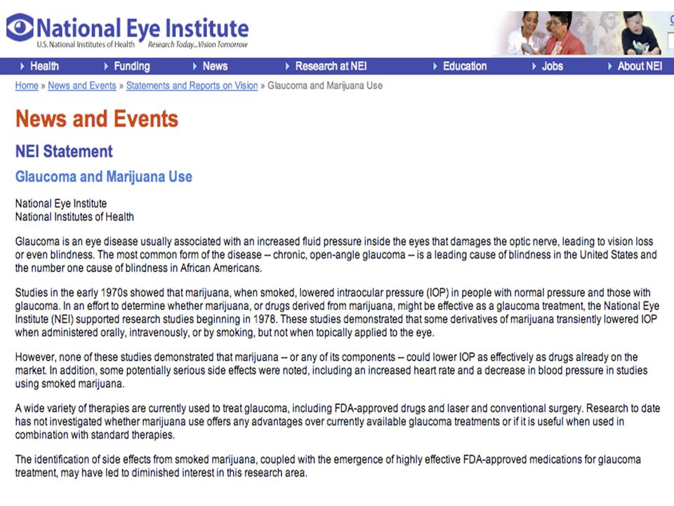
Use of marijuana to treat glaucoma

77
Next time Gene therapy How do you replace a faulty gene?

Similar presentations


>")


 is decreased vision that results from abnormal visual development in infancy and early childhood.>")
