Download presentation
Presentation is loading. Please wait.

1
The Essentials for Paraoptometric Personnel in Understanding What We Tell Our Patient’s About Eye Surgery Jeff D. Miller, O.D. Stillwater, Oklahoma millereyedoc@cockrelleyecare.com

2
Anatomy of the Eye Cornea Retina Lens Optic Nerve Iris Pupil Macula

3
Cataract Surgery A cataract is a clouding or opacification of the natural crystalline lens of the eye. As the lens becomes darker and darker the patients visual acuity drops to levels that limit them from performing their daily activities. A cataract is a clouding or opacification of the natural crystalline lens of the eye. As the lens becomes darker and darker the patients visual acuity drops to levels that limit them from performing their daily activities. Surgical removal allows restoration of vision and in most cases the opportunity to choose the patient’s final prescription through the lens implant power. Surgical removal allows restoration of vision and in most cases the opportunity to choose the patient’s final prescription through the lens implant power. Patients can choose to be clear in the distance, have mono vision or consider a multi-focal (bifocal) lens implant. Patients can choose to be clear in the distance, have mono vision or consider a multi-focal (bifocal) lens implant.
 lens implant. Patients can choose to be clear in the distance, have mono vision or consider a multi-focal (bifocal) lens implant..")
4
Cataract Surgery Cataract surgery has evolved tremendously over the last 25 years. Incisions made on the eye (scleral)have gone from 10-12 mm to 2-3mm and are now made on the cornea. Lens implants are now foldable to allow smaller incisions. These changes improve healing time, result in less swelling, produce less astigmatism, allow surgery to be completed without sutures, and can be done with topical anesthetic (drops) alone. That is, the patient is awake with only mild sedation from an IV. Cataract surgery has evolved tremendously over the last 25 years. Incisions made on the eye (scleral)have gone from 10-12 mm to 2-3mm and are now made on the cornea. Lens implants are now foldable to allow smaller incisions. These changes improve healing time, result in less swelling, produce less astigmatism, allow surgery to be completed without sutures, and can be done with topical anesthetic (drops) alone. That is, the patient is awake with only mild sedation from an IV.
have gone from mm to 2-3mm and are now made on the cornea. Lens implants are now foldable to allow smaller incisions. These changes improve healing time, result in less swelling, produce less astigmatism, allow surgery to be completed without sutures, and can be done with topical anesthetic (drops) alone. That is, the patient is awake with only mild sedation from an IV. Cataract surgery has evolved tremendously over the last 25 years. Incisions made on the eye (scleral)have gone from mm to 2-3mm and are now made on the cornea. Lens implants are now foldable to allow smaller incisions. These changes improve healing time, result in less swelling, produce less astigmatism, allow surgery to be completed without sutures, and can be done with topical anesthetic (drops) alone. That is, the patient is awake with only mild sedation from an IV..")
6
Cataract extraction is the most common outpatient surgery performed in the United States today. Cataracts are the leading cause of blindness in the world however, because of access and healthcare in the USA they are not a significant cause of “permanent” vision loss

7
Cataract Illustration and Video

8
The Future of Implants

9
Corneal Transplant The cornea is the clear dome shaped tissue on the front of the eye directly in front of the iris. The cornea is the clear dome shaped tissue on the front of the eye directly in front of the iris. The cornea is referred to as the window of the eye and must remain crystal clear or vision can be reduced. The cornea is referred to as the window of the eye and must remain crystal clear or vision can be reduced. The cornea is responsible for a significant portion of the “power” or prescription of the eye. The cornea is responsible for a significant portion of the “power” or prescription of the eye. The cornea can become cloudy or opacified for several reasons including trauma, infection, or corneal disease. The cornea can become cloudy or opacified for several reasons including trauma, infection, or corneal disease. Common reasons for a corneal transplant include herpetic eye disease, scarring from corneal ulcers, keratoconus, corneal dystrophies like Fuch’s endothelial dystrophy and trauma, Common reasons for a corneal transplant include herpetic eye disease, scarring from corneal ulcers, keratoconus, corneal dystrophies like Fuch’s endothelial dystrophy and trauma,

10
Anatomy of the Eye Cornea Retina Lens Optic Nerve Iris Pupil Macula

11
Corneal Transplant

12
Corneal Transplant Video Presentation

13
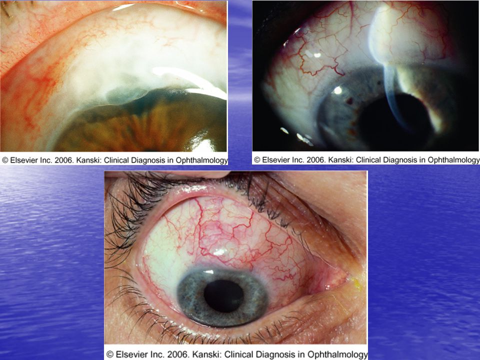
Pterygium Removal A Pinguecula and Pterygium – what’s the difference? A pinguecula (Latin for “fatty”)is a yellowish, raised growth located on the conjunctiva at 3:00 or 9:00 o’clock. It is commonly thought to be the precursor of a pterygium. As the mound of tissue (pinguecula) grows in size and onto the cornea it is then termed a pterygium. A Pinguecula and Pterygium – what’s the difference? A pinguecula (Latin for “fatty”)is a yellowish, raised growth located on the conjunctiva at 3:00 or 9:00 o’clock. It is commonly thought to be the precursor of a pterygium. As the mound of tissue (pinguecula) grows in size and onto the cornea it is then termed a pterygium. These are considered degenerative lesions causally related to chronic long term exposure to sunlight. These are considered degenerative lesions causally related to chronic long term exposure to sunlight. If allowed to grow in front of the pupil, scarring can result in a permanent decrease in vision in addition to If allowed to grow in front of the pupil, scarring can result in a permanent decrease in vision in addition to induced astigmatism. induced astigmatism.
is a yellowish, raised growth located on the conjunctiva at 3:00 or 9:00 o’clock. It is commonly thought to be the precursor of a pterygium. As the mound of tissue (pinguecula) grows in size and onto the cornea it is then termed a pterygium. A Pinguecula and Pterygium – what’s the difference. A pinguecula (Latin for fatty )is a yellowish, raised growth located on the conjunctiva at 3:00 or 9:00 o’clock. It is commonly thought to be the precursor of a pterygium. As the mound of tissue (pinguecula) grows in size and onto the cornea it is then termed a pterygium. These are considered degenerative lesions causally related to chronic long term exposure to sunlight. These are considered degenerative lesions causally related to chronic long term exposure to sunlight. If allowed to grow in front of the pupil, scarring can result in a permanent decrease in vision in addition to If allowed to grow in front of the pupil, scarring can result in a permanent decrease in vision in addition to induced astigmatism. induced astigmatism..")
15
Pterygium Removal Video Presentation

16
Glaucoma Surgery Trabeculectomy A trabeculectomy involves removing a tiny piece of the eye right at the place where the cornea connects to the sclera (the white part), and creating a flap to allow fluid to escape the anterior chamber without deflating the eye. A trabeculectomy involves removing a tiny piece of the eye right at the place where the cornea connects to the sclera (the white part), and creating a flap to allow fluid to escape the anterior chamber without deflating the eye. Along with that tiny piece of cornea and sclera comes a piece of iris. The whole area is called the trabeculum. Along with that tiny piece of cornea and sclera comes a piece of iris. The whole area is called the trabeculum. Fluid can then flow out onto the surface of the eye but under the conjunctiva, the clear tissue on top of the white part of the eye or sclera. Fluid can then flow out onto the surface of the eye but under the conjunctiva, the clear tissue on top of the white part of the eye or sclera. The fluid forms a “bleb” on the surface of the eye just under the top eyelid. The fluid is absorbed by the conjunctiva. The fluid forms a “bleb” on the surface of the eye just under the top eyelid. The fluid is absorbed by the conjunctiva.
, and creating a flap to allow fluid to escape the anterior chamber without deflating the eye. Along with that tiny piece of cornea and sclera comes a piece of iris. The whole area is called the trabeculum. Along with that tiny piece of cornea and sclera comes a piece of iris. The whole area is called the trabeculum. Fluid can then flow out onto the surface of the eye but under the conjunctiva, the clear tissue on top of the white part of the eye or sclera. Fluid can then flow out onto the surface of the eye but under the conjunctiva, the clear tissue on top of the white part of the eye or sclera. The fluid forms a bleb on the surface of the eye just under the top eyelid. The fluid is absorbed by the conjunctiva. The fluid forms a bleb on the surface of the eye just under the top eyelid. The fluid is absorbed by the conjunctiva..")
17
TRABECULECTOMY

19
Trabeculectomy vs. Shunts or Valves Trabeculectomies can scar shut such that the drainage of fluid is reduced or stopped and eye pressure goes back up to uncontrolled levels. Trabeculectomies can scar shut such that the drainage of fluid is reduced or stopped and eye pressure goes back up to uncontrolled levels. Various drugs have been used to control healing and scarring over time. Some work on certain individuals some don’t. (mitomycin C) Various drugs have been used to control healing and scarring over time. Some work on certain individuals some don’t. (mitomycin C) Valves or shunts have now become the choice of many glaucoma specialists because it is less likely in some patient populations these will fail over time. Valves or shunts have now become the choice of many glaucoma specialists because it is less likely in some patient populations these will fail over time.
 Various drugs have been used to control healing and scarring over time. Some work on certain individuals some don’t. (mitomycin C) Valves or shunts have now become the choice of many glaucoma specialists because it is less likely in some patient populations these will fail over time. Valves or shunts have now become the choice of many glaucoma specialists because it is less likely in some patient populations these will fail over time..")
20
The Ex-PRESS Mini Glaucoma Shunt Excessive Pressure REgulation Shunt System

22
Ex-PRESS TM in a Nutshell Consistent Predictable Results Reproducible Minimally invasive Consistent lumen Less inflammation Less complications Low diffuse blebs High success rate

24
Refractive Surgery LASIK or Laser Assisted In-Situ Keratomileusis – is a laser procedure that simply put, sculpts the curve of your glasses or contact lens right on the surface of your cornea. LASIK or Laser Assisted In-Situ Keratomileusis – is a laser procedure that simply put, sculpts the curve of your glasses or contact lens right on the surface of your cornea. LASIK is the most popular refractive surgery performed LASIK is the most popular refractive surgery performed today in the US. today in the US. This procedure first creates a protective flap on the cornea that is folded over and then the Excimer Laser is programmed for that persons prescription. This procedure first creates a protective flap on the cornea that is folded over and then the Excimer Laser is programmed for that persons prescription. The protective flap can be made with an automated device called a microkeratome or can be made with a laser. The protective flap can be made with an automated device called a microkeratome or can be made with a laser. The procedure takes minutes to perform and is painless. The procedure takes minutes to perform and is painless. Visual acuity recovery is usually rapid with in days to 1 week Visual acuity recovery is usually rapid with in days to 1 week

26
The Precision of the Excimer Laser 1000 microns is equal to 1mm. The average corneal thickness is 550 microns. The Excimer laser can remove ¼ of one micron with every pulse of the laser. The Accuracy of this laser is unparalleled throughout the medical field.

27
The corneal flap is created and then carefully folded back. THE LASIK PROCEDURE

28
Laser of cornea THE LASIK PROCEDURE

29
Corneal flap is replaced over treated cornea THE LASIK PROCEDURE Final corneal shape with corrected corneal curvature

30
LASIK VIDEO PRESENTATION

31
PRK – Photorefractive Keratectomy PRK was first performed in the USA in 1987. After the success of that patient the Excimer Laser was studied for 8 more years before the FDA approved its use. PRK was first performed in the USA in 1987. After the success of that patient the Excimer Laser was studied for 8 more years before the FDA approved its use. LASIK has surpassed PRK as the procedure of choice because of LASIK’s rapid recovery and lack of discomfort. LASIK has surpassed PRK as the procedure of choice because of LASIK’s rapid recovery and lack of discomfort. PRK is still performed in the US and remains the procedure of choice in some patient populations PRK is still performed in the US and remains the procedure of choice in some patient populations Thin cornea’s, patients with corneal dystrophies, and those at risk for flap complications have PRK performed. Thin cornea’s, patients with corneal dystrophies, and those at risk for flap complications have PRK performed. Healing is longer thus visual recovery is slower however; still occurs with in 1 week to a month for good vision. There is more potential for discomfort and because medications, specifically steroids, are used longer side effects are more likely. (increased IOP) Healing is longer thus visual recovery is slower however; still occurs with in 1 week to a month for good vision. There is more potential for discomfort and because medications, specifically steroids, are used longer side effects are more likely. (increased IOP)
 Healing is longer thus visual recovery is slower however; still occurs with in 1 week to a month for good vision. There is more potential for discomfort and because medications, specifically steroids, are used longer side effects are more likely. (increased IOP).")
32
PRK The procedural difference between PRK and LASIK is that no flap is created. The top layer of tissue, the corneal epithelium, is removed and the laser is then applied to the cornea. A bandage contact lens is then placed on the cornea until complete re-epithelialization has occurred. Vision is blurry during this period. Once the CL is removed vision begins to clear and improves over 3-4 weeks. The procedural difference between PRK and LASIK is that no flap is created. The top layer of tissue, the corneal epithelium, is removed and the laser is then applied to the cornea. A bandage contact lens is then placed on the cornea until complete re-epithelialization has occurred. Vision is blurry during this period. Once the CL is removed vision begins to clear and improves over 3-4 weeks.

33
PRK VIDEO PRESENTATION

34
Phakic IOL’s or ICL’s Phakic – this term refers to the fact that the natural crystalline lens is still present. Phakic – this term refers to the fact that the natural crystalline lens is still present. Aphakic – is what a patient is when they have no crystalline lens in their eye. 35 yrs ago patients were left aphakic after cataract surgery. They would use very thick +13 and higher prescription glasses or contacts instead. Aphakic – is what a patient is when they have no crystalline lens in their eye. 35 yrs ago patients were left aphakic after cataract surgery. They would use very thick +13 and higher prescription glasses or contacts instead. Pseudophakic – is what a patient is after they have had cataract surgery and have an intraocular lens implant in their eye. Pseudophakic – is what a patient is after they have had cataract surgery and have an intraocular lens implant in their eye. Phakic IOL – phakic intraocular lens Phakic IOL – phakic intraocular lens (placed in front of the natural crystalline lens behind the iris) (placed in front of the natural crystalline lens behind the iris) VeriSyse Phakic IOL VeriSyse Phakic IOL ICL’s – Implantable Contact Lens ICL’s – Implantable Contact Lens Visian ICL – Implantable Collamer Lens by Visian Visian ICL – Implantable Collamer Lens by Visian
 (placed in front of the natural crystalline lens behind the iris) VeriSyse Phakic IOL VeriSyse Phakic IOL ICL’s – Implantable Contact Lens ICL’s – Implantable Contact Lens Visian ICL – Implantable Collamer Lens by Visian Visian ICL – Implantable Collamer Lens by Visian.")
35
PHAKIC IOL’s

36
VeriSyse Phakic IOL

38
Visian - ICL

39
Phakic IOL Video Presentation

40
Chalazion

41
Chalazion A chalazion (meibomian gland cyst) is a chronic, sterile granulomatous inflammatory lesion caused by retained sebaceous secretion leaking from the meibomian glands or other sebaceous glands. A chalazion (meibomian gland cyst) is a chronic, sterile granulomatous inflammatory lesion caused by retained sebaceous secretion leaking from the meibomian glands or other sebaceous glands. Internal Hordeolum is a chalazion that is infected usually by Staph. Aureus bacteria. Internal Hordeolum is a chalazion that is infected usually by Staph. Aureus bacteria. Simply put, these are big, deep, pimples that you can’t express by squeezing them. Simply put, these are big, deep, pimples that you can’t express by squeezing them.
 is a chronic, sterile granulomatous inflammatory lesion caused by retained sebaceous secretion leaking from the meibomian glands or other sebaceous glands. Internal Hordeolum is a chalazion that is infected usually by Staph. Aureus bacteria. Internal Hordeolum is a chalazion that is infected usually by Staph. Aureus bacteria. Simply put, these are big, deep, pimples that you can’t express by squeezing them. Simply put, these are big, deep, pimples that you can’t express by squeezing them..")
42
MEIBOMIAN GLANDS

43
Chalazion Removal Video Presentation

Similar presentations


 3.Farsightedness.>")

