Download presentation
Presentation is loading. Please wait.

1
Introduction to the NERVOUS SYSTEM
Dr Abubakr H Mossa Anatomy Instructor MBBS, 15/9/2011 Introduction to the NERVOUS SYSTEM

2
Divisions of nervous system:
Topic outline: Divisions of nervous system: Anatomical (structural): Cellular components: Neurons: parts and types Neuroglia Central nervous system (CNS) Brain and spinal cord White and grey matter Protection Peripheral nervous system (PNS) Nerves and ganglia Functional: Somatic nervous system Motor components Sensory components Autonomic nervous system (ANS) Sympathetic NS Parasympathetic NS
: Cellular components: Neurons: parts and types. Neuroglia. Central nervous system (CNS) Brain and spinal cord. White and grey matter. Protection. Peripheral nervous system (PNS) Nerves and ganglia. Functional: Somatic nervous system. Motor components. Sensory components. Autonomic nervous system (ANS) Sympathetic NS. Parasympathetic NS.")
4
Cellular components of NS
Neuron; is the functional unit of NS and has the ability to generate and transmit impulses. Consists of: Cell body (soma or perikaryon): contains the nucleus and other cellular organelles Processes: Can be myelinated (if surrounded by myelin sheath) or unmyelinated. Dendrites (to receive input) and an axon (to convey the impulse Supporting cells (neuroglia): as the name denotes they support and “serve” the neurons but they do not have the ability to generate or conduct impulses.
: contains the nucleus and other cellular organelles. Processes: Can be myelinated (if surrounded by myelin sheath) or unmyelinated. Dendrites (to receive input) and an axon (to convey the impulse. Supporting cells (neuroglia): as the name denotes they support and serve the neurons but they do not have the ability to generate or conduct impulses.")
5
Myelinated fiber: The myelinating cell membrane folds on itself many times to create a phospholipid coat Unmyelinated fibers: Group of axons invaginate into the cell membrane of Schwan cell or oligodendrocyte

6
Serves and supports the neurons (structurally)
Blood brain barrier Myelination in the CNS In the PNS we have Schwan cells Protection of the NS Cerebrospinal fluid (CSF) secretion
 secretion.")
8
Central nervous system (CNS)
Parts of nervous system protected by the skull and the vertebral column. Integrate and coordinate body functions and responsible for the higher mental functions. Comprises the; Brain: cerebrum, cerebellum and brain stem Spinal cord

9
Central nervous system (CNS)
White and grey matter Cell bodies of the neurons tend to aggregate in the CNS to form areas which appear darker than the rest of the brain tissue and so called GREY MATTER. Grey matter in the CNS is found in the cerebral cortex and cerebellar cortex superficially, while in the spinal cord the grey matter lies internally. There are other collections of cell bodies (grey matter) deep in the brain called nuclei.
 deep in the brain called nuclei.")
10
Central nervous system (CNS)
White and grey matter The areas occupied by the myelinted processes of the neurons in the CNS appear yellow-white and thus called WHITE MATTER. The processes run in bundles called tracts (ascending or descending) within the CNS. White matter lies internally in the brain and externally in the spinal cord.
 within the CNS. White matter lies internally in the brain and externally in the spinal cord.")
11
Grey matter in the cortex of the brain
Spinal cord Grey matter in the Basal nuclei

12
Protection As we mentioned before, the CNS is protected in the skull and the vertebral column. Furthermore, the brain and the spinal cord are surrounded by three protective layers called meninges: Dura matter: outer most, related to the periosteum of the internal aspect of the skull and vertebral canal. (in spinal cord this layer is separated from the vertebral canal by fat) Arachnoid matter: fibrous layer, deep to the dura Pia matter: inner most and applied directly to the brain and spinal cord surface. CSF runs between this layer and the arachnoid.
 Arachnoid matter: fibrous layer, deep to the dura. Pia matter: inner most and applied directly to the brain and spinal cord surface. CSF runs between this layer and the arachnoid.")
13
Coverings of the brain (meninges)
")
14
Peripheral nervous system

15
Peripheral nervous system
Formed by the nervous tissue parts found outside the skull and the vertebral column. Here also we have collections of cell bodies and bundles of processes. The collections of neuronal cell bodies in the PNS are called GANGLIA. The bundles of neuronal processes (fibers) are called NERVES.
 are called NERVES.")
16
Ganglia and peripheral nerves
Peripheral nervous system Ganglia and peripheral nerves Nerves forming the PNS are: 12 pairs of cranial nerves: arise from the brain and supply structures in the head and neck. 31 pairs of spinal nerves: 8 cervical, 12 thoracic, 5 lumbar, 5 sacral and 1 sacral. Other autonomic nerves and plexuses Ganglia: Ganglia related to the cranial nerves Spinal (dorsal root) ganglia Autonomic ganglia
 ganglia. Autonomic ganglia.")
17
Peripheral nervous system
peripheral nerves Structure of a peripheral nerve: Myelinated nerve axons supported by endoneurium. A bundle of axons is surrounded by another connective tissue envelope called perineurium to form a fascicle. Fascicles and the supplying blood vessels are enclosed by the epineurium to form a nerve

18
Peripheral nervous system
peripheral nerves

19
Peripheral nervous system
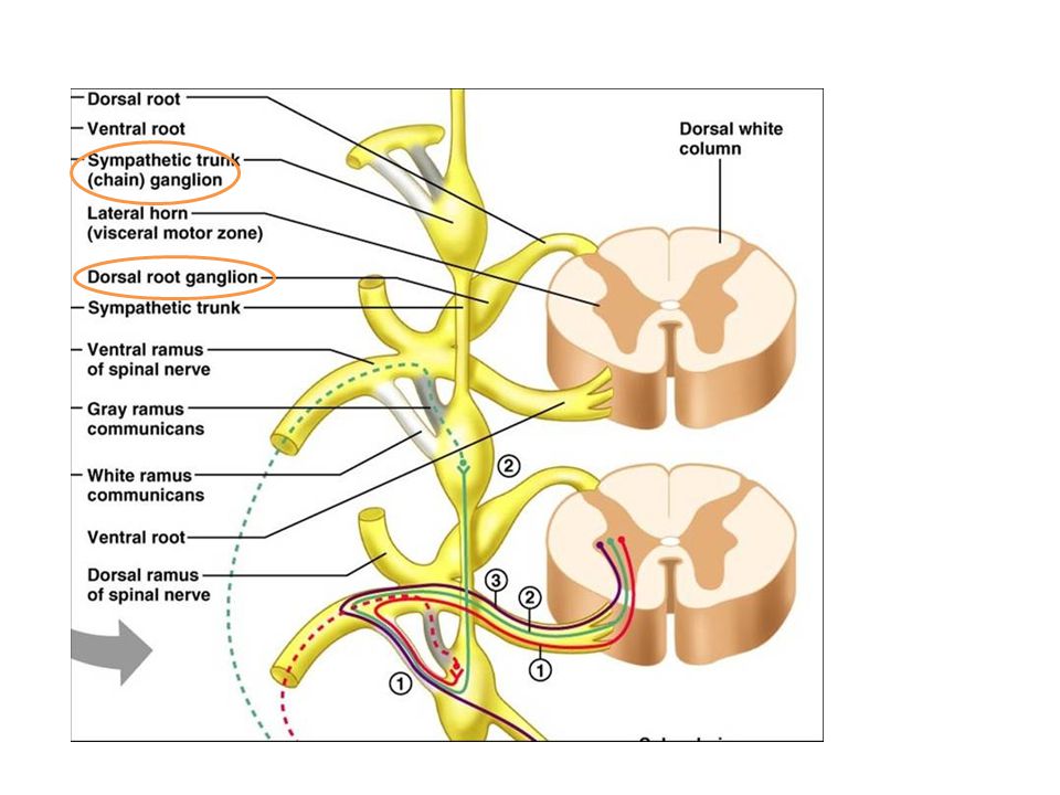
peripheral nerves Structure of spinal nerve: Ventral (motor) and dorsal (sensory) rootlets Ventral and dorsal root Dorsal root ganglia Spinal nerve: mixing of fibers happen here Sympathetic (paravertebral) ganglion is connected to the spinal nerve by grey and white rami communicans The spinal nerve, as it leaves the vertebral canal divides into Anterior (ventral) and posterior (dorsal) rami, both are mixed: Dorsal rami innervate the muscles and the skin over the vertebral column. Anterior rami supply the lateral and anterior aspects of the trunk as well as the limbs. The anterior rami also intermingle with each other at certain levels to form plexuses (cervical p. to the neck, brachial p. to the upper limb and lumbosacral p. to the lower limb)
 and dorsal (sensory) rootlets. Ventral and dorsal root. Dorsal root ganglia. Spinal nerve: mixing of fibers happen here. Sympathetic (paravertebral) ganglion is connected to the spinal nerve by grey and white rami communicans. The spinal nerve, as it leaves the vertebral canal divides into Anterior (ventral) and posterior (dorsal) rami, both are mixed: Dorsal rami innervate the muscles and the skin over the vertebral column. Anterior rami supply the lateral and anterior aspects of the trunk as well as the limbs. The anterior rami also intermingle with each other at certain levels to form plexuses (cervical p. to the neck, brachial p. to the upper limb and lumbosacral p. to the lower limb)")
20
Peripheral nervous system
peripheral nerves Dorsal rami innervate the muscles and the skin over the vertebral column. Anterior rami supply the lateral and anterior aspects of the trunk as well as the limbs. The anterior rami also intermingle with each other at certain levels to form plexuses (cervical p. to the neck, brachial p. to the upper limb and lumbosacral p. to the lower limb)
")
23
Peripheral nervous system
Ganglia Ganglia: Ganglia related to the cranial nerves: They host the cell bodies of the sensory neurons of the cranial nerves and they lie within the skull. Spinal (dorsal root) ganglia: Contains the cell bodies of the sensory neurons receiving information from the body to the CNS (spinal cord). Autonomic ganglia: are either: Sympathetic : paravertebral or prevertebral Parasympathetic: close to the target organ
 ganglia: Contains the cell bodies of the sensory neurons receiving information from the body to the CNS (spinal cord). Autonomic ganglia: are either: Sympathetic : paravertebral or prevertebral. Parasympathetic: close to the target organ.")
25
Functional division

26
Functional division Functionally we can not separate the CNS from the PNS. For example if we consider a motor order for a certain muscle, this should involve parts in both; the CNS and PNS. Generally the NS can be divided according to the functions into: Somatic nervous system: under our conscious control Motor sensory Autonomic (visceral or involantry) nervous system
 nervous system.")
27
Somatic nervous system
Functional division Somatic nervous system

28
Somatic nervous system
Functional division Somatic nervous system Motor system: This includes the parts of the CNS which generate, conduct and coordinate the motor orders (cerebral cortex, basal nuclei, anterior grey horn of spinal cord, descending tracts…) AND the nerve fibers (anterior roots of spinal nerves) which convey these orders to the target muscle via their peripheral nerves.
 AND the nerve fibers (anterior roots of spinal nerves) which convey these orders to the target muscle via their peripheral nerves.")
29
Somatic nervous system
Functional division Somatic nervous system Sensory system: Consists of: The peripheral receptor which are distributed all over the body to receive different stimuli. Sensory nerves and their cell bodies in the dorsal root ganglia Ascending tracts in the CNS Diencephalon and cerebral cortex which understand and process these information

30
Autonomic nervous system
Functional division Autonomic nervous system

31
Autonomic nervous system
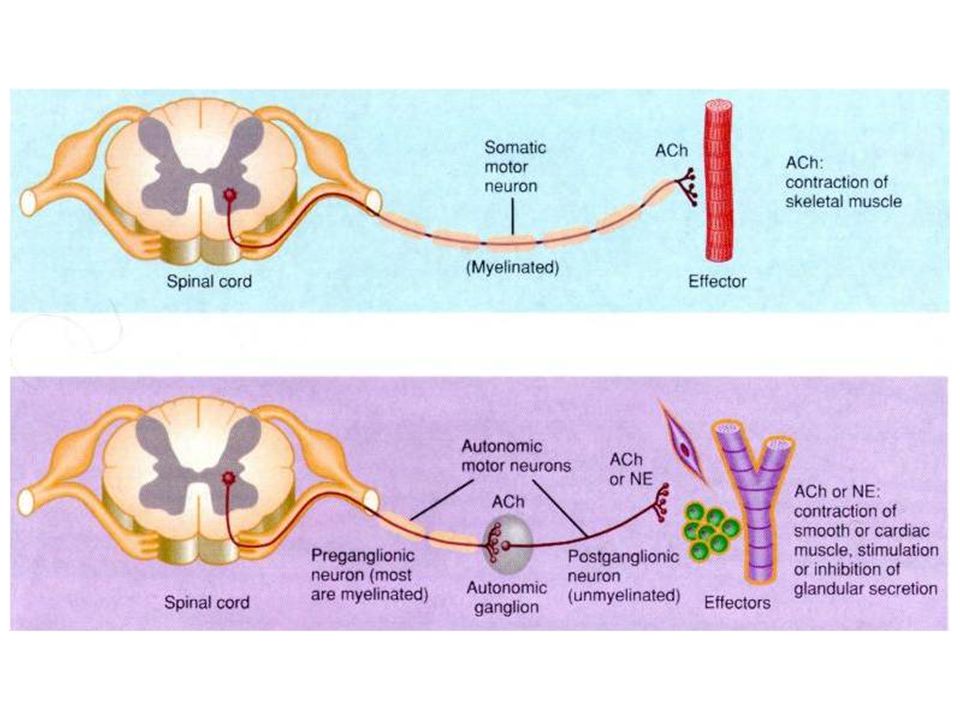
Functional division Autonomic nervous system Controls the involuntary function of the internal body organs. Controlled by higher centers in the brain Is divided into sympathetic and parasympathetic nervous systems Each system of the two consists of two sets of neurons and ganglia at which the two neurons synapse. The cell body of the first neuron is found in the CNS while the cell body of the second is located in peripheral ganglia. The first neuron is called preganglionic neuron The second is called postganglionic neuron

32
Sympathetic nervous system
Functional division Sympathetic nervous system Structure of the sympathetic nervous system: Preganglionic neuronal cell body is found in the lateral horn of the spinal cord segments from T1-L2 The ganglia are either: Paravertebral; on both sides of the vertebral column forming two sympathetic chains Prevertebral: anterior to the vertebral column related to the major branches of the abdominal aorta From these ganglia the postganglionic neuronal fiber reaches the target organs through the branches of the spinal nerves or along the blood vessels or by their own nerves (visceral or splanchnic plexuses)
")
34
Parasympathetic nervous system
Functional division Parasympathetic nervous system Structure of the parasympathetic nervous system: Preganglionic neuronal cell bodies are found in the: Nuclei of cranial nerves III, VII, IX and X Grey matter of sacral spinal cord segments 2,3 and 4 Hence they constitute the craniosacral flow The ganglia are: Four in the region of head Plenty of parasymathetic ganglia on the wall of target organ (i.e. the parasympathetic ganglia for the stomach lies in the wall of the stomach and so on) Thus, the postganglionic neuron is short. The parasympathetic fibers are distributed to the body via the above-mentioned cranial nerves and through the pelvic plexus.
 Thus, the postganglionic neuron is short. The parasympathetic fibers are distributed to the body via the above-mentioned cranial nerves and through the pelvic plexus.")
35
Cranio-sacral flow

39
Thanks

Similar presentations


>")

>")


 and Nerves. NERVOUS SYSTEM 1.Collect sensory input 2.Integrate sensory input 3.Motor output Functions of Nervous System.>")
 neurons.>")

