Download presentation
Presentation is loading. Please wait.

1
Prof. Ahmed Fathalla Ibrahim Professor of Anatomy College of Medicine King Saud University E-mail: ahmedfathala@gmail.com

2
OBJECTIVES At the end of the lecture, students should: Define Define the autonomic nervous system. structure Describe the structure of autonomic nervous system preganglionic & postganglionic neurons Trace the preganglionic & postganglionic neurons in both sympathetic & parasympathetic nervous system. the main effects Enumerate in brief the main effects of sympathetic & parasympathetic system

3
DEFINITION both central & peripheral nervous system innervation of involuntary structures: Nerve cells located in both central & peripheral nervous system that are concerned with innervation of involuntary structures: viscera, smooth & cardiac muscles, glands. Function: homeostasis Function: maintains homeostasis of internal environment. Regulation: Regulation: by hypothalamus.

4
STRUCTURE OF AUTONOMIC NERVOUS SYSTEM

5
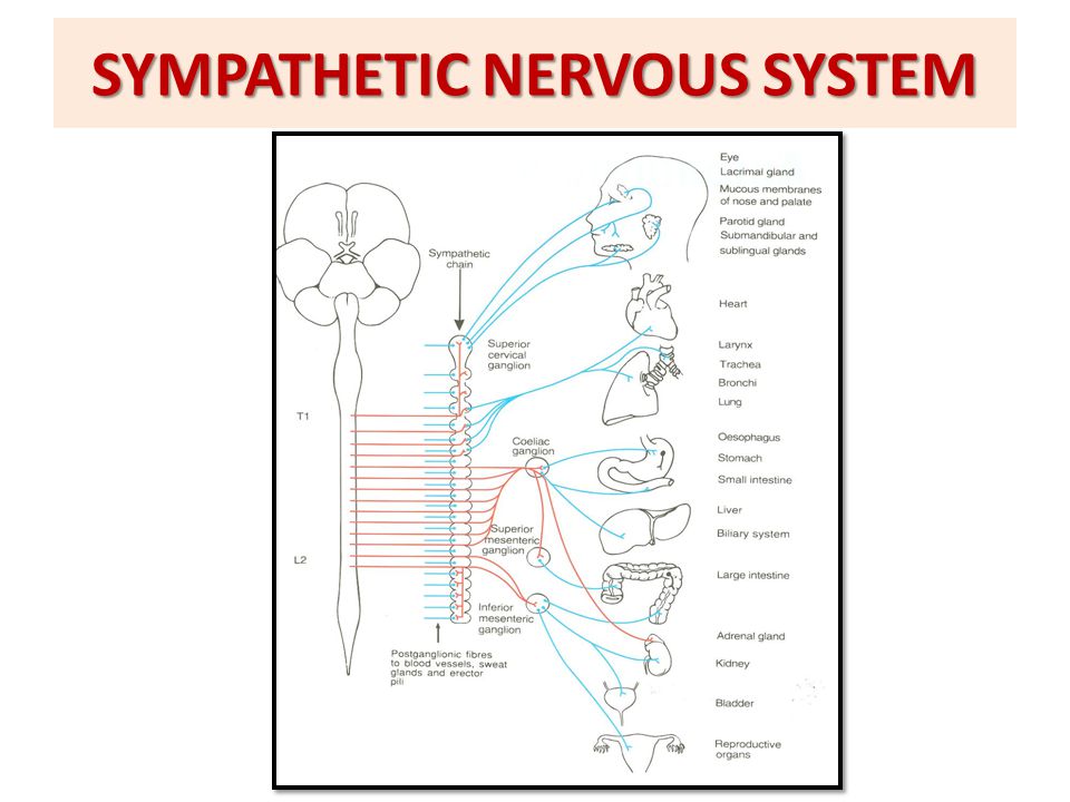
SYMPATHETIC NERVOUS SYSTEM Cells of lateral horn of spinal cord (T1 – L3) Cells of sympathetic chain Cells of plexuses surrounding abdominal aorta (Coeliac, superior & inferior mesenteric) (Coeliac, superior & inferior mesenteric) Short axon Long axon
 Cells of sympathetic chain Cells of plexuses surrounding abdominal aorta (Coeliac, superior & inferior mesenteric) (Coeliac, superior & inferior mesenteric) Short axon Long axon")
6
SYMPATHETIC NERVOUS SYSTEM

8
Preganglionic sympathetic neurons: cells of the lateral horn of spinal cord in all thoracic + upper 3 lumbar segments. Preganglionic axons (via the white ramus communicans) Preganglionic axons leave the spinal cord, join corresponding spinal nerves & reach the sympathetic chain (via the white ramus communicans). They either: postganglionic axons (via grey ramus communicans) 1.Synapse with cells of paravertebral ganglia located in sympathetic chain (postganglionic neurons are cells of paravertebral ganglia: postganglionic axons leave the sympathetic chain & join again the spinal nerve (via grey ramus communicans) to supply structures in head & thorax + blood vessels & sweat glands.
 Preganglionic axons leave the spinal cord, join corresponding spinal nerves & reach the sympathetic chain (via the white ramus communicans). They either: postganglionic axons (via grey ramus communicans) 1.Synapse with cells of paravertebral ganglia located in sympathetic chain (postganglionic neurons are cells of paravertebral ganglia: postganglionic axons leave the sympathetic chain & join again the spinal nerve (via grey ramus communicans) to supply structures in head & thorax + blood vessels & sweat glands..")
9
SYMPATHETIC NERVOUS SYSTEM (without synapse) Postganglionic axons 2. Leave the sympathetic chain (without synapse) to reach coeliac & mesenteric plexuses (around branches of abdominal aorta) to synapse with their cells. Postganglionic neurons are cells of coeliac & mesenteric plexuses. Postganglionic axons supply abdominal & pelvic viscera.
 to reach coeliac & mesenteric plexuses (around branches of abdominal aorta) to synapse with their cells. Postganglionic neurons are cells of coeliac & mesenteric plexuses. Postganglionic axons supply abdominal & pelvic viscera..")
10
PARAVERTEBRAL GANGLIA 2 sympathetic chains, one on each side of vertebral column. They are interconnected to form 2 sympathetic chains, one on each side of vertebral column. Number of ganglia: 1.Three 1.Three ganglia in cervical part of chain 2.Eleven to twelve 2.Eleven to twelve ganglia in thoracic part 3.Four 3.Four in lumbar & sacral parts. ‘ganglion impar’ The chains end into a common ‘ganglion impar’ in front of coccyx

11
PARASYMPATHETIC NERVOUS SYSTEM Cranial: cells in brain stem: nuclei of 3 rd, 7 th 9 th & 10 th Sacral: cells in S2 – S4 segments of spinal cord Long axon Cranial: cells of ciliary, pterygopalatine, submandibular, otic & peripheral ganglia & peripheral ganglia Sacral: cells of peripheral ganglia Short axon Nucleus: group of neurons inside CNS Ganglion: group of neurons outside CNS

12
PARASYMPATHETIC NERVOUS SYSTEM

13
Preganglionic parasympathetic neurons: Preganglionic axons 3 rd, 7 th, 9 th & 10 th cranial nerves Postganglionic axons 1.Cells located in brain stem: Preganglionic axons leave the brain stem, join 3 rd, 7 th, 9 th & 10 th cranial nerves & reach ciliary, pterygopalatine, submandibular, otic & peripheral ganglia (Postganglionic neurons are cells of those ganglia). Postganglionic axons supply structures in head, thorax & abdomen. Preganglionic axons join corresponding sacral spinal nerves Postganglionic axons 2.Cells located in 2 nd, 3 rd & 4 th sacral segments of spinal cord. Preganglionic axons leave the spinal cord, join corresponding sacral spinal nerves to reach peripheral ganglia in pelvis where they synapse. Postganglionic neurons are cells of peripheral ganglia. Postganglionic axons supply pelvic viscera.

15
QUESTION 1 At which one of the following sites are located preganglionic neurons of the sympathetic nervous system ? 1.Brain stem 2.Thoracic segments of spinal cord 3.Sacral segments of spinal cord 4.Sympathetic chain

16
QUESTION 2 Regarding the parasympathetic nervous system, which one of the following statements is correct? 1.Its preganglionic axons are short. 2.It supplies sweat glands. 3.Its preganglionic neurons are located in the sacral segments of spinal cord. 4.Its postganglionic neurons are located in the coeliac & mesenteric plexuses.

17
THANK YOU

Similar presentations


 Lec 8 & 9. Differences between Somatic & Autonomic Nervous system.>")


 neurons.>")

 Introduction to ANS: It is the part of the peripheral nervous system (Cranial and spinal nerves ). It composed of nerves,>")


 Assoc. Prof. Wantanee Trakulrungsi Department of Anatomy, Faculty of Science, Mahidol University.>")