Download presentation
Presentation is loading. Please wait.

1
Development of the Circulatory System
Development of cardiovascular system Development of lymphatic vascular system

2
overview The cardiovascular system is derived from the mesoderm.
It appears in the middle of the 3rd week. Blood circulation starts by the end of the 3rd week. Some changes take place at birth and in the 1st postnatal month.

3
Key points Development of primitive cardiovascular system
Development of Heart Formation and modification of aortic arches Circulation before and after birth Congenital Malformations ※

5
Primitive Cardiovascular System
Oropharyngeal membrane Cardiogenic area

6
Primitive cardiovascular system
Yolk sac mesenchyme cells blood islands Central C Peripheral C Primitive Blood cell Endothelia C Blood C Vessels Endothelial tubes of intraembryo and extraembryo are connected with each other by body stalk.

7
primitive cardiovascular system
Yolk sac mesenchyme cells blood islands Central C Peripheral C Primitive Blood cell Endothelia C Blood C Vessels Vessels net primitive cardiovascular system

8
① heart tube:2 tubes 1 tube Primitive heart ② arteries ③ veins
Heart tubes 20d 4w End of 4w ① heart tube:2 tubes 1 tube Primitive heart ② arteries ③ veins

9
① heart tube ② arteries ③ veins 2 dorsal A 1 aorta,many branches
Aortic arches Dorsal aorta 20d 4w End of 4w Vitelline A Umbilical A ① heart tube ② arteries ③ veins 2 dorsal A 1 aorta,many branches Few pairs of vitelline A 1 pair of umbilical A 6 pairs of aortic arches

10
① heart tube ② arteries ③ veins 1 pair of anterior cardinal V
A cardinal V Posterior Vitelline V Common cardinal V Umbilical V 20d 4w End of 4w ① heart tube ② arteries ③ veins 1 pair of anterior cardinal V 1 pair of posterior cardinal V 1 pair of vitelline V 1 pair of umbilical V Common cardinal V heart

11
Three separate circulations
vitelline, umbilical and embryonic circulation.

12
Development of the Heart
►Development of the heart tube ► Morphogenesis of the heart ► Partitioning of Heart Chambers ► Development of sinus venosus and differentiation of veins

13
Development of the heart tube
Cardiogenic area Oropharyngeal membrane Cardiogenic area is anterior to the oropharyngeal membrane and the neural plate.

14
Development of the heart tube
Pericardial cavity Buccopharyngeal M cardiaogenic plate About 18~19d, a cavity appears in the cardiogenic area --pericardiac cavity B. Ventral of the cavity is cardiaogenic cords --cardiaogenic plate

15
Development of the heart tube
Pericardial cavity cardiac tube The 20th d C. cardiaogenic plate becomes hollow--cardiac tube

16
Development of the heart tube
Pericardial cavity cardiac tube The 22nd d D. As the embryo folds cephalocaudally, the developing heart tube bulges more and more into the pericardial cavity.

17
Development of the heart tube
Pericardial cavity cardiac tube The 28th d E. The paired heart tubes merge except at their caudalmost ends. F. The tube remains attached to the dorsal side of the pericardial cavity by the dorsal mesocardium.

18
Development of the heart tube
Caudal end Cephalic end Heart tube Pericardial cavity G. Cephalic end Arteries,Caudal end Veins

20
Pericardial cavity

21
Wall of primitive heart tube
Endocardial heart tube → endocardium Myoepicardial mantle → myocardium, epicardium Cardiac jelly → subendocardial tissue

22
Morphogenesis of the heart
Vein end Cardiac tube Artery end A. Part of the cardiac tubes merged B. Cephalic end A Caudal end V The 21st d

23
Morphogenesis of the heart
atrium ventricle bulbus cordis C. Heart tubes almost merged D. Three expansions bulbus cordis(心球) Ventricle(心室) Atrium(心房) The 22nd d
 Ventricle(心室) Atrium(心房) The 22nd d.")
24
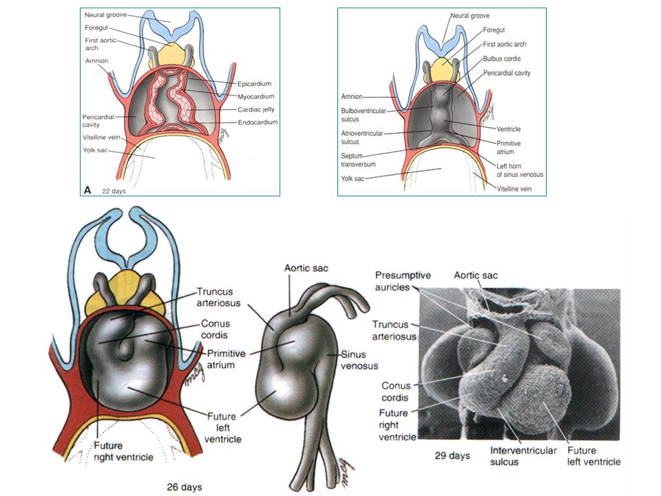
Morphogenesis of the heart
truncus arteriosus E. The 4th expansion, the sinus venosus(静脉窦) appears F. The truncus arteriosus(动脉干)appears G. The heart tube starts to bend bulbus cordis ventricle atrium sinus venosus The 23rd d
 appears. F. The truncus arteriosus(动脉干)appears. G. The heart tube starts to bend. bulbus. cordis. ventricle. atrium. sinus. venosus. The 23rd d.")
25
Morphogenesis of the heart
The cephalic portion bends ventrally, caudally, and to the right. The caudal part shifts dorsocranially and to the left. Form a ‘U’ like structure, the cardiac loop (bulboventricular loop). truncus arteriosus Bulboventricular loop Sinus venosus The 24th d
. truncus. arteriosus. Bulboventricular. loop. Sinus venosus. The 24th d.")
26
Morphogenesis of the heart
I. The bulboventricular loop keeps turning, the atrium shifts to the cephalodorsal of the ventricle G. Two atria and two ventricles Aortic arches atrium ventricle The 35th d The normal heart shape was established, but partitioning has not completed

29
Development of the Heart
►Development of the heart tube ► Morphogenesis of the heart ► Partitioning of Heart Chambers ► Development of sinus venosus and differentiation of veins

30
Partitioning of Heart Chambers
►Division of atrioventricular canal ► Partitiioning of the primitive atrium ► Partitioning of the primitive ventricle ► Division of truncus and bulbus

31
Division of atrioventricular canal
Subendocardial tissue → dorsal and ventral endocardial cushions → fuse → right and left canals

33
Septum formation in part arises from development of endocardial cushion. Many malformations are related to abnormal cushion morphogenesis. atria Endocardiac cushion ventricle The 4th w

34
The 5th w Fusion of the opposing superior and inferior
cushions divides the orifice into R and L aterioventricular canals. Endocardiac cushion L AV orifice R AV orifice The 5th w

35
The 4th month Left biscuspid Right tricuspid bicuspid tricuspid
Endocardiac cushion Left Right biscuspid tricuspid bicuspid tricuspid The 4th month

36
Partitioning of Heart Chambers
►Division of atrioventricular canal ► Partitiioning of the primitive atrium ► Partitioning of the primitive ventricle ► Division of truncus and bulbus

Similar presentations