Download presentation
Presentation is loading. Please wait.

1
Chapter 27 Development of circulatory system

2
Formation of primitive cardiovascular system

3
1) extra-embryonic blood vessels
---blood island: at the middle of 3rd week, wall of the yolk sac mesenchyma proliferate and form isolated cell clusters, the peripheral cell become flattened and differentiate into endothelial cell to from endothelial tube; central located cells are detached and develop into primitive blood cells(blood stem cell)
")
4
---endothelial tube approach and fuse with each other to form an endothelial tube network
---endothelial tube network appears in chorionic membrane and body stalk, and connect to vitelline circulation

5
2) intra-embryonic blood vessels
---by the 18-20th days, endothelial tube network appears in intraembryonic mesenchyma to form intraembryonic endothelial tube network ---by the end of 3rd week, intraembryonic and extraembryonic endothelial tube networks connect to each other to form a diffuse endothelial tube network ---endothelial tube networks fuse or disappear to form primitive cardiovascular system

6
---primitive cardiovascular system:
/heart tube: paired, by the 4th week, which fuse /arteries: -dorsal aorta: -vitelline artery -umbilical artery -aortic arch: 6 pairs /veins: -anterior cardinal vein -posterior cardinal vein -common cardinal vein -vitelline vein -umbilical vein

7
2. Development of heart 1) Formation of primitive heart
---cardiogenic plate: proliferation of splanchnic mesodermal cell to form a pair of longitudinal cell cord, then differentiate into cardiac tube ---pericardiac coelom: space in mesoderm

8
---rapid growth of brain vesicles pulls the buccopharyngeal membrane, the cardiogenic plate and pericardiac coelom forward

9
---lateral folds of the embryo make the two cardiac tube approach and fuse with each other

10
---dorsal mesocardium: cardiac tube invaginate into pericardiac coelom and connect to it by dorsal mesocardium

11
---the mesoderm adjacent to cardiac tube differentiates into the myoepicardial mantle
---cardiac jelly: gelatinous substance between endothelial wall and myoepicardial mantle, then invaded by mesenchymal cell ---heart wall: endocardium; myocardium and epicardium

13
2) Further development of the heart
---single cardiac tube connected caudally to the umbilical, vitelline and common cardinal vein; cephalically connected to the dorsal aortae by means of aortic arches

14
---three dilatations: /bulbus cordis /ventricle
/atrium

15
---sinus venousus: dilatation which receives the umbilical, vitelline, and common cardinal veins
---truncus arteriosus: distal part of the bulbus cordis, connect with aortic sac cephalically

16
---bulboventricular loop: bulboventricular portion of heart tube grows rapidly, bends forming a loop
---“U”-shaped heart; bulboventricular loop bends in ventral and caudal directions and slightly to the right

17
---“S”-shaped heart: the heart tube continues to grow and bend, atrium shifts in dorso-cranial direction; sinus venousus located at caudal portion of atrium

18
---atrioventricular canal: atrioventricular junction remains narrow
---the bulbus cordis divided into three portions: /distal portion: truncus arteriosus /midportion: bulbus arteriosus cordis /proximal portion: develops into the right ventricle ---primary ventricle develop into the left ventricle

20
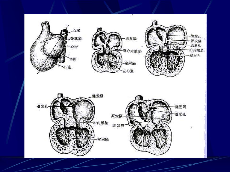
3) Partitioning of the heart chambers
---from 27th day to 37th day

21
① division of the atrioventricular canal
---endocardiac cushion: develop as thickenings of subendocardial tissue in the dorsal and ventral walls of the heart ---the endocardial cushions grow toward each other and fuse ---lateral atrioventricular cushion: form atrioventricular valve

22
② partitioning of the primitive atrium
---septum primum: a thin sickle-shaped membrane appearing from dorsocranial wall of atrium ---foramen primum: septum primum grows toward the endocardial cushions, leaving an opening between its lower edge and the endocardial cushions

23
---foramen secundum: the upper part of the septum primum perforated and form an opening

24
---septum secundum: another membrane appears in the ventrocranial wall in the right of the septum primum ---foramen ovale: oval passage between atrium, covered by valve of foramen ovale

25
---before birth, blood can flow from right atrium toward the left atrium
---after birth, two septums fuse to separate atrium completely

27
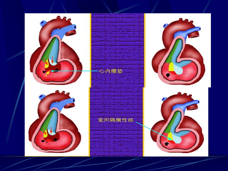
③partitioning of the primitive ventricle
---muscular part of interventricular septum: by the end of 4th week, tissue of ventricular wall grows toward endocardial cushions, but left an opening so called interventricular foramen

28
---membranous part of interventricular septum: made up of right bulbar ridge, left bulbar ridge and dorsal endocardial cushion

30
④division of truncus arteriosus and bulbus cordis
---right and left bulbar ridges: two spiral mesodermal ridges grow from the inner walls of the truncus arteriosus and bulbus cordis, twist around each other and fuse to form a spiral aortico-pulmonary septum

31
---bulbus arteriosus cordis and truncus arteriosus are divides into the aorta and the pulmonary trunk

32
3. Blood circulation of fetus and circulatory changes after birth

33
1) blood circulatio of fetus
umbilical V→ducts venosus or hepatic sinus →inferior vena cava → right atrium →foramen ovale →left atrium →left ventricle →head and neck region →superior vena cava →right atrium→right ventrical → ductus arteriosus → aorta→gut and lower region of the body→umbilical A → placenta

34
2) circulatory changes after birth
a. umbilical V and ducts venousus: constrict and becomes into ligamentum teres hepatis and ligamentum venosus b.umbilical A: becomes into medial umbilical ligament, but proximal portion persist as superior vesical arteries c. ductus arteriosus: constrict and become arterial ligament d. foramen ovale closed

36
4. Congenital malformations
1) atrial septal defect: ---patency of the foramen ovale ---foramen secundum defect ---foramen primum effect ---endocardial cushion defect
 atrial septal defect: ---patency of the foramen ovale. ---foramen secundum defect. ---foramen primum effect. ---endocardial cushion defect.")
37
2) ventricular septal defect:
---membranous part of the ventricular septum defect ---muscular part of the ventricular septum

38
3) abnormalities of the truncus arteriosus and bulbus arteriosus cordis
---unequal division of truncus arteriosus ---presistent truncus arteriosus ---transposition of the great vessels

39
4) tetralogy of Fallot: ---unequal division of truncus arteriosus ---four defects: /pulmonary stenosis /overriding aorta /membranous part fails to develop /hypertrophy of right ventricle

40
5) patent ductus arteriosus
 patent ductus arteriosus")
Similar presentations


, UNSEPARATED VENTRICLE (18), LIVER (12), UMBILICAL VEIN (17), TRANSVERSE.>")


