Download presentation
Presentation is loading. Please wait.

1
presented by: Ms Asmaa A Basonbul

2
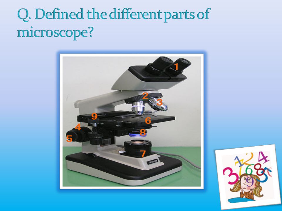
1. Recognize different parts of a compound light microscope. 2. Learn how using appropriate objective lenses e.g 10x for focusing 40x for wet preparation 100x oil immersion 3. Learn how using condenser and iris diaphragm to adjust the light source to the optimum.

3
Microscopy: The use of microscopes in all their various forms. Q. Why we use microscope in haematology lab? To study various blood cells and inclusion bodies.

4
How? By using a system of lenses and illumination sources it’s make object visible. Q. How many times the microscope can magnify that object? Microscope can magnify an object from 100-1000 times of it’s original size.

5
To magnify the object the LM use a system of lenses (objectives and oculars) to manage the path of light beam that travel b/w object which we’re studied to the eye.
 to manage the path of light beam that travel b/w object which we’re studied to the eye.")
6
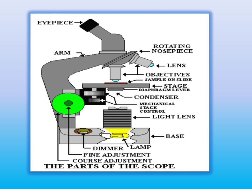
1. Eye picec (ocular lenses): Magnify lens with magnification power 10x.
: Magnify lens with magnification power 10x.")
7
2. Body tube: Contains mirrors and prisms that transmit the image from the objective lens to the ocular lens.

8
3. Objective lenses: Primary lenses that magnify specimen a) Low power 10x b) High power 40x c) Oil immersion 100x
 Low power 10x b) High power 40x c) Oil immersion 100x.")
9
4. Stage: Holds slide in position.

10
5. Condenser: Lens system that condenses light before it passes through the specimen.

11
6. Iris diaphragm: Control the amount of light entering the condenser.

12
7. Coarse and fine adjustment knobs: Used for focusing the specimen.

13
8. Light: Source of illumination, bulb.

15
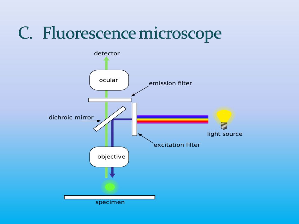
A. Bright field microscopy. B. Dark field microscopy. C. Fluorescence microscopy. D. Phase contrast microscopy.

16
Light bulb Day light

18
Special objective U Special condenser in mico Light source Object in the specimen “Microorganism”

22
Acridine orange Auramine/rhodamine

24
Bright(denser) Dark(lighter) special objectives Light source Special condenser Object in the specimen contrast
 Dark(lighter) special objectives Light source Special condenser Object in the specimen contrast")
26
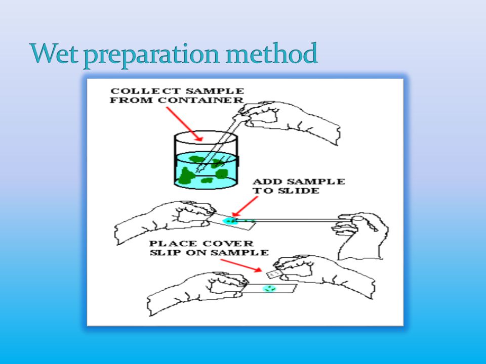
Compound microscope. Lens cleaning paper/ cloth. Immersion oil. Wet preparation. Stained preparation.

28
Oil immersion objective(100x): Put drop of oil immersion on the slide. Turn the oil immersion objective 1oox and put above the slide. The 100x objective will immersed in the oil. The picture is transmitted from objective lens to ocular lens.

29
The oil helps to keep light rays together as they pass b/w the specimen and objective lens.

31
= power of the objective lens X power magnification of eye piece e.g : using oil immersion objective 100x ocular lens (eye piece) 10x ??!! So 100x X 10x =1000

32
Is measure of its ability to discriminate b/w two adjacent objects. Absolute limit of the resolving power = wave length of LM = 400-800nm

33
1) The microscope is delicate instrument which is must be: Handle gently. Put in a clean environment away from chemical, direct sunlight, evaporation, heating source or moisture. Cleaned immediately if the stage is contaminated with saline to avoid corrosion. 2) The temperate climate humidity and high temperature causes growth of fungus which can damage optical surfaces
 The temperate climate humidity and high temperature causes growth of fungus which can damage optical surfaces.")
34
A. Optics: The low and high power lens paper tissue. Oil immersion lens paper tissue and few drop from xyelen (if needed). B. Non-optics: Eyepices: Using the soft camel-hair brush to remove the dust from in and out side.
. B. Non-optics: Eyepices: Using the soft camel-hair brush to remove the dust from in and out side..")
35
Condenser and iris diaphragm: Using soft cloth or tissue moistened with toluene and the mirror with 5% alcohole. Other parts cleaned with mild detergent and remove grease or oil with petroleum ether followed by 45% ethanol in water.

37
Let us to start practical part

Similar presentations


>")


