Download presentation
Presentation is loading. Please wait.

1
Skeletal System

2
Chapter 7.1 Objective- Read 7.1 and understand that bones are alive and multifunctional. Objective- Read 7.1 and understand that bones are alive and multifunctional.

3
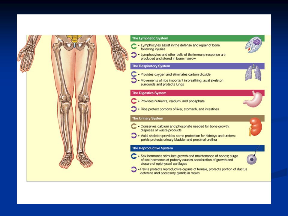
Introduction: Introduction: A.Bones are very active tissues A.Bones are very active tissues B.Each bone is made up of several types of tissues and so is an organ. C. Bone functions include: muscle attachment, protection and support, blood cell production and storage of minerals Copyright The McGraw-Hill Companies, Inc. Permission required for reproduction or display.

6
Section 7.2 Objective- Read 7.2 and appreciate the various size and shape of bone and be able to describe in detail the parts of long bone and microscopic structures. Objective- Read 7.2 and appreciate the various size and shape of bone and be able to describe in detail the parts of long bone and microscopic structures.

7
Bone Structure A.Bones differ in size and shape, yet are similar in several ways. Copyright The McGraw-Hill Companies, Inc. Permission required for reproduction or display.

9
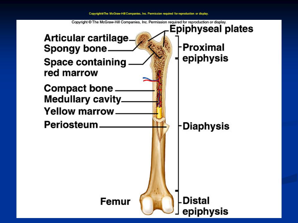
B.Parts of a Long Bone 1.Expanded ends of bones that form joints with adjacent bones are called epiphyses. 2.Articular cartilages (hyaline cartilage) cover the epiphyses. Copyright The McGraw-Hill Companies, Inc. Permission required for reproduction or display.
 cover the epiphyses. Copyright The McGraw-Hill Companies, Inc. Permission required for reproduction or display..")
11
3.The shaft of the bone is the diaphysis. 4.A tough layer of vascular connective tissue, called the periosteum, covers the bone and is continuous with ligaments and tendons. 5.A bone's shape makes possible its function; bony processes or grooves indicate places of attachment for muscles. Copyright The McGraw-Hill Companies, Inc. Permission required for reproduction or display.

12
6.Compact bone makes up the wall of the diaphysis; the epiphyses are filled with spongy bone to reduce the weight of the skeleton. 7.The diaphysis contains a hollow medullary cavity that is lined with endosteum and filled with marrow. Copyright The McGraw-Hill Companies, Inc. Permission required for reproduction or display.

15
Do now- List five major parts of long bone.

16
1.Bone cells (osteocytes) are located within lacunae that lie in concentric circles around osteonic canals. 2.Osteocytes pass nutrients and gasses in the matrix through canaliculi. 3.Intercellular material consists of collagen and inorganic salts. C.Microscopic Structure

17
4.In compact bone, osteocytes and intercellular material are organized into osteons that are cemented together. 5.Osteonic canals contain blood vessels and nerve fibers, and extend longitudinally through bone. Copyright The McGraw-Hill Companies, Inc. Permission required for reproduction or display.

18
6.Osteonic canals are interconnected by transverse perforating canals. 7.Unlike compact bone, the osteocytes and intercellular material in spongy bone are not arranged around osteonic canals. Copyright The McGraw-Hill Companies, Inc. Permission required for reproduction or display.

20
Do now- What is structures are found in the central canal of osteons and how do osteocytes get there nutrients?

21
Section 7.3 Objective- Read 7.3 and distinguish between intramembranous and endochondral describe the bone development of long (endochondral) bone. Objective- Read 7.3 and distinguish between intramembranous and endochondral describe the bone development of long (endochondral) bone.
 bone..")
22
Bone Development and Growth A.Bones form by replacing connective tissue in the fetus. B.Some form within sheetlike layers of connective tissue (intramembranous bones), while others replace masses of cartilage (endochondral bones). Copyright The McGraw-Hill Companies, Inc. Permission required for reproduction or display.
, while others replace masses of cartilage (endochondral bones). Copyright The McGraw-Hill Companies, Inc. Permission required for reproduction or display..")
25
C.Intramembranous Bones 1.The flat bones of the skull form as intramembranous bones that develop from layers of connective tissue. 2.Osteoblasts deposit bony tissue around themselves. Copyright The McGraw-Hill Companies, Inc. Permission required for reproduction or display.

26
3.Once osteoblasts deposit bone are located in lacunae, they are called osteocytes. 4.Cells of the membranous connective tissue that lie outside the developing bone give rise to the periosteum. Copyright The McGraw-Hill Companies, Inc. Permission required for reproduction or display.

27
1.Most of the bones of the skeleton fall into this category. 2.They first develop as hyaline cartilage models and are then replaced with bone. C.Endochondral Bones

28
3.Cartilage is broken down in the diaphysis and progressively replaced with bone while the periosteum develops on the outside. 4.Cartilage tissue is invaded by blood vessels and osteoblasts that first form spongy bone at the primary ossification center in the diaphysis. Copyright The McGraw-Hill Companies, Inc. Permission required for reproduction or display.

29
5.Osteoblasts beneath the periosteum lay down compact bone outside the spongy bone. 6.Secondary ossification centers appear later in the epiphyses. Copyright The McGraw-Hill Companies, Inc. Permission required for reproduction or display.

30
7.A band of hyaline cartilage, the epiphyseal plate, forms between the two ossification centers. 8.Layers of cartilage cells undergoing mitosis make up the epiphyseal plate. 9.Osteoclasts break down the calcified matrix and are replaced with bone- building osteoblasts that deposit bone in place of calcified cartilage. Copyright The McGraw-Hill Companies, Inc. Permission required for reproduction or display.

31
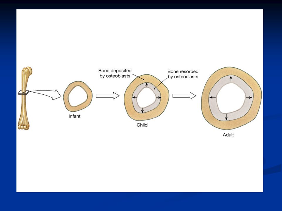
10.Epiphyseal plates are responsible for lengthening bones while increases in thickness are due to intramembranous ossification underneath the periosteum. 11.A medullary cavity forms in the region of the diaphysis due to the activity of osteoclasts. Copyright The McGraw-Hill Companies, Inc. Permission required for reproduction or display.

34
E.Homeostasis of Bone Tissue 1.Osteoclasts tear down and osteoblasts build bone throughout the lifespan with the processes of resorption and deposition, with an average of 3% to 5% of bone calcium exchanged annually. Copyright The McGraw-Hill Companies, Inc. Permission required for reproduction or display.

35
Do Now- Explain how endochondral bone develops. Do Now- Explain how endochondral bone develops.

36
Section 7.4 Objective- Read 7.4 and discuss the major functions of bones. Objective- Read 7.4 and discuss the major functions of bones.

37
Bone Function A.Support and Protection 1.Bones give shape to the head, thorax, and limbs. 2.Bones such as the pelvis and lower limbs provide support for the body. 3.Bones of the skull protect the brain, ears, and eyes. Copyright The McGraw-Hill Companies, Inc. Permission required for reproduction or display.

38
B.Body Movement 1.Bones can act as levers. A lever has four components: a rigid bar, a pivot or fulcrum, an object that is move against resistance, and a force that supplies energy. Copyright The McGraw-Hill Companies, Inc. Permission required for reproduction or display.

39
1.Two kinds of marrow occupy the medullary cavities of bone. a.Red marrow functions in the formation of red blood cells, white blood cells, and platelets, and is found in the spongy bone of the skull, ribs, sternum, clavicles, vertebrae, and pelvis. b.Yellow marrow, occupying the cavities of most bones, stores fat. C.Blood Cell Formation

40
D.Storage of Inorganic Salts 1.The inorganic matrix of bone stores inorganic mineral salts in the form of calcium phosphate that is important in many metabolic processes. 2.Calcium in bone is a reservoir for body calcium; when blood levels are low, osteoclasts release calcium from bone. Copyright The McGraw-Hill Companies, Inc. Permission required for reproduction or display.

41
3.Calcium is stored in bone under the influence of calcitonin when blood levels of calcium are high. 4. 4. Bone also stores magnesium, sodium, potassium, and carbonate ions. 5. 5. Bones can also accumulate harmful elements, such as lead, radium, and strontium. Copyright The McGraw-Hill Companies, Inc. Permission required for reproduction or display.

42
Do Now- Name three major functions of bones. Do Now- Name three major functions of bones.

43
Section 7.5 Objective- Read 7.5 and distinguish between the axial and appendicular skeletons and name the major parts of each. Objective- Read 7.5 and distinguish between the axial and appendicular skeletons and name the major parts of each.

44
Skeletal Organization A.The axial skeleton consists of the skull, hyoid bone, vertebral column (vertebrae and intervertebral disks and sacrum), and thorax (ribs and sternum). B.The appendicular skeleton consists of the pectoral girdle (scapulae and clavicles), upper limbs (humerus, radius, ulna, carpals, metacarpals, and phalanges), pelvic girdle (coxal bones), and lower limbs (femur, tibia, fibula, patella, tarsals, metatarsals, phalanges). Copyright The McGraw-Hill Companies, Inc. Permission required for reproduction or display.
, upper limbs (humerus, radius, ulna, carpals, metacarpals, and phalanges), pelvic girdle (coxal bones), and lower limbs (femur, tibia, fibula, patella, tarsals, metatarsals, phalanges). Copyright The McGraw-Hill Companies, Inc. Permission required for reproduction or display..")
50
Do Now- List the bones of the axial skeleton and the appendicular skeleton. Do Now- List the bones of the axial skeleton and the appendicular skeleton.

51
Section 7.6-7.12 Objective- Read 7.6 through 7.12 and locate and identify the bones and the major features of the skull, vertebral column, thoracic cage, pectoral girdle, upper limb, pelvic girdle, and lower limbs. Objective- Read 7.6 through 7.12 and locate and identify the bones and the major features of the skull, vertebral column, thoracic cage, pectoral girdle, upper limb, pelvic girdle, and lower limbs.

52
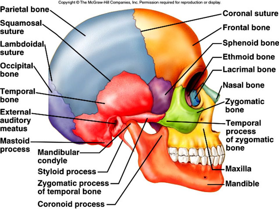
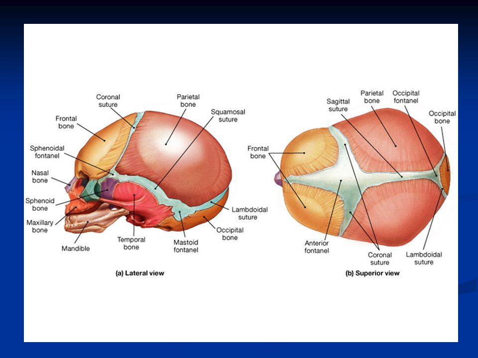
Copyright The McGraw-Hill Companies, Inc. Permission required for reproduction or display. Skull A.The skull is made up of 22 bones, including 8 cranial bones, 13 facial bones, and the mandible.

60
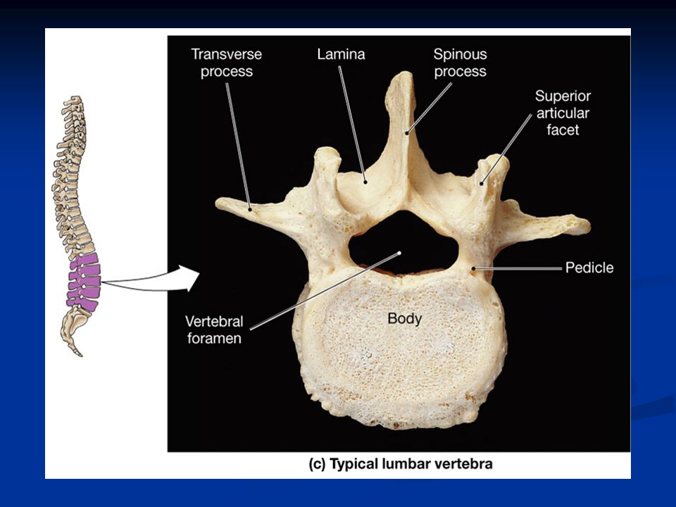
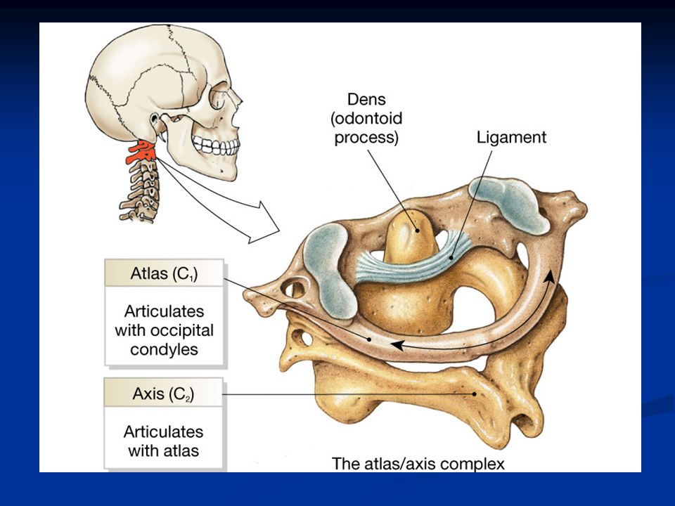
The Vertebral Column 7 cervical 7 cervical 12 Thoracic 12 Thoracic 5 Lumbar 5 Lumbar Sacrum (5 fused) Sacrum (5 fused) 3-5 Coccygeal vertebrae 3-5 Coccygeal vertebrae
 Sacrum (5 fused) 3-5 Coccygeal vertebrae 3-5 Coccygeal vertebrae")
61
Know the primary and secondary structures of the vertebral column!

66
Copyright The McGraw-Hill Companies, Inc. Permission required for reproduction or display. Thoracic Cage Thoracic Cage A.The thoracic cage includes the ribs, thoracic vertebrae, sternum, and costal cartilages. B.It supports the pectoral girdle and upper limbs, functions in breathing, and protects thoracic and upper abdominal organs.

69
Copyright The McGraw-Hill Companies, Inc. Permission required for reproduction or display. Pectoral Girdle A.The pectoral girdle makes an incomplete ring that supports the upper limbs. B.It is made up of two scapulae and two clavicles.

71
Copyright The McGraw-Hill Companies, Inc. Permission required for reproduction or display. Upper Limb A.Bones of the upper limb form the framework for the arm, forearm, and hand.

75
Copyright The McGraw-Hill Companies, Inc. Permission required for reproduction or display. Pelvic Girdle A.The pelvic girdle consists of the two coxal bones and the sacrum; it supports the trunk of the body on the lower limbs. B.The pelvic girdle supports and protects the lower abdominal and pelvic organs.

79
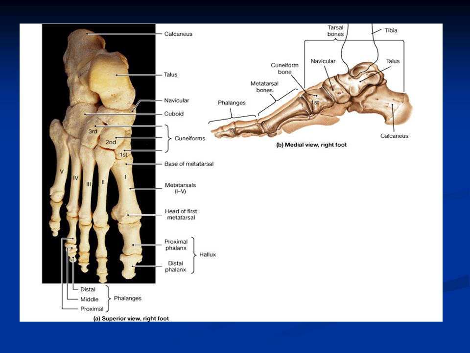
Copyright The McGraw-Hill Companies, Inc. Permission required for reproduction or display. Lower Limb A.The bones of the lower limb provide the framework for the thigh, lower leg, and foot.

83
Section 7.13 Objective- Read 7.3 and list the classes of joints and the six types of synovial joints and action of each. Also be able to identify several types of joint movements. Objective- Read 7.3 and list the classes of joints and the six types of synovial joints and action of each. Also be able to identify several types of joint movements.

84
Copyright The McGraw-Hill Companies, Inc. Permission required for reproduction or display. Joints A.Joints (articulations) are the functional junctions between bones. B.Joints enable a wide variety of body movements. C.Joints can be classified according to the degree of movement possible and can be immovable, slightly movable, or freely movable.
 are the functional junctions between bones. B.Joints enable a wide variety of body movements. C.Joints can be classified according to the degree of movement possible and can be immovable, slightly movable, or freely movable..")
86
Copyright The McGraw-Hill Companies, Inc. Permission required for reproduction or display. D.Joints can also classified according to the type of tissue that binds them together. E.Fibrous Joints 1.Fibrous joints are held close together by dense connective tissue and are immovable (sutures of skull) or only slightly movable(joint between the distal tibia and fibula).
 or only slightly movable(joint between the distal tibia and fibula)..")
87
Copyright The McGraw-Hill Companies, Inc. Permission required for reproduction or display.

88
F.Cartilaginous Joints 1.Hyaline cartilage or disks of fibrocartilage unite the bones in cartilaginous joints. 2.Intervertebral disks between vertebrae help absorb shock and are slightly movable. 3.Other examples of cartilaginous joints include the symphysis pubis and the first rib with the sternum.

89
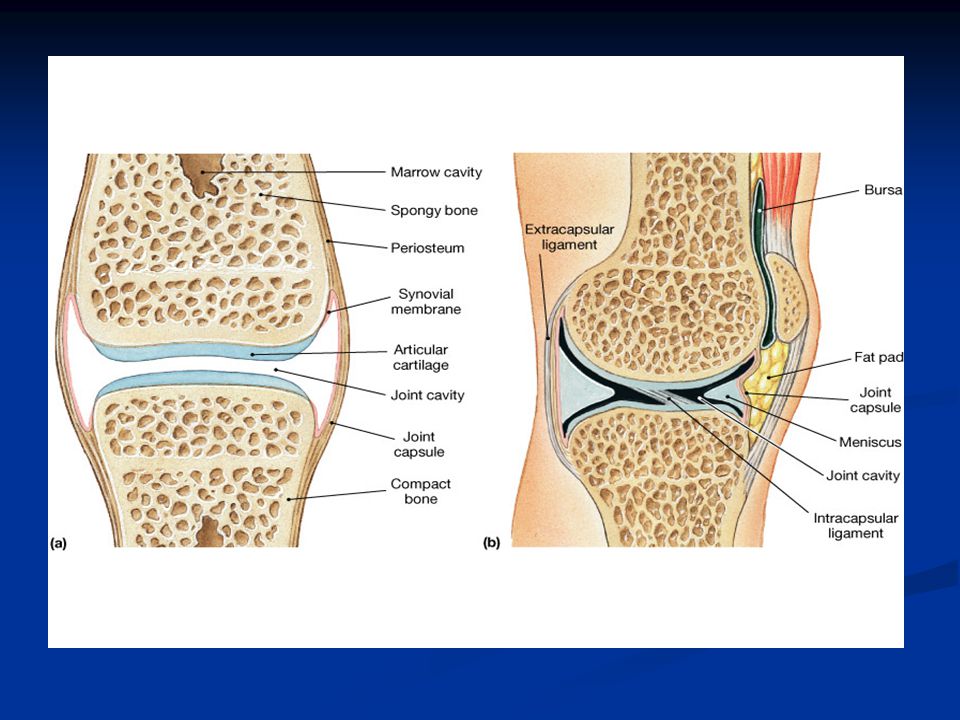
Copyright The McGraw-Hill Companies, Inc. Permission required for reproduction or display. G.Synovial Joints 1. Most joints of the skeleton are synovial joints, which are more complex than fibrous or cartilaginous joints. 2.The articular ends of bone in a synovial joint are covered with hyaline cartilage.

90
Copyright The McGraw-Hill Companies, Inc. Permission required for reproduction or display. 3.A joint capsule consists of an outer layer of dense connective tissue that joins the periosteum, and an inner layer made up of synovial membrane. a.Synovial fluid has the consistency of egg whites and lubricates articulating surfaces within the joint.

95
Copyright The McGraw-Hill Companies, Inc. Permission required for reproduction or display. 4.Some synovial joints contain shock-absorbing pads of fibrocartilage called menisci. 5.Some synovial joints have fluid-filled sacs called bursae.

96
Copyright The McGraw-Hill Companies, Inc. Permission required for reproduction or display.

97
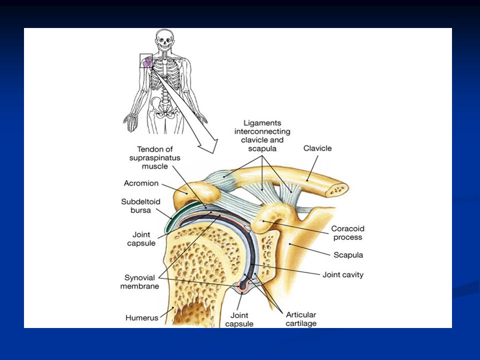
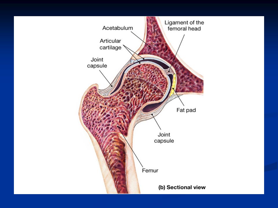
6.Based on the shapes of their parts and the movements they permit, synovial joints can be classified as follows: a.A ball-and-socket joint consists of a bone with a globular or egg-shaped head articulating with the cup-shaped cavity of another bone; a very wide range of motion is possible; examples include the hip and shoulder joint.

101
Copyright The McGraw-Hill Companies, Inc. Permission required for reproduction or display. b.A condyloid joint consists of an ovoid condyle fitting into an elliptical cavity, permitting a variety of motions; an example is the joint between a metacarpal and a phalange.

102
Copyright The McGraw-Hill Companies, Inc. Permission required for reproduction or display. c.Gliding joints occur where articulating surfaces are nearly flat or slightly curved, allowing a back-and- forth motion; the joints of the wrist and ankle, as well as those between vertebrae, are gliding joints.

104
Copyright The McGraw-Hill Companies, Inc. Permission required for reproduction or display. d.In a hinge joint, a convex surface fits into a concave surface, as is found in the elbow and phalange joints; movement is in one plane only. e.In a pivot joint, a cylindrical surface rotates within a ring of bone and fibrous tissue; examples include the joint between the proximal ends of the radius and ulna.

108
Copyright The McGraw-Hill Companies, Inc. Permission required for reproduction or display. f.A saddle joint forms where articulating surfaces have both concave and convex areas, permitting a wide range of movements; the joint between the trapezium and the metacarpal of the thumb is of this type.

109
Copyright The McGraw-Hill Companies, Inc. Permission required for reproduction or display. H.Types of Joint Movements 1.When a muscle contracts, its fibers pull its movable end (insertion) toward its stationary end (origin), causing movement at a joint.
 toward its stationary end (origin), causing movement at a joint..")
110
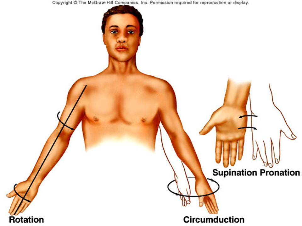
Copyright The McGraw-Hill Companies, Inc. Permission required for reproduction or display. 2.These terms describe movements that occur at joints: flexion, extension, dorsiflexion, plantar flexion, hyperextension, abduction, adduction, rotation, circumduction, pronation, supination, eversion, inversion, retraction, protraction, elevation, and depression.

114
Do Now- name the types of synovial joints and give example of where they are found in the body. Do Now- name the types of synovial joints and give example of where they are found in the body.

Similar presentations


 Mineral Storage of Calcium and Phosphate Red Blood Cell Production (long.>")
 fibers along with water and mineral salts (calcium hydroxide & calcium.>")


