Download presentation
Presentation is loading. Please wait.

4
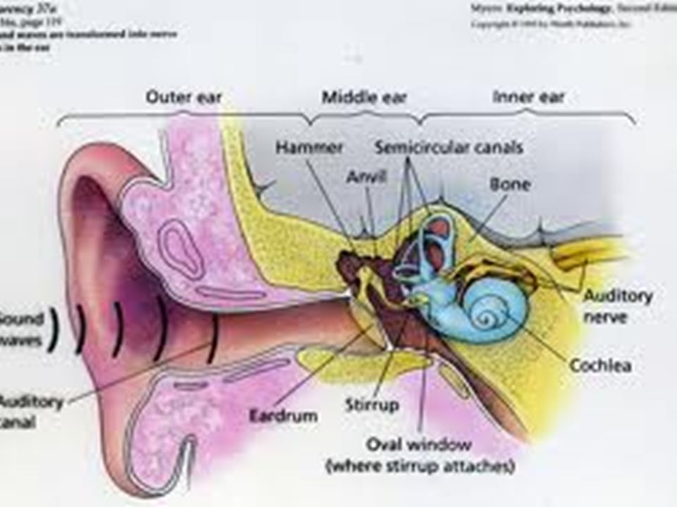
The external ear funnels sound waves to the external auditory meatus
The external ear funnels sound waves to the external auditory meatus.tsound. From the meatus, the external auditory canal passes inward to the tympanic membrane (eardrum(- The middle ear is an air-filled cavity in the temporal bone that opens via the auditory (eustachian) tube into the nasopharynx and through the nasopharynx to the exterior. The tube is usually closed, but during swallowing, chewing, and yawning it opens, keeping the air pressure on the two sides of the eardrum equalized. The three auditory ossicles, the malleus, incus, and stapes, are located in the middle ear. The manubrium (handle of the malleus) is attached to the back of the tympanic membrane. Its head is attached to the wall of the middle ear, and its short process is attached to the incus, which in turn articulates with the head of the stapes. . Its foot plate is attached by an annular ligament to the walls of the ovalwindow . Two small skeletal muscles, the tensor tympani and the stapedius, are also located in the middle ear. Contraction of the former pulls the manubrium of the malleus medially and decreases the vibrations of the tympanic membrane; contraction of the latter pulls the footplate of the stapes out of the oval window.
 tube into the nasopharynx and through the nasopharynx to. the exterior. The tube is usually closed, but during swallowing, chewing, and yawning it opens, keeping the air pressure on the two sides of the eardrum. equalized. The three auditory ossicles, the malleus, incus, and stapes, are located in the middle ear. The manubrium (handle of the malleus) is attached. to the back of the tympanic membrane. Its head is attached to the wall of the middle ear, and its short process is attached to the incus, which in turn articulates. with the head of the stapes. . Its foot plate is attached by an annular ligament to the walls of the ovalwindow . Two small skeletal muscles, the tensor tympani and the stapedius, are also located in the middle ear. Contraction of the former pulls. the manubrium of the malleus medially and decreases the vibrations of the tympanic membrane; contraction of the latter pulls the footplate of the stapes out of. the oval window.")
5
Inner Ear The inner ear (labyrinth) is made up of two parts, one within the other. The bony labyrinth is a series of channels in the petrous portion of the temporal bone. Inside these channels, surrounded by a fluid called perilymph, is the membranous labyrinth This membranous structure more or less duplicates the shape of the bony channels. It is filled with a fluid called endolymph, and there is no communication between the spaces filled with endolymph and those filled with perilymph.
 is made up of two parts, one within the other. The bony labyrinth is a series of channels in the petrous portion of the temporal bone. Inside these channels, surrounded by a fluid called perilymph, is the membranous labyrinth This membranous structure more or less. duplicates the shape of the bony channels. It is filled with a fluid called endolymph, and there is no communication between the spaces filled with endolymph and those filled with perilymph.")
6
Cochlea The cochlear portion of the labyrinth is a coiled tube which in humans is 35 mm long and makes many turns. it is devide into three chambers (scalae) The upper scala vestibuli and the lower scala tympani contain perilymph and communicate with each other at the apex of the cochlea through a small opening called the helicotrema. At the base of the cochlea, the scala vestibuli ends at the oval window,. The scala tympani ends at the round window, The scala media, the middle cochlear chamber does not communicate with the other two (scalae. It contains endolymph.
 The upper scala vestibuli and the lower scala tympani contain perilymph and communicate with each other at the apex of the cochlea through a small opening called the helicotrema. At the base of the cochlea, the scala vestibuli ends at. the oval window,. The scala tympani ends at the round window, The scala media, the middle cochlear chamber does not communicate with the other two (scalae. It contains endolymph.")
7
Organ of Corti Located on the basilar membrane is the organ of Corti, the structure that contains the hair cells which are the auditory receptors. This organ extends from the apex to the base of the cochlea and consequently has a spiral shape. The processes of the hair cells pierce the tough, membrane-like reticular lamina that is supported by the rods of Corti (Figure 9-4). The hair cells are arranged in four rows: three rows of outer hair cells lateral to the tunnel formed by the rods of Corti, and one row of inner hair cellsThe axons of the afferent neurons that innervate the hair cells form the auditory (cochlear) division of the vestibulocochlear acoustic nerve and terminate in the dorsal and ventral cochlear nuclei of the medulla oblongata Central Auditory Pathways From the cochlear nuclei, auditory impulses pass via a variety of pathways to the inferior colliculi, the centers for auditory reflexes, and via the medial geniculate body in the thalamus to the auditory cortex. Others enter the reticular formation (Figure 9-5). Information from both ears converges on each superior olive, and at all higher levels most of the neurons respond to inputs from both sides. The primary auditory cortex, Brodmann's area 41,
. The hair cells are arranged in four rows: three rows of outer hair cells lateral to the tunnel formed by the rods of. Corti, and one row of inner hair cellsThe axons of the afferent neurons that innervate the hair cells form the auditory (cochlear) division of the vestibulocochlear acoustic nerve and terminate in the dorsal and ventral cochlear nuclei of the medulla oblongata. Central Auditory Pathways. From the cochlear nuclei, auditory impulses pass via a variety of pathways to the inferior colliculi, the centers for auditory reflexes, and via the medial. geniculate body in the thalamus to the auditory cortex. Others enter the reticular formation (Figure 9-5). Information from both ears converges on each. superior olive, and at all higher levels most of the neurons respond to inputs from both sides. The primary auditory cortex, Brodmann s area 41,")
8
HEARING Sound Waves Sound is the sensation produced when longitudinal vibrations of the molecules in the external environment, ie, alternate phases of condensation and rarefaction of the molecules, strike the tympanic membrane. A plot of these movements as changes in pressure on the tympanic membrane per unit of time is a series of waves (Figure 9-10), and such movements in the environment are generally called sound waves. The waves travel through air at a speed of approximately 344 m/s (770 miles/h) at 20 °C at sea level. The speed of sound increases with temperature and with altitude Generally speaking, the loudness of a sound is correlated with the amplitude of a sound wave and its pitch with the frequency (number of waves per unit of time).. The amplitude of a sound wave can be expressed in terms of the maximum pressure change at the eardrum,
, and such movements in the environment are generally called sound waves. The waves travel through air at a speed of. approximately 344 m/s (770 miles/h) at 20 °C at sea level. The speed of sound increases with temperature and with altitude. Generally speaking, the loudness of a sound is correlated with the amplitude of a sound wave and its pitch with the frequency (number of waves per unit of. time).. The amplitude of a sound wave can be expressed in terms of the maximum pressure change at the eardrum,")
9
Functions of the Tympanic Membrane & Ossicles
In response to the pressure changes produced by sound waves on its external surface, the tympanic membrane moves in and out. The membrane therefore functions as a resonator that reproduces the vibrations of the sound source. It stops vibrating almost immediately when the sound wave stops; ie, it is very nearly critically damped. The motions of the tympanic membrane are imparted to the manubrium of the malleus. The malleus rocks on an axis through the junction of its long and short processes, so that the short process transmits the vibrations of the manubrium to the incus. The incus moves in such a way that the vibrations are transmitted to the head of the stapes. Movements of the head of the stapes swing its footplate to and fro like a door hinged at the posterior edge of the oval window. The auditory ossicles thus function as a lever system that converts the resonant vibrations of the tympanic membrane into movements of the stapes against the perilymph-filled scala vestibuli of the cochlea This system increases the sound pressure that arrives at the oval window, because the lever action of the malleus and incus multiplies the force 1.3 times and the area of the tympanic membrane is much greater than the area of the footplate of the stapes. There are losses of sound energy as a result of resistance sound energy incident on the tympanic membrane is transmitted to the fluid in the cochlea.

10
Tympanic Reflex When the middle ear muscles—the tensor tympani and the stapedius—contract, they pull the manubrium of the malleus inward and the footplate of the stapes outward. This decreases sound transmission. Loud sounds initiate a reflex contraction of these muscles generally called the tympanic reflex. Its function is protective, preventing strong sound waves from causing excessive stimulation of the auditory receptors. Bone & Air Conduction Conduction of sound waves to the fluid of the inner ear via the tympanic membrane and the auditory ossicles, the main pathway for normal hearing, is called ossicular conduction. Sound waves also initiate vibrations of the secondary tympanic membrane that closes the round window. This process, unimportant in normal hearing, is air conduction. A third type of conduction, bone conduction, is the transmission of vibrations of the bones of the skull to the fluid of the inner ear. Considerable bone conduction occurs when tuning forks or other vibrating bodies are applied directly to the skull. This route also plays a role in transmission of extremely loud sounds.

11
Functions of the Inner & Outer Hair Cells
The inner hair cells are the primary sensory cells that generate action potentials in the auditory nerves, The outer hair cells, on the other hand, have a different function. These respond to sound, like the inner hair cells, but depolarization makes them shorten and hyperpolarization makes them lengthen. They do this over a very flexible part of the basal membrane, and this action somehow increases the amplitude and clarity of sounds.

Similar presentations


 SYSTEM>")


: External ear Middle ear Internal ear Equilibrium sense (Parts involved): Internal ear.>")


