Download presentation
Presentation is loading. Please wait.

1
LECTURE 3 MICROSCOPY AND Basic Cell Structure

2
TODAYS MENU MICROSCOPY BASIC CELL STRUCTURE SUMMARY #1 and ASSIGNMENT 2 DUE PAGES TO READ: 77-85

3
MICROSCOPY The microscope is used to magnify objects Two types A) LIGHT MICROSCOPES (LMs) – use a visible light source and lenses to magnify image. Two kinds: a compound microscope and a dissecting microscope. B) ELECTRON MICROSCOPES (Ems): use an electron beam instead of a light microscope [Transmission and scanning electron microscope]
 ELECTRON MICROSCOPES (Ems): use an electron beam instead of a light microscope [Transmission and scanning electron microscope].")
4
Differences between LMs and EMs LMs Use light Resolving Power ≥0.2 µm Magnification max 1000X Specimens can be dead or alive Ems Use electron beam Resolving Power ≥0.1 nm Magnification >1000X Specimens are dead and preparation method introduce artifacts.

5
Light Microscope

6
Electron Microscope

7
A comparison of microscope images

8
Light Microscope Two important parameters Magnification: how large to make image of specimen or ratio of an object’s image to it’s real size. Resolving Power: measure of clarity of image.

9
Parts of a compound microscope Eyepiece: houses ocular lens (Magnification 10X) Nosepiece: rotates to allow use of a particular objective lens. Objective lenses: Have various magnifications Stage: holds microscope slide for viewing Stage Manipulator rods: allow movement of slide while viewing Light Source: illuminates object on slide

10
Parts of a compound microscope Diaphragm: regulates amount of light transmitted through object. Condenser: focuses the light beam. Coarse Adjustment Knob: for focussing changes Fine Adjustment Knob: for very fine focussing changes Base and Arm: the supporting structures of the microscope

11
How to Use a Compound Microscope Remove the cover, plug the microscope in Always start and end with Low Power! Place the slide on the microscope stage, with the specimen directly over the center of the glass circle on the stage (directly over the light). NOTE: If you wear glasses, take them off; if you see only your eyelashes, move closer. Be sure to close, or cover your other eye!! NOTE: If you see a dark line that goes part way across the field of view, try turning the eyepiece. That dark line is a pointer eyepiece
. NOTE: If you wear glasses, take them off; if you see only your eyelashes, move closer. Be sure to close, or cover your other eye!. NOTE: If you see a dark line that goes part way across the field of view, try turning the eyepiece. That dark line is a pointer eyepiece.")
12
How to Use a Compound Microscope If, and ONLY if, you are on LOW POWER, lower the objective lens to the lowest point, then focus using first the coarse knob, then the fine focus knob. Adjust the Diaphragm as you look through the Eyepiece, and you will see that MORE detail is visible when you allow in LESS light! Once you have found the specimen on Low Power, unless specifically asked to draw it on low power, center the specimen in your field of view, then, without changing the focus knobs, switch it to High Power.

13
How to Use a Compound Microscope Once you have it on High Power remember that you only use the fine focus knob!

14
Care of the Instrument – Do’s and Don’ts Transport: Always carry the microscope with one hand on the Arm and one hand on the Base. Carry it close to your body. Clutter: Keep workstation uncluttered Electric Cord: Place the excess cord on the table! The High Power Objective is very close to the slide. Use of the coarse focus knob will scratch the lens, and crack the slide. More expensive sounds... Only use the fine adjustment

15
Care of the Instrument – Do’s and Don’ts The 100X objective is an oil immersion lens. Without the oil to lubricate the lens, you will destroy it! More expensive sounds... Also, the oil is needed to help gather enough light to actually see through the lens! Cleaning Tissues: Use only lint-free, optically safe tissues to clean lenses. Solvents: Be cautious. Do not place microscope under running water.

16
Exercises Total Magnification: Ocular lens * Objective lens Image Orientation: reverse of specimen Size of object: Diameter field of view * estimate of space taken up by image Diameter field of view: 4X=4.3 mm; 10X = 1.7mm; 40X = 0.4mm Working Distance and the field of view is inverse proportional to length of Objective lens Working distance: 4X = 25mm; 10X=8.3mm; 40X =0.5 mm

17
Exercises Total Magnification: Ocular lens 10X and Objective lens 40X = 10 * 40 = 400X Size of an object whose image covers half the field of view using the 10X objective length. = ½ * 8.3 mm = 4.15 mm

18
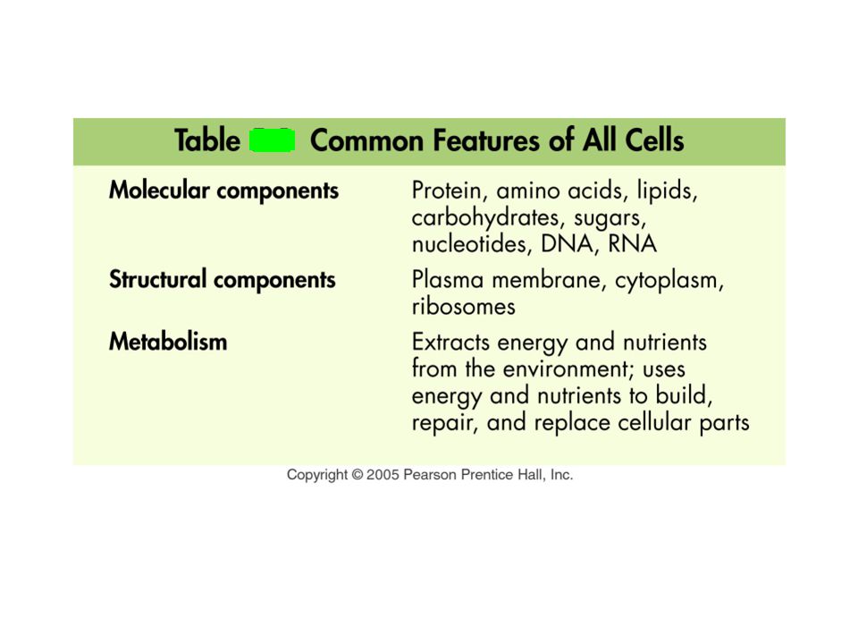
Basic Cell Structure All Living Things Are Composed of One or More Cells The term was coined by Robert Hooke (1665) All Cells Share Certain Common Features
 All Cells Share Certain Common Features")
20
What Are the Basic Features of Cells? –The Plasma Membrane Encloses the Cell and Mediates Interactions Between the Cell and Its Environment –All Cells Use DNA as a Hereditary Blueprint and Contain Cytoplasm –All Cells Obtain Energy and Nutrients from Their Environment –Cell Function Limits Cell Size

21
frog embryo most eukaryotic cells mitochondrion most bacteria virus proteins diameter of DNA double helix chicken egg adult human tallest trees atoms 1 centimeter (cm) = 1/100 m 1 millimeter (mm) = 1/1000 m Units of measurement: 1 meter (m) = 39.37 inches Diameter visible with conventional electron microscope visible with special electron microscopes visible with llight microscope visible with unaided human eye 1 micrometer (µm) = 1/1,000,000 m 1 nanometer (nm) = 1/1,000,000,000 m 100 µm 10 µm 1 µm 100 nm 10 nm 1 nm 0.1 nm 1 mm 1 cm 10 cm 100 m 10 m 1 m
 = 1/100 m 1 millimeter (mm) = 1/1000 m Units of measurement: 1 meter (m) = inches Diameter visible with conventional electron microscope visible with special electron microscopes visible with llight microscope visible with unaided human eye 1 micrometer (µm) = 1/1,000,000 m 1 nanometer (nm) = 1/1,000,000,000 m 100 µm 10 µm 1 µm 100 nm 10 nm 1 nm 0.1 nm 1 mm 1 cm 10 cm 100 m 10 m 1 m")
22
most eukaryotic cells mitochondrion most bacteria virus proteins diameter of DNA double helix atoms 1 centimeter (cm) = 1/100 m 1 millimeter (mm) = 1/1000 m Units of measurement: 1 meter (m) = 39.37 inches visible with conventional electron microscope visible with special electron microscopes visible with llight microscope 1 micrometer (µm) = 1/1,000,000 m 1 nanometer (nm) = 1/1,000,000,000 m 100 µm 10 µm 1 µm 100 nm 10 nm 1 nm 0.1 nm
 = 1/100 m 1 millimeter (mm) = 1/1000 m Units of measurement: 1 meter (m) = inches visible with conventional electron microscope visible with special electron microscopes visible with llight microscope 1 micrometer (µm) = 1/1,000,000 m 1 nanometer (nm) = 1/1,000,000,000 m 100 µm 10 µm 1 µm 100 nm 10 nm 1 nm 0.1 nm")
23
What Are the Basic Features of Cells? There Are Two Basic Types of Cells: Prokaryotic and Eukaryotic Prokaryotic cells – represented by the Domains Archaea (archaebacteria) and Bacteria Eukaryotic cells – represented by the Domain Eukarya (algae, protozoans, fungi, plants and animals)
 and Bacteria Eukaryotic cells – represented by the Domain Eukarya (algae, protozoans, fungi, plants and animals).")
24
chromosome (nucleoid region) pili ribosomes food granule prokaryotic flagellum capsule or slime layer cell wall plasma membrane cytoplasm plasmid (DNA) A generalized prokaryotic cell
 pili ribosomes food granule prokaryotic flagellum capsule or slime layer cell wall plasma membrane cytoplasm plasmid (DNA) A generalized prokaryotic cell")
26
mitochondrion vesicle cytoplasm flagellum lysosome centriole Golgi complex vesicle nuclear pore nuclear envelope chromatin (DNA) nucleolus nucleus ribosome free ribosome microtubules rough endoplasmic reticulum Smooth endoplasmic reticulum plasma membrane intermediate filaments A generalized animal cell
 nucleolus nucleus ribosome free ribosome microtubules rough endoplasmic reticulum Smooth endoplasmic reticulum plasma membrane intermediate filaments A generalized animal cell")
27
central vacuole plastid mitochondrion vesicle plasmodesma cell wall plasma membrane intermediate filaments free ribosome ribosomes nucleus nucleolus nuclear pore chromatin nuclear envelope Golgi complex chloroplast Microtubules (part of cytoskeleton) smooth endoplasmic reticulum rough endoplasmic reticulum A generalized plant cell
 smooth endoplasmic reticulum rough endoplasmic reticulum A generalized plant cell")
28
What Are the Major Features of Prokaryotic Cells? Prokaryotic Cells Are Small Prokaryotic Cells Have Fewer Specialized Structures Within Their Cytoplasm Have no membrane-enclosed organelles within their cytoplasm DNA is located in the “nucleoid”

29
What Are the Major Features of Eukaryotic Cells? Eukaryotic Cells Contain Organelles The Nucleus Is the Control Center of the Eukaryotic Cell (Have the DNA) Have membrane-enclosed organelles within the cytoplasm
 Have membrane-enclosed organelles within the cytoplasm.")
30
Functions of Cell Structures Eukaryotic Cells Contain a Complex System of Membranes –The Plasma Membrane Both Isolates the Cell and Allows Selective Interactions Between the Cell and Its Environment –The Endoplasmic Reticulum Forms Membrane- Enclosed Channels Within the Cytoplasm –The Golgi Complex Sorts, Chemically Alters, and Packages Important Molecules

31
Functions of Cell Structures Lysosomes Serve as the Cell’s Digestive System –Plant Cells Have Central Vacuoles

32
smooth endoplasmic reticulum nuclear envelope Golgi complex exocytosis plasma membrane phagocytosis lysosome fused with food vacuole food vacuole rough endoplasmic reticulum The flow of membr ane within the cell

33
cytoplasm central vacuole cell wall plasma membrane The central vacuole and turgor pressure in plant cells

34
Functions of Cell Structures Mitochondria Extract Energy from Food Molecules, and Chloroplasts Capture Solar Energy –Mitochondria Use Energy Stored in Food Molecules to Produce ATP –Chloroplasts Are the Sites of Photosynthesis

35
Differences between plant and animal cells Plant cells are surrounded by a rigid cell wall Plant cells have chloroplast Plant cells have a large central vacuole Plant cells contain plasmodesmata-openings in the cell wall of adjacent cells Animal cells have lysosomes Animal cells have Centrioles

36
KEY WORDS Main features of Prokaryotic and Eukaryotic cells Functions of cell structures Differences between Plant and animal cells Types of cells Types of Microscopes Magnification Resolving Power Calculations- Magnification, Size of Object Do’s and Don’t’s of microscopes

37
LAB EXERCISES EX 1 – page 78. Fill out the labels EX 2- page 80. The Letter E. Place the slide with the letter E on the stage of the microscope, secure with the clips. Focus using low magnification before moving on to higher magnification. Answer the question on pg 80/81. EX 3 – Crossed threads, use to determine the depth of view. EX – 4 – Dissecting Microscope EX – 5, pg 82 Plant Cells and Ex 6 pg 85- Animal Cells Assignment 3: EX 2 – Qu 1-8 and EX 5 – Qu 1-3

38
Next Class Menu Mitosis & Meiosis (Watch video) Read pages 193
 Read pages 193")
Similar presentations


 What is your nucleus? How many people or beings could be supported by your.>")


, visible light passes through a specimen and then through glass lenses, which magnify the image The quality of an.>")

 Robert Hooke -- 1665: examined thinly sliced cork and coined term “cell”>")


 Arm Stage Coarse Adjustment Knob Fine Adjustment Knob Always carry a microscope with one.>")


