Download presentation
Presentation is loading. Please wait.

1
Skeletal System Drg. Fidya, M.Si Departemen of Anatomy Histology

2
Part of Skeletal System
Skeleton Cartilages Ligaments Joint Consist of : Birth skeletal 270 Adult skeletal 207/206 Why different?

3
Function of Skeletal Support Storage of minerals Blood cell production
Protection Body movement

4
FUNCTIONS OF THE SKELETON
Supports the body. The bones of the lower limbs support the entire body when we are standing, and the pelvic girdle supports the the abdominal cavity Protects soft body part The bones of the skull protect the brain; The rib cage protects the heart and lungs. Produces blood cells All bones in the fetus have red bone marrow that produces blood cells. In the adult, only certain bones produce blood cells.

5
Stores minerals and fat
All bones have a matrix that contains calcium phosphate, a source of calcium ions and phosphate ions in the blood. Fat is stored in yellow bone marrow Along with the muscles, permits flexible body movement While articulations ( joints ) occur between all the bones, we associate body movement in particular with the bones of the limbs
 occur between all the bones, we associate body movement in particular with the bones of the limbs.")
6
Skeletal System Divide into 2 division: Axial skeleton
Appendicular skeleton

7
Skeletal System Axial Skeleton Appendicular skeleton Skull Hyoid Bone
Auditory ossicle Vertebral Column Rib cage Pectoral gridle Upper extremitas Pelvic gridle Lower extremitas

9
Skull Consist of: Cranial bone (8) Facial bone (14) frontal (1)
parietal (2) occipital (1) temporal (2) sphenoid (1) ethmoid (1) Facial bone (14) maxilla (2) palatine (2) zygomatic (2) lacrimal (2) nasal (2) vomer (1) inferior nasal concha (2) mandible (1)
 occipital (1) temporal (2) sphenoid (1) ethmoid (1) Facial bone (14) maxilla (2) palatine (2) zygomatic (2) lacrimal (2) nasal (2) vomer (1) inferior nasal concha (2) mandible (1)")
13
Hyoid Bones The hyoid bone above the larynx below the mandible
Supports the tongue Assists in swallowing

14
Auditory ossicle Three auditory ossicles (“ear bones”) : present in the middle-ear chamber of each ear Serve to transmit sound impulses. Three small paired bones (auditory ossicles) located: within the middle-ear cavities in the petrous part of the temporal bones Malleus (hammer), incus (anvil), stapes (stirrup) (outer-inner)
 located: within the middle-ear cavities in the petrous part of the temporal bones. Malleus (hammer), incus (anvil), stapes (stirrup) (outer-inner)")
15
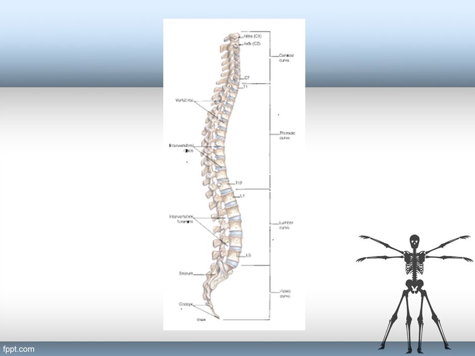
Vertebral Column Composed of 33 individual vertebrae, some of which are fused. Consist of 7 cervical, 12 thoracic, 5 lumbar, 4 to 5 fused sacral, and 3 or 5 fused coccygeal Adult vertebral column composed of a total of 26 movable parts. Vertebrae are separated by fibrocartilaginous intervertebral discs. Secured to each other by interlocking processes and binding ligaments. Between the vertebrae are openings called intervertebral foramina that allow passage of spinal nerves.

17
Rib Cage The cone-shaped, flexible rib cage, consists of the thoracic vertebrae, 12 paired ribs, costal cartilages, and the sternum. Encloses and protects the thoracic viscera Directly involved in the mechanics of breathing.

19
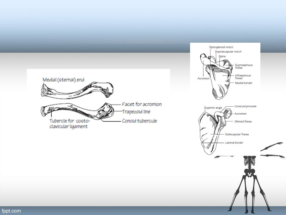
Pectoral Gridle Two scapulae and two clavicles
having only an anterior attachment to the axial skeleton sternoclavicular joint at the sternum. Lacking a posterior attachment to the axial skeleton, the pectoral girdle has a wide range of movement. Function: - To provide attachment areas for the numerous muscles that move the shoulder and elbow joints.

21
Upper Extremitas Brachium : - Humerus Antebrachium: - Radius - Ulna
Manus: - Carpus - Metacarpus - Phalanges

23
Pelvic Gridle Formed by two ossa coxae. together with sacrum and coccyx pelvis United anteriorly at the symphysis pubis Attached posteriorly to the sacrum of the vertebral column. Function: - support the weight of the body from the vertebral column (with assosiated ligament) - supports and protects the lower viscera, (urinary bladder, reproductive organs, developing fetus)
 - supports and protects the lower viscera, (urinary bladder, reproductive organs, developing fetus)")
25
Lower Extermitas Femoralis Genu Crus : -Tibia Fibula Pes

26
2.3 JOINT ( ARTICULATION ) There are two systems for classification :
According amount of movement they allow According to their structure ( the convention followed here ) A joint called : SYNARTHROSIS ( IMMOVABLE ) AMPHIARTHROSIS ( SLIGHT MOVEMENT ) DIARTHROSIS ( FREELY MOVABLE )
 A joint called : SYNARTHROSIS ( IMMOVABLE ) AMPHIARTHROSIS ( SLIGHT MOVEMENT ) DIARTHROSIS ( FREELY MOVABLE )")
27
SYNARTHROSIS ( IMMOVABLE )
Occur where fibrous connective tissue joints bone to bone fibrous joint Sutures, in the cranial vault and face; and are immovable

28
AMPHIARTHROSIS ( SLIGHT MOVEMENT )
Where bones are joined by hyalin cartilage or fibrocartilage cartilaginous joints Slightly movable The pubic symphysis, the joint between the two pubic bones

29
DIARTHROSIS ( FREELY MOVABLE )
Bone ends do not contact each other, but are enclosed in a capsule Generally freely movable Two bones are separated by a joint cavity is lined by a synovial membrane, which produces synovial fluid, a lubricant for the joint Ligament, which are composed of dense regular connective tissue, bind the two bones and add even more stability

31
MOVEMENT PERMITTED BY SYNOVIAL JOINTS
Skeletal muscles are attached to bones by tendons that cross joint. When a muscle contracts, one bone moves in elation to another bone Types of movement : ANGULAR MOVEMENT CIRCULAR MOVEMENT SPECIAL MOVEMENT

32
ANGULAR MOVEMENT FLEXION EXTENTION ADDUCTION ABDUCTION
Decreases the joint angle EXTENTION Increases the joint angle ADDUCTION the movement of body part toward the midline ABDUCTION The movement of the body part laterally, away from midline

33
CIRCULAR MOVEMENT CIRCUMDUCTION ROTATION SUPINATION PRONATION
The movement of the body part in a wide, makes arm circle ROTATION The movement of a body part around its own axis: The arm is twisted toward the trunk ( MEDIAL ROTATION The arm away from the trunk ( LATERAL ROTATION ) SUPINATION The rotation of the forearm the palm in upward PRONATION Is the opposite, the movement of the forearm the palm is downward
 SUPINATION. The rotation of the forearm the palm in upward. PRONATION. Is the opposite, the movement of the forearm the palm is downward.")
34
SPECIAL MOVEMENT INVERSION EVERSION ELEVATION and DEPRESSION
Turning the foot so that the sole faces inward EVERSION Turning the foot so that the sole faces outward ELEVATION and DEPRESSION Refer to the lifting up and down, respectively, of a body part Shrug shoulder Move jaw up and down

35
Terima Kasih

Similar presentations


 Weakest parts of the skeleton Weakest parts of the skeleton Articulation – site where two or more bones meet Articulation – site.>")

identifying the four bone types. 6) Identify bones that compose the skeletal system. 6.2) identifying.>")


