Download presentation
Presentation is loading. Please wait.

1
STRESS ECHO DEEPAK NANDAN

2
Stress echo is a family of examinations in which 2D echocardiographic monitoring is undertaken before, during & after cardiovascular stress Cardiovascular stress exercise pharmacological agents Stress echo is a family of examinations in which 2D echocardiographic monitoring is undertaken before, during & after cardiovascular stress Cardiovascular stress exercise pharmacological agents

3
PHYSIOLOGY Coronary blood flow – pulsatile & phasic Precapillary arterioles – resistance vessels * principal contributor of resistance * main controller of coronary blood flow ↑ CBF on ↑ demand occurs through reduction in resistance at this level

4
CORONARY BLOOD FLOW RESERVE Maximal CBF / basal CBF Magnitude of bf ↑ secondary to any stress relative to resting flow In discrete stenosis – CFR begins to ↓ when stenosis reaches 50% dm CFR is abolished when stenosis reaches 90% Resting bf remains constant up to 85- 90% of the stenosis

5
Cellular Mechanism of Ischemia Consequence(s) of Mechanical Dysfunction Mechanical Dysfunction Abnormal Contraction and Relaxation Diastolic Tension Diastolic Tension O 2 Consumption (to maintain tonic contraction) ATP Hydrolysis Diastolic Wall Tension (Stiffness) O 2 Demand O 2 Supply Extravascular Compression Blood Flow to Microcirculation ( O 2 delivery to Myocytes) Modified from: Belardinelli et al. Eur Heart 8 (Suppl. A):A10-A13, 2006
:A10-A13,")
7
BASIC PRINCIPLES OF STRESS ECHO BASIC PRINCIPLES OF STRESS ECHO ↑ Cardiac work load - ↑O2 demands- demand supply mismatch- ischemia Impairment of myocardial thickening and endocardial motion

9
Treadmill protocol

10
Stress echo-Standard-format

11
Supine bicycle ergometry

13
Supine bicycle standard format Supine bicycle standard format

14
Treadmill vs supine bicycle advantage Add information Wide spread availability Simple protocol High work load > Sensitive Disadvantage Imaging post ex only advantage Add information Wide spread availability Simple protocol High work load > Sensitive Disadvantage Imaging post ex only Advantage Image through out the exercise- peak Onset of RWMA Better image quality Contrast stress echo > Specific Disadvantage Lower work load Supine position affects ex.physio Advantage Image through out the exercise- peak Onset of RWMA Better image quality Contrast stress echo > Specific Disadvantage Lower work load Supine position affects ex.physio

15
Information obtained from Exercise Stress but not available with Pharmacological Test Exercise Duration/Tolerance Reproducibility of Symptoms with Activity Heart rate response to exercise Blood Pressure response Detection of Stress Induced Arrhythmias Assess control of angina with medical therapy Prognosis

16
Indication pharmacological stress echocardiography Inadequate exercise Left bundle branch block Paced ventricular rhythm pre-excitation or conduction abnormality Medication: beta-blocker, calcium channel blocker Evaluation of patients very early after MI(<3 days) or angioplasty stent(<2weeks) Poor image degradation with exercise Poor patient motivation to exercise
 or angioplasty stent(<2weeks) Poor image degradation with exercise Poor patient motivation to exercise")
17
Pharmacologic Stress Agents Stress agents Coronary vasodilator Dipyridamole Adenosine Inotropic agents Dobutamine Arbutamine

18
DOBUTAMINE STRESS ECHO Dobutamine- synthetic catecholamine Inotropic & chronotropic- β1,β2 & α Action: onset – 2 min half life – 2 min: continous IV Metabolizd by cathechol-o-methyl transferase Excretion: hepatobiliary system and kidney Dobutamine- synthetic catecholamine Inotropic & chronotropic- β1,β2 & α Action: onset – 2 min half life – 2 min: continous IV Metabolizd by cathechol-o-methyl transferase Excretion: hepatobiliary system and kidney

19
Dobut-protocol

20
Protocol for Dobutamine Stress Echo.

21
End points to terminate

22
Works by inducing myocardial ischemia Modest ↑ SBP and ↓ DBP May be arrhythmogenic (0.7% rate in 8500 consecutive studies performed at Mayo Clinic) Usually ineffective in patients on beta blockers High rate of side effects Hypotension induced does not have prognostic value unlike TMT Does not interact with dipyridamole
 Usually ineffective in patients on beta blockers High rate of side effects Hypotension induced does not have prognostic value unlike TMT Does not interact with dipyridamole")
23
Dipyridamole Potent coronary vasodilator Provoke anginal attack in angina patients Vasodilation effect inhibition of reuptake of adenosine by the endothelial cell CBF increases 4 to 5 times in normal vessel Reduction of subendocardial blood flow in stenotic coronary artery

24
Dipyridamole Coronary steal phenomenon Standard protocol: 0.54 mg/kg for 4 min High dose protocol: 0.84mg/kg Antidote: theophylline

25
CORONARY STEAL CORONARY STEAL Myocardial area Supplied by severe epi stenosis Collateral from remote cor art Blood flow Depends on prefusion pressure Cor collaterals Steal - ischemia Vasodilator ↓pp & flow ↓in collaterals Flow through stenotic vessel ↓

26
Dipyridamole Contraindication active wheezing high degree AV block hypotension(SBP<90 mmHg) recent use of dipyridamole(<24 hours) Relative contraindication Hx of reactive airway disease sick sinus syndrome severe sinus bradycardia
 recent use of dipyridamole(<24 hours) Relative contraindication Hx of reactive airway disease sick sinus syndrome severe sinus bradycardia")
27
Adenosine Adenosine Naturally occuring agent Types of receptors A1: slowing HR and conduction A2a: c-AMP – decrease calcium uptake by SR -- smooth muscle relaxation vasodilation Half life: 2 seconds need constant IV infusion Rapidly removed from RBC and endothelial cell

28
Adenosine – side effect Flushng: 37% Dyspnea: 35% GI discomfort: 15% Headache:14% Light-headedness 9% Most side effect – short-lived and mild

29
Myocardial contrast in stress echo Left vent opacification for border enhancement Myocardial perfusion imaging Perfusion at resting state-stress is performed and perfusion imaging is done at peak stress

30
Vasodilator stress echo-perfusion imaging Vasodilator stress echo-perfusion imaging

31
Stress Echo Stress Echocardiography Diagnosis PrognosisViability Treatment

33
Exercise –preferred-add information > sensitive in CAD compared to dobutamine Treadmill >sensitive, Bicycle>specific Bicycle –during stress-> accurate presence and extend of dis vs pat choice,availability etc. Dobutamine is limited to pats who cant exert adequately & when the Q of viability is addressed In pharmaclogical stress dobutamine is the agent- produces true ischemia than a flow mismatch

34
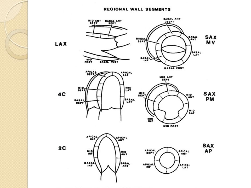
INTERPRETATION OF STRESS ECHO Subjective assessment of regional wall motion Compares wall thickening & endocardial excursion at baseline and stress Limitation- subjective & nonquantitative Measures like EF, ESV change, and strain rate to overcome limitations

35
Strain rate-myocardial velocity gradient -postsystolic shortening Strain rate-myocardial velocity gradient -postsystolic shortening TDI/Strain imaging> sensitive Ischemia delays onset & rate of regional myo relaxation Time quantified using TDI QRS-onset of relaxation-350-400ms Interval ↓ by 34+/_10% in nl segments in response to high dose dopamine ↓in interval is <12+/-18% in ischemic seg

37
Grade 1-normal 2-hypokinesis 3-akinesis 4-dyskinesis Nl WMSI-1 at baseline and stress Any score>1-abnormal Good prognostic value

38
Hypokinesia-<5 mm of endocardial excursion Akinesis - -ve syst thickening & endo excursion Dyskinesis –systolic thinning & outward motion nl resp-hyperkinesis Absence –low work load, β blockade, cardiomyopathy & delayed post stress imaging Localisation>specific in multivessel dis & in LAD than RCA/LCX

40
Normal- hyperkinesis during stress test Normal- hyperkinesis during stress test

41
DYSKINESIA OF THE APEX IN STRESS DYSKINESIA OF THE APEX IN STRESS

43
Prognostic value A new wall motion abnormality,rest & exercise WMSI,ESV response-correlated with risk

44
Chamber dilatation in resp to stress Chamber dilatation in resp to stress

45
Prognostic value of stress echo Prognostic value of stress echo Independent predictors of cardiac events a)WMSI with exercise b) ST ↓≥1 mm c) treadmill time Risk Index(RI)=1.02(WMSI)+1.04(ST change)− 0.14(Treadmilltime) RI in upper quartile(+0.66 to+2.02)– risk was highest(30%) Prognostic value is comparable in women and men
WMSI with exercise b) ST ↓≥1 mm c) treadmill time Risk Index(RI)=1.02(WMSI)+1.04(ST change)− 0.14(Treadmilltime) RI in upper quartile(+0.66 to+2.02)– risk was highest(30%) Prognostic value is comparable in women and men")
46
Stress echo after revascularisation Stress echo after revascularisation

47
PRE-OPERATIVE RISK STRATIFICATION WITH DOBUTAMINE STRESS ECHO *Mayo Clinic, 530 Patients

48
Perioperative marker of coronary event patients with a positive electrocardiographic response to treadmill stress test but no inducible wall motion abnormality on stress echocardiogram have a very low rate of adverse cardiovascular events during follow-up

49
VIABILITY OF MYOCARDIUM VIABILITY OF MYOCARDIUM That has the potential for functional recovery;- either stunned/hibernating myocardium >6mm thickness -viable segment Stunned or hibernating improved contractility with dobutamine, not in infarcted myocardium Biphasic response – low dose ↑ contractility(10 to 20 mcg/kg), at higher dose CBF ↓ -- contractility ↓
, at higher dose CBF ↓ -- contractility ↓")
50
Biphasic response is the most predictive of the functional recovery after revascularisation Sustained improvement/no change-nonviable For viability assessment – nuclear techniques are more sensitive dobut stress echo more specific PPV-similar NPV- favours dobut stress echo

51
Myocardial viability-Biphasic response Myocardial viability-Biphasic response

52
Sensitivity and specificity of exercise and pharmacologic stress test Sensitivity(%) Specificity(%) Dobutamine 71 – 96 66 - 83 Dipyridamole 43 – 74 92 - 100 Exercise 74 – 97 64 - 88
 Specificity(%) Dobutamine 71 – Dipyridamole 43 – Exercise 74 –")
53
Advantages of Stress Echocardiography Compared to Nuclear Stress Testing Higher Specificity Visualization of cardiac valves Evaluate for presence of pericardial effusion Ability to measure RV Systolic Pressure More accurate assessment of LV ejection fraction Doppler interrogation to determine Diastolic Function Lower Cost Lack of Radiation Exposure

54
Sensitivity Comparison of Different Testing Modalities

55
Situations Where Stress Echo Preferred Situations Where Stress Echo Preferred Younger patients with lower likelihood of symptomatic coronary artery disease Pericardial Disease suspected Valvular heart disease needs to also be evaluated Need to evaluate for pulmonary hypertension Exertional dyspnea is the predominant complaint

56
Thank you

Similar presentations


 Ashraf Hamdan, Ingo Paetsch, Eike Nagel German Heart Institute Berlin and www.cmr-academy.com Created.>")


 F.R.C.P.(E) F.R.C.P.(LONDON) F.A.C.C. DESIGNED AT A.V. DEPTT F.J.M.C. BY RABIA KAZMI.>")
