Download presentation
Presentation is loading. Please wait.

1
Karam Paul MS, MD, MBA, FACC Community Heart and Vascular

2
Know why to undertake a stress test Know who should have one Know how it is performed Understand the limitations Understand which to choose Know what to do with the result

3
Why do a stress test?

4
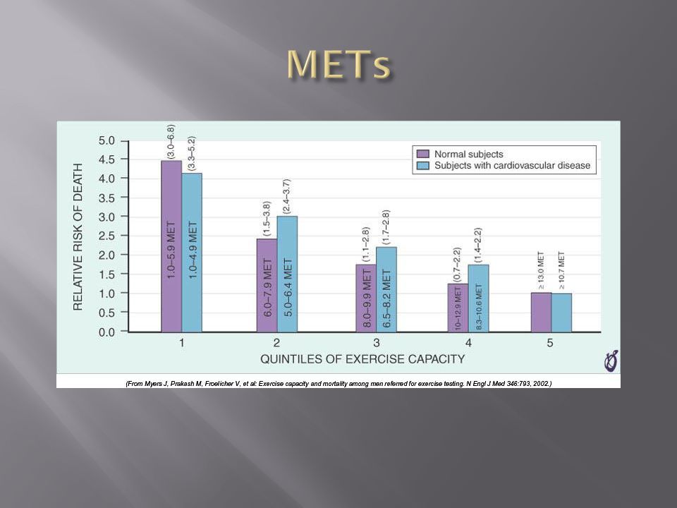
Elicit abnormalities not present at rest Estimate functional capacity Estimate prognosis Likelihood of coronary artery disease Extent of coronary artery disease Effect of treatment

5
Who should have one?

6
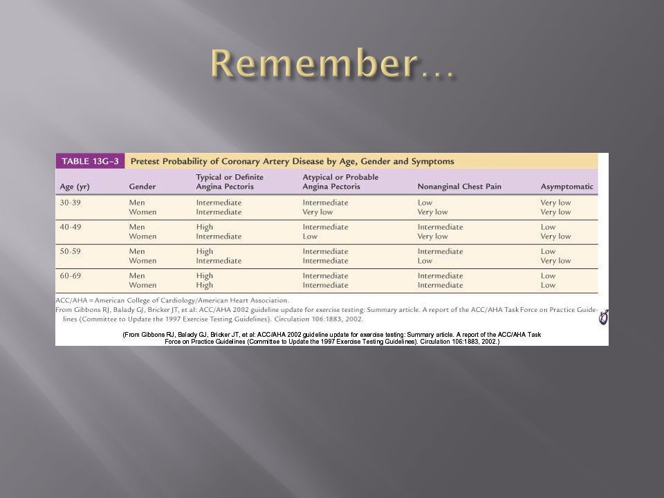
Bayes Theorem Consider the pre-test risk Sensitivity & specificity of the test Post-test probability of CAD Diagnostic power of EST is maximal when the pre-test probability is intermediate.

9
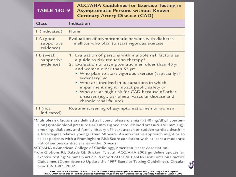
Pre-existing coronary artery disease Diabetes Hypertension Smoking history Family history Renal disease

10
Pre-existing coronary artery disease Diabetes Hypertension Hyperlipidemia Smoking history Family history Renal disease

12
Pre-existing coronary artery disease Diabetes Hypertension Hyperlipidemia Smoking history Family history Renal disease

17
How is it done?

20
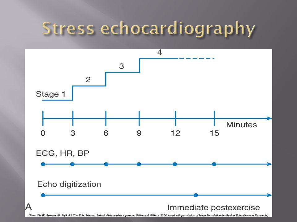
ECG Exercise capacity (METS – metabolic equivalent) Symptoms Blood pressure Heart rate response & recovery
 Symptoms Blood pressure Heart rate response & recovery")
21
1mm planar ST depression 3 consecutive beats

22
The normal and rapid upsloping ST segment responses are normal responses to exercise. Minor ST depression can occur occasionally at submaximal workloads in patients with coronary disease. The slow upsloping ST segment pattern often demonstrates an ischemic response in patients with known coronary disease or those with a high pretest clinical risk of coronary disease. Downsloping ST segment depression represents a severe ischemic response. ST segment elevation in an infarct territory (Q wave lead) indicates a severe wall motion abnormality and, in most cases, is not considered an ischemic response. (From Chaitman BR: Exercise electrocardiographic stress testing. In Beller GA [ed]: Chronic Ischemic Heart Disease. In Braunwald E [series ed]: Atlas of Heart Diseases. Vol 5. Chronic Ischemic Heart Disease. Philadelphia, Current Medicine, 1995, pp 2.1-2.30
 indicates a severe wall motion abnormality and, in most cases, is not considered an ischemic response. (From Chaitman BR: Exercise electrocardiographic stress testing. In Beller GA [ed]: Chronic Ischemic Heart Disease. In Braunwald E [series ed]: Atlas of Heart Diseases. Vol 5. Chronic Ischemic Heart Disease. Philadelphia, Current Medicine, 1995, pp")
23
Influenced by: Body position Respiration Hyperventilation Drug Rx Myocardial ischemia Necrosis Pseudonormalisation: Usually non-diagnostic Consider ancillary imaging

25
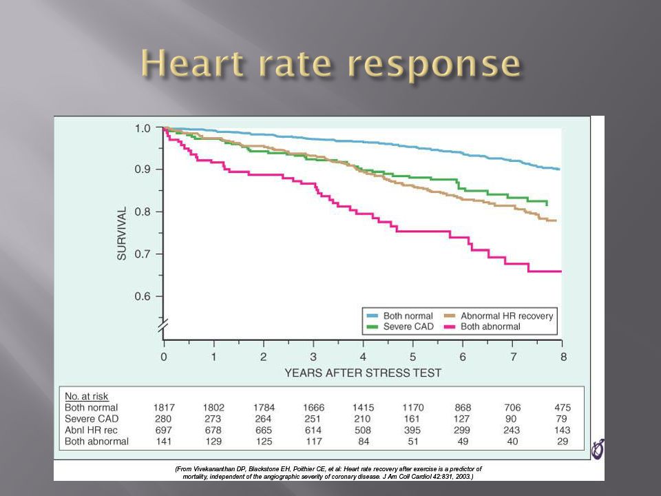
Peak HR > 85% of maximal predicted for age HR recovery >12 bpm (erect) HR recovery >18 bpm (supine)
 HR recovery >18 bpm (supine)")
27
Parameters associated with adverse prognosis or multi- vessel disease Duration of symptom-limiting exercise <5 METs Failure to increase sBP 120mmHg, or a sustained decreased 10mmHg, or below rest levels, during progressive exercise ST segment depression 2mm, downsloping ST segment, starting at <5 METs, involving 5 leads, persisting 5 min into recovery Exercise-induced ST segment elevation (aVR excluded) Angina pectoris at low exercise workloads Reproducible sustained (>30 sec) or symptomatic ventricular tachycardia
 Angina pectoris at low exercise workloads Reproducible sustained (>30 sec) or symptomatic ventricular tachycardia")
28
Non-diagnostic ECG changes False positives/false negatives Women – false positives Elderly – more sensitive/less specific Diabetics – autonomic dysfunction Hypertension Inability to exercise Drugs – digoxin; anti-anginals

29
Anemia Cardiomyopathy Digoxin Glucose load Hyperventilation Hypokalemia Intraventricular conduction disturbance Mitral valve prolapse Pre-excitation syndrome Severe aortic stenosis Severe hypertension Severe hypoxia Severe volume overload (aortic or mitral regurgitation) Sudden excessive exercise Supraventricular tachycardia's
 Sudden excessive exercise Supraventricular tachycardia s")
30
Sensitivity 68% Specificity 77%

32
Echocardiography Radionuclide imaging

34
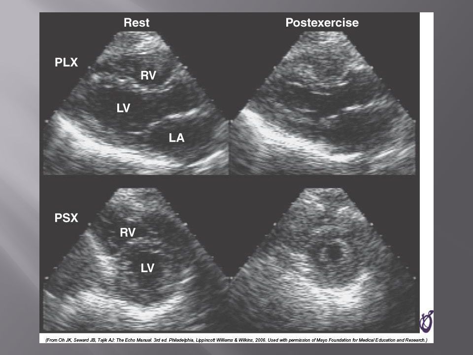
Compares pre & post: Regional contractility Overall systolic function Volumes Pressure gradients Filling pressures Pulmonary pressures Valvular function

36
Factors which effect image quality: Body habitus Lung disease Breast implants

40
54 year old bank project manager Exertional chest pain & dyspnea Ex-smoker TC = 6.7mmol/L Stress ECG – 2mm ST segment depression in 5 leads

43
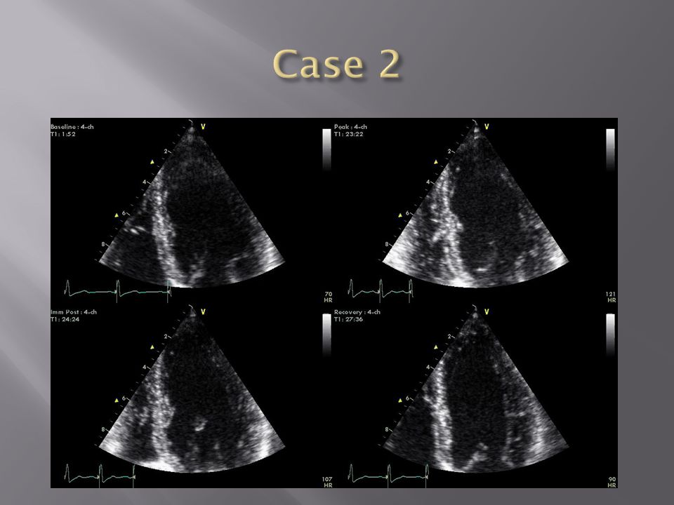
62 year old female Chest pain & dyspnea Treadmill exercise test – non-diagnostic sub-maximal Hypertension No ECG changes

44
Exercised 7½ minutes (9.4 METS) No chest pain ECG changes
 No chest pain ECG changes")
48
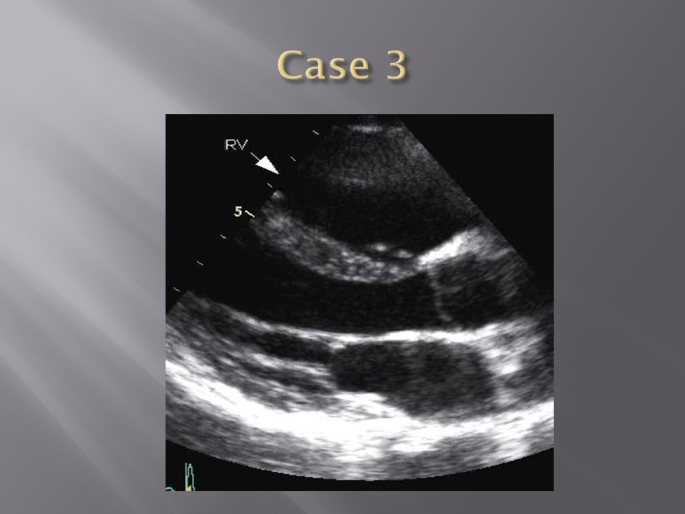
24 year old female engineer Exertional dyspnea Palpitations

49
Inducible dyspnea ECG partial right bundle branch block no ischemic changes

53
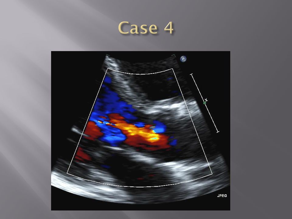
43 year old male - airline catering Chest pain Dyspnea

54
Inducible dyspnea Non-specific T wave changes No ST segment shift Global deterioration in left ventricular function

58
Radio-tracer injection Isotopes: Thallium-201 Technetium 99m (sestamibi) Myocardial uptake Photon emission captured by gamma camera Rest & redistribution phases Pharmacologic protocols available Digital presentation
 Myocardial uptake Photon emission captured by gamma camera Rest & redistribution phases Pharmacologic protocols available Digital presentation")
62
Reversible inferior wall defect Milder reversible inferior wall defect

63
Time-consuming Artifacts Balanced ischemia Radiation

64
Normal apical thinning.

65
A. Breast attenuationB. Anterior ischemia

67
Risk of iatrogenic malignancy Linear no-threshold model Consider: age gender background

68
Einstein, A. J. et al. Circulation 2007;116:1290-1305

69
Useful for: Patients unable to exercise ECG uninterpretable Unsuitable for DSE And…. No radiation But… Not currently available

70
45 year old diabetic man Anterior chest discomfort with exertion Exercised for 2 mins 30 secs (4.6 METs) 95% maximal predicted heart rate Mild chest pain BP increased from baseline to 180/80mmHg 1mm ST depression in leads II, III, aVF, V4-6
 95% maximal predicted heart rate Mild chest pain BP increased from baseline to 180/80mmHg 1mm ST depression in leads II, III, aVF, V4-6")
71
1. Pre-test risk is intermediate 2. Post-test probability for cardiac events is high 3. The ECG changes are non-diagnostic 4. The ECG changes are false-positive in the setting of hypertension 5. Chest pain is not a useful symptom in diabetics

72
1. Pre-test risk is intermediate 2. Post-test probability for cardiac events is high 3. The ECG changes are non-diagnostic 4. The ECG changes are false-positive in the setting of hypertension 5. Chest pain is not a useful symptom in diabetics

74
Parameters associated with adverse prognosis or multi- vessel disease Duration of symptom-limiting exercise <5 METs Failure to increase sBP 120mmHg, or a sustained decreased 10mmHg, or below rest levels, during progressive exercise ST segment depression 2mm, downsloping ST segment, starting at <5 METs, involving 5 leads, persisting 5 min into recovery Exercise-induced ST segment elevation (aVR excluded) Angina pectoris at low exercise workloads Reproducible sustained (>30 sec) or symptomatic ventricular tachycardia
 Angina pectoris at low exercise workloads Reproducible sustained (>30 sec) or symptomatic ventricular tachycardia")
75
Pre-test risk of disease Sensitivity & specificity of the test Value of supplementary data AND JUST ONE MORE TIP……..

77
So….which one to choose?

78
Remember Bayes theorem Consider the pre-test risk Be aware of the sensitivity & specificity of the test Apply the post test probability

80
Correlates with presence & extent of CAD Strong negative predictive value Cannot predict functional significance Higher scores can predict events Recommended for asymptomatic with intermediate risk

81
Calcification of the left anterior descending coronary artery ( large arrow ) and left circumflex coronary artery (small arrow).
 and left circumflex coronary artery (small arrow).")
82
Score description RR 0 nil 1 – 99 mild 1.9 100 – 399 moderate 4.3 400 – 999 severe 7.2 >1000 extensive 10.8

83
Indicated – asymptomatic with intermediate risk Not for low risk/population screening High risk – use current guidelines Do not reduce Rx if intermediate risk & 0 score

84
2-dimensional & 3- dimensional reconstructions Relies on slow, regular heart rate High negative predictive value (rule out ability)
")
85
Lower positive predictive value (over-estimation tendency) Grading of stenosis limited Does not evaluate functional significance Radiation exposure
 Grading of stenosis limited Does not evaluate functional significance Radiation exposure")
86
Role not yet clearly defined Potential for those with intermediate likelihood of disease: Where stress testing not possible Stress test equivocal/uninterpretable Acute chest pain/no ECG changes/normal enzymes Role in anomalous anatomy

Similar presentations


>")


 stress testing Standard Echocardiographic (echo) stress testing Pharmacologic.>")
>")
 ASSOCIATE PROFESSOR OF ISFAHAN UNIVERSITY.>")