Download presentation
Presentation is loading. Please wait.

1
Renal Board Review Brenda Shinar, MD

2
Question 1. Answer: A: Combinaton drug therapy

3
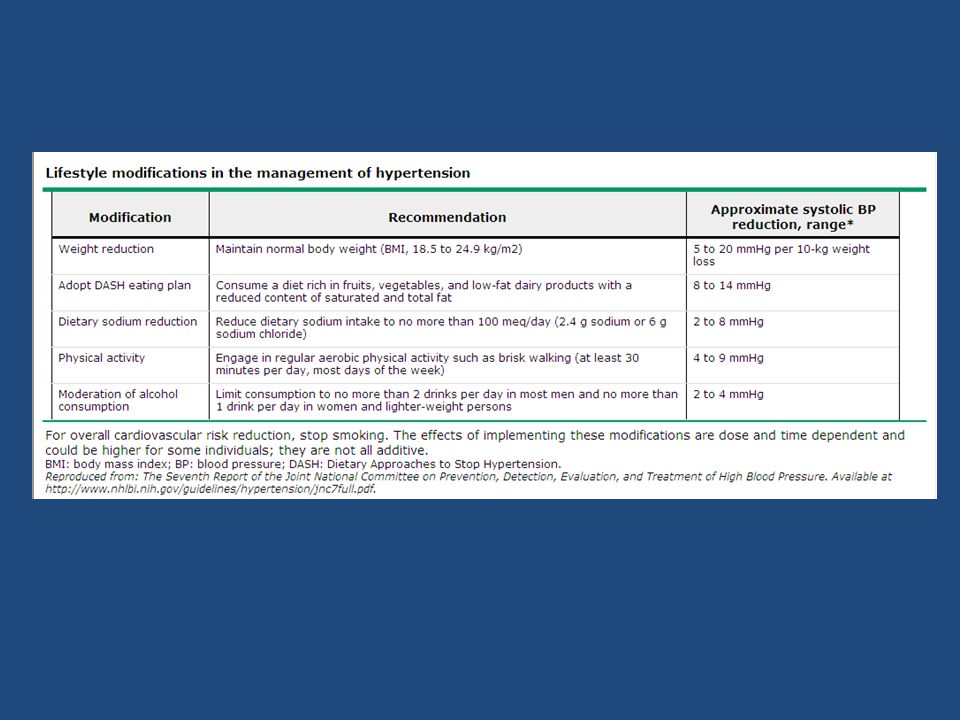
Manage Newly Diagnosed Stage 2 Hypertension

4
Untreated stage 1 hypertension with onset at age 35 will decrease a person’s lifespan by 16 years!

7
Question 2. Answer: D: Repeat blood pressure measurement

8
Identify the cause of a patient’s change in blood pressure

9
Question 3. Answer: D: No change in management

10
Manage HTN in a patient who is over age 80.

11
Question 4. Answer: D: Obtain a plasma aldosterone- plasma renin activity ratio

12
Understand the differential diagnosis of resistant hypertension Definition: Blood pressure measurements consistently exceed goal on 3 antihypertensive medications, one of which is a diuretic BP controlled on > or = to four medications 12.8% of adults being treated for HTN Non-adherence to therapy White coat resistant hypertension Renal parenchymal disease Renal artery stenosis Pheochromocytoma Primary hyperaldosteronism OSAS Drug- induced Volume overload

13
Primary Hyperaldosteronism: 60% of patients have NORMAL K level ALDOSTERONE HIGH RENIN LOW AR RATIO >25 is suggestive BUT NOT diagnostic Metabolic alkalosis and hypokalemia MAY OR MAY NOT be present 8 AM draw OFF spironolactone or eplerenone for 6 weeks Possibly off ACEI NO CONFIRM test needed: – Spontaneous hypokalemia – Undetectable renin level – Aldosterone >30 ng/dL

14
Question 5. Answer: D: Type B lactic acidosis

15
Diagnose type B lactic acidosis Type A Lactic Acidosis Tissue hypoperfusion due to shock Reduced systolic blood pressure may be minimized by severe vasoconstriction Altered mental status Cool extremities Oligoanuria Lactic acid level predicts mortality Type B Lactic Acidosis Normal systemic perfusion Impaired cellular metabolism or regional ischemia Metformin Linezolid (IV, prolonged) HIV medications Liver disease Malignancy D-lactic acidosis
 HIV medications Liver disease Malignancy D-lactic acidosis")
16
Question 6. Answer: C: Urine chloride level

17
Evaluate a Patient with Hypokalemic Metabolic Alkalosis Chloride Responsive 90% Vomiting, Low effective circulating volume Volume depleted NORMAL increase in renin, angiotensin, aldosterone Distal tubule wastes H+ and K+ and hold onto Na+ Urine chloride LOW <15 Cannot replace K until volume is replaced Chloride UN-responsive 10% Hypertensive Hypervolemic ABNORMAL increase in aldosterone or renin Distal tubule wastes H+ and K+ to hold onto Na+ Urine chloride HIGH >15

18
Question 7. Answer: B: Estimate GFR using the Chronic Kidney Disease-Epidemiology Collaboration equation (CKD-Epi)
.")
19
Estimate the glomerular filtration rate in a low-risk, healthy person Equations Preferred to estimate GFR Creatinine MUST be stable for 24-48 hours MDRD (Modification of Diet in Renal Disease) CKD-EPI (Chronic Kidney Disease Epidemiology) Cockcroft-Gault 24 hour measurement Extremes in age Extremes in weight Pregnancy Cirrhosis Amputations Radionuclide Scanning Most precise method Used for kidney donor eval if GFR is borderline
 CKD-EPI (Chronic Kidney Disease Epidemiology) Cockcroft-Gault 24 hour measurement Extremes in age Extremes in weight Pregnancy Cirrhosis Amputations Radionuclide Scanning Most precise method Used for kidney donor eval if GFR is borderline")
20
Question 8. Answer: B: Hypokalemic distal (type 1) renal tubular acidosis
 renal tubular acidosis")
21
Diagnose hypokalemic distal (type 1) renal tubular acidosis Type 1 RTA (distal) Type 2 RTA (proximal) Type 4 RTA (distal) Chloride↑↑↑ Bicarbonate↓↓↓ Potassium↓NL↑ Urine pHHigh ( > 5.5)Low (<5.5) except with bicarb load Low (<5.5) Etiologies Chronic hepatitis Amphotericin B Toluene Lithium Sjogren’s; SLE Multiple Myeloma Metal poisoning Acetazolamide Diabetes mellitus Sickle cell Spironolactone Associations Nephrolithiasis due to hypercalcuria
 renal tubular acidosis Type 1 RTA (distal) Type 2 RTA (proximal) Type 4 RTA (distal) Chloride↑↑↑ Bicarbonate↓↓↓ Potassium↓NL↑ Urine pHHigh ( > 5.5)Low (<5.5) except with bicarb load Low (<5.5) Etiologies Chronic hepatitis Amphotericin B Toluene Lithium Sjogren’s; SLE Multiple Myeloma Metal poisoning Acetazolamide Diabetes mellitus Sickle cell Spironolactone Associations Nephrolithiasis due to hypercalcuria")
22
Question 9. Answer: B: Chlorthalidone

23
Manage hypercalciuria in a patient with nephrolithiasis Hypercalciuria: – >300 mg/24 hr MEN – >250 mg/24 hr WOMEN – >200 mg/24 hr BOTH WORSENED by: – High sodium diet – High animal protein diet – Loop diuretics DO NOT advise a calcium restricted diet, as this increases GI absorption of oxalate, increasing oxaluria DO ADVISE: – Thiazide diuretic – Fluids > 2 liters/day

24
Question 10. Answer: C: Split urine collection

25
Orthostatic proteinuria : Uncommon over age 30 (2-5 % of adolescents) <1-2 grams per day Supine collection is NORMAL BENIGN condition
 <1-2 grams per day Supine collection is NORMAL BENIGN condition")
26
Question 11. Answer: D: Serum and Urine Electrophoresis

27
Diagnose Multiple Myeloma as a cause of acute kidney injury Clinical Features of MM: Anemia (NCNC) (80%) Bone pain (70%) Recurrent infections – 25% presenting – 75% during disease Renal complications (50%) Hypercalcemia (25%) Renal failure (25%) Renal Complications: Tubular Damage – Adult Fanconi’s syndrome – RTA Proximal Type 2 Cast Nephropathy * – Proteinaceous casts clog the tubules resulting in tubule atrophy Glomerulopathy – Light chain disease deposition – Resulting in albuminuria! Exquisite sensitivity to IV contrast! *most common

28
Question 12. Answer: A: Acute interstitial nephritis

29
Diagnose Acute (Allergic) Interstitial Nephritis Idiosyncratic drug-induced hypersensitivity Fever, rash, eosinophilia, and elevated creatinine (10%) UA: WBC, WBC casts Eosinophiluria NEGATIVE CULTURE (sterile pyuria) < 2 gm/ 24 hr proteinuria DRUGS: – Antibiotics (B-lactam) – NSAIDS * (nephrotic proteinuria) with minimal change disease on biopsy – Thiazides – Proton Pump Inhibitors – Phenytoin – Allopurinol
 Interstitial Nephritis Idiosyncratic drug-induced hypersensitivity Fever, rash, eosinophilia, and elevated creatinine (10%) UA: WBC, WBC casts Eosinophiluria NEGATIVE CULTURE (sterile pyuria) < 2 gm/ 24 hr proteinuria DRUGS: – Antibiotics (B-lactam) – NSAIDS * (nephrotic proteinuria) with minimal change disease on biopsy – Thiazides – Proton Pump Inhibitors – Phenytoin – Allopurinol")
30
Question 13. Answer: B: IgA nephropathy

31
Diagnose IgA nephropathy: T he most common cause of nephritic syndrome Ig A Nephropathy (25-30%) CONCOMMITANT pharyngitis Normal complements DIFFERENTIAL (NL C3): Pauci-immune glomerulonephritis (15-25%) Anti-GBM antibody disease (3%) Post-Strep Gnitis (4-8%) 7-10 days AFTER pharyngitis Antibodies to: – Streptolysin O – DNAse B Low complements (C3) DIFFERENTIAL (LOW C3): Lupus nephritis (20-30%) Membranoproliferative Gnitis (MPGN) (6-10%)
 CONCOMMITANT pharyngitis Normal complements DIFFERENTIAL (NL C3): Pauci-immune glomerulonephritis (15-25%) Anti-GBM antibody disease (3%) Post-Strep Gnitis (4-8%) 7-10 days AFTER pharyngitis Antibodies to: – Streptolysin O – DNAse B Low complements (C3) DIFFERENTIAL (LOW C3): Lupus nephritis (20-30%) Membranoproliferative Gnitis (MPGN) (6-10%)")
32
Question 14. Answer E: Supportive Care

33
Manage Post-Infectious Glomerulonephritis (PIGN) MANY bacteria, viruses and parasites can cause PIGN Most common nephritogenic strains of strep and staph Rapid onset of hypertension, oliguria, erythrocyte casts, and edema, LOW C3 MANAGEMENT is SUPPORTIVE: Early treatment of the bacterial infection Diuretics for volume overload Antihypertensives for elevated BP Dialysis if necessary NO evidence for immunosuppression, steroids, plasmapheresis
 MANY bacteria, viruses and parasites can cause PIGN Most common nephritogenic strains of strep and staph Rapid onset of hypertension, oliguria, erythrocyte casts, and edema, LOW C3 MANAGEMENT is SUPPORTIVE: Early treatment of the bacterial infection Diuretics for volume overload Antihypertensives for elevated BP Dialysis if necessary NO evidence for immunosuppression, steroids, plasmapheresis")
34
Question 15. Answer: A: Cryoglobulinemia associated with Hepatitis C

35
Diagnose Hepatitis C virus associated glomerulonephritis Occurs in up to 20% of patients with chronic Hepatitis C Presents as membranoproliferative gnitis or mixed cryoglobulinemia (skin, kidney, and nerve involvement) 1/3 relapsing dz with progression to ESRD Low complement (C4) + Rheumatoid factor TREAT Hep C virus EXTRA-HEPATIC MANIFESTATIONS OF HEPATITIS C INFECTION: 1. Membranoproliferative GNitis 2. Mixed Essential Cryoglobulinemia 3. Lichen Planus – (the 5 Ps) 4. Porphyria Cutanea Tarda – (vesicles, milia, photosensitivity)
 4. Porphyria Cutanea Tarda – (vesicles, milia, photosensitivity).")
36
Question 16. Answer: D: Pigment nephropathy

37
Diagnose Pigment Nephropathy from Rhabdomyolysis 2 types of pigment induced nephropathy: hemolysis or rhabdomyolysis Urine dip + for blood with urine micro negative for red cells Ischemia, blockage, or injury to tubules Associated lab abnormalities with rhabdo: – Elevated CK level, aldolase – Hyperkalemia, hyperuriciemia, hyperphosphatemia, hypocalcemia Treatment: – IV fluid, UOP 200-300/hour – Bicarbonate not proven – Dialysis not helpful unless indicated

38
Question 17. Answer A: Focal Segmental Glomerulosclerosis

39
Diagnose Focal Segmental Glomerulosclerosis : the most common renal cause of nephrotic syndrome in US (36-80%) Primary Disease: Podocyte damage similar to minimal change dz Secondary Disease Associations: 1.Morbid obesity 2.Heroin use 3.HIV 4.Vesicoureteral reflux Risk factors for progression to ESRD: 1.Black race 2.>2 gm/24 hr proteinuria 3.Low GFR 4.BMI>27 5.Hypertension Clinical Manifestations: Acute onset of nephrotic syndrome with hematuria, renal failure and hypertension in primary disease; Asymptomatic subnephrotic to nephrotic proteinuria in secondary disease Management: Immunosuppression with steroids or calcineurin inhibitors in primary disease ACEI +/- ARB in primary and secondary disease BP goal < 125/75 mm Hg
 Primary Disease: Podocyte damage similar to minimal change dz Secondary Disease Associations: 1.Morbid obesity 2.Heroin use 3.HIV 4.Vesicoureteral reflux Risk factors for progression to ESRD: 1.Black race 2.>2 gm/24 hr proteinuria 3.Low GFR 4.BMI>27 5.Hypertension Clinical Manifestations: Acute onset of nephrotic syndrome with hematuria, renal failure and hypertension in primary disease; Asymptomatic subnephrotic to nephrotic proteinuria in secondary disease Management: Immunosuppression with steroids or calcineurin inhibitors in primary disease ACEI +/- ARB in primary and secondary disease BP goal < 125/75 mm Hg")
40
Question 18. Answer: B: Membranous nephropathy

41
Diagnose Membranous Nephropathy : the second most common renal cause of nephrotic syndrome Primary disease: Immune complexes of IgG react with antigens in the outer aspect of GBM Secondary disease associations: Malignancy Hep B and C NSAIDS Risk factors for progression to ESRD: Male gender Age>50 Persistent proteinuria > 4g/24 h over 6 months Declining GFR Clinical manifestations: Nephrotic syndrome with preserved GFR Renal vein thrombosis Management: ACE and/or ARB BP goal < 125/75 mm Hg Persistent proteinuria > 4g/24 hr cyclophosphamide, corticosteroids, or calcineurin inhibitor with possible need for rituxan

42
Question 19. Answer: B: 3% saline infusion

43
Treat a patient who has symptomatic hyponatremia

44
Question 20. Answer: A: Chronic kidney disease and hypertension

45
Diagnose underlying chronic kidney disease and hypertension in a pregnant patient Normal physiology in pregnancy Blood pressure lowers Plasma volume increases GFR increases Renal pelvis, calices, and ureters dilate Increased risk for pyelo Hyperventilation causes resp alkalosis with increased renal bicarb excretion Pearls in Pregnancy: Hypertension BEFORE 20 th week indicates new dx of chronic hypertension Safe BP agents in pregnancy are methyldopa and labetalol: ACEI, ARBs and renin inhibitors are NOT safe Gestational hypertension develops AFTER 20 weeks: no proteinuria or end-organ damage Pre-eclampsia hypertension develops AFTER 20 weeks and is associated with proteinuria LOW dose aspirin reduces the risk of preeclampsia

Similar presentations


>")


. Comparison of the effects of losartan and enalapril on renal function in adults with chronic kidney disease at.>")
