Download presentation
Presentation is loading. Please wait.

1
Pediatric Hearing Loss and Testing Techniques
Diego A Preciado MD PhD Pediatric Otolaryngology Children’s National Medical Center George Washington University Washington, DC

2
11 month child seems to ‘hear well’ at home, but daycare provider concerned with ability to respond to verbal stimuli at times

3
A) Child can only be tested asleep B) Child can be tested awake C) Child can only be screened for hearing loss at his age D) Child can only be tested by the old ‘rub/snap your fingers’ next to the ear trick
 Child can only be tested asleep B) Child can be tested awake C) Child can only be screened for hearing loss at his age D) Child can only be tested by the old ‘rub/snap your fingers’ next to the ear trick")
4
Childhood Hearing Loss
Moderate to profound congenital hearing impairment occurs in 4 per 1000 live births Recommendations specify that All children screened at birth All children diagnosed by 3 months of age All children treated by 6 months 43 states have mandated Universal Newborn Hearing Screening (UNHS) programs
 programs.")
5
Types of Hearing Tests Screening (PASS OR FAIL) (electrophysiological)
Otoacoustic Emissions (OAE) Automated ABR Diagnostic (NOT PASS OR FAIL) ABR (electrophysiological; all ages) Pure Tone Audiometry (>4 yrs of age) Infant Audiometry (>6 mo of age)
 Automated ABR. Diagnostic (NOT PASS OR FAIL) ABR (electrophysiological; all ages) Pure Tone Audiometry (>4 yrs of age) Infant Audiometry (>6 mo of age)")
6
Here’s the clinical challenge we are faced with…..
Language behaviors UNHS Infants with HL ENT HA, Rehab, SLP Optimal Age for CI Target Age age in months 6 12 18

7
Electrophysiologic Testing
Use in neonates, uncooperative patients, brain injury Otoacoustic emissions (OAEs) Originates in cochlea, evoked with sound stimulation Absent suggests > 30 dB HL
 Originates in cochlea, evoked with sound stimulation. Absent suggests > 30 dB HL.")
8
Electrophysiologic Testing
Evoked auditory brainstem response (ABR or BAER) Auditory electrical responses Diagnoses presence, degree and type of HL
 Auditory electrical responses. Diagnoses presence, degree and type of HL.")
9
Hearing Loss Types Conductive Sensorineural Mixed
measured by “air” stimulation on audiogram Sensorineural measured by “bone” stimulation on audiogram Mixed

10
Conductive Loss Conductive loss (CHL) results from increase in impedance (resistance) Audiometric profile of conductive hearing loss is threshold for air conduction is worse than for bone conduction i.e. large “air-bone gap”

11
Conductive Hearing Loss
Generally reversible Middle or External Ear Pathology External auditory canal (EAC) obstruction Cerumen impactions, foreign body, otitis externa, EAC atresia Abnormality of ear drum Perforation, retraction ME conditions AOM, OM with effusion, cholesteatoma, tumor Ossicular chain anomalies Disruption - associated with trauma Fixation – often congenital
 obstruction. Cerumen impactions, foreign body, otitis externa, EAC atresia. Abnormality of ear drum. Perforation, retraction. ME conditions. AOM, OM with effusion, cholesteatoma, tumor. Ossicular chain anomalies. Disruption - associated with trauma. Fixation – often congenital.")
12
Tympanometry Not a hearing test!
Objective measure of middle ear (ME) compliance Complements ear exam
 compliance. Complements ear exam.")
13
COMPLIANCE/Admitance
Tympanograms Volume COMPLIANCE/Admitance

14
Tympanograms Type A- Normal

15
Tympanograms Type B- High Volume Type B- Low Volume
Perforation or PE tube Type B- Low Volume Fluid

16
Tympanograms Type C- Negative Pressure Retraction

17
Sensorineural Loss Sensorineural hearing loss (SNHL) results because of lesions in the auditory nerves and/or cochlea Audiometric profile of sensorineural hearing loss demonstrates air conduction and bone conduction reduced without an air-bone gap

18
Etiology of SNHL 70% recessive 25% dominant 5% X-linked

19
SNHL – Associated Conditions
Loop diuretics, aminoglycosides, aspirin Hyperbilirubinemia Severe depression at birth (asphyxia) Anomalies of external and middle ear Usually irreversible Family history – congenital, delayed onset childhood SNHL Congential infections – CMV, rubella Bacterial meningitis
 Anomalies of external and middle ear. Usually irreversible. Family history – congenital, delayed onset childhood SNHL. Congential infections – CMV, rubella. Bacterial meningitis.")
20
Mixed Hearing Loss Mixed hearing loss results from BOTH a conductive and sensorineural hearing loss Audiometric profile shows a drop in air and bone conduction with an air-bone gap

21
Audiogram Listening to spoken language during early life is a critical prerequisite for the typical development of speech. Each of these terms is often accompanied by specific threshold levels of loss in the frequency region for speech. These terms are meant to convey the extent of hearing loss and are useful in explaining to parents how much of speech their child can expect to hear.

22
Behavioral Audiometry
Test Techniques: Behavioral Observation Audiometry (BOA) Visual reinforcement audiometry (VRA) Conditioned play audiometry (CPA) Conventional hand-raising procedures Gold Standard Achieve auditory thresholds at all test frequencies for both ears. Ongoing age-specific activity – the methodology and specific techniques are modified for the developmental age of the child. Utilize cross-check principle using available objective tests
 Visual reinforcement audiometry (VRA) Conditioned play audiometry (CPA) Conventional hand-raising procedures. Gold Standard. Achieve auditory thresholds at all test frequencies for both ears. Ongoing age-specific activity – the methodology and specific techniques are modified for the developmental age of the child. Utilize cross-check principle using available objective tests.")
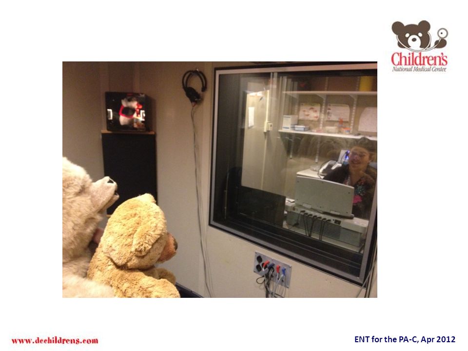
23
Behavioral Observation Audiometry

24
Behavioral Observation Audiometry (BOA)
Children aged ~5 months to 2 years Individuals with neurological/developmental involvements Primarily sound field testing Subjective observation by the clinician Stimuli may include speech, warble tones, narrowband noise (NBN), parent’s voice Soundfield = air conduction/sometimes bone conduction *children are placed on parent’s/caregiver’s lap, or in a high chair, 450 azimuth to loudspeakers Warbled tones = modulated pure tone (frequency and amplitude) Narrowband noise = passing a broad band white noise signal through narrowband filters Observed responses: Early attending: Sucking, Eye widening, Eyebrow raise, ↑ or ↓ in motor activity Later attending: Searching behavior (without true localization), Listening posture, Localized head turn-4-7 mo: Side-to-side, 6-9 mo: Sides, above & below
, parent’s voice. Soundfield = air conduction/sometimes bone conduction *children are placed on parent’s/caregiver’s lap, or in a high chair, 450 azimuth to loudspeakers. Warbled tones = modulated pure tone (frequency and amplitude) Narrowband noise = passing a broad band white noise signal through narrowband filters. Observed responses: Early attending: Sucking, Eye widening, Eyebrow raise, ↑ or ↓ in motor activity. Later attending: Searching behavior (without true localization), Listening posture, Localized head turn-4-7 mo: Side-to-side, 6-9 mo: Sides, above & below.")
25
Audiometry during infancy
Symbols Sound field S S S S Speech = 20 NBN or warbled tones = 20-50 S Speech does not occur at a single intensity or frequency. In general vowel sounds are low frequency in nature and more intense “carry more power”. Whereas consonants, particularly voiceless consonants /s/, /sh/, /t/ “fish” thin”, are composed of higher frequencies and are least intense. Each child’s capacities do vary as a consequence of listening circumstances. Descriptive terms only partially explain the listening experiences of a particular child. Age-appropriate responses for infants aged ~5 – 9 months

26
Visual reinforcement audiometry (VRA)
Employs lighted transparent-boxed toys to reinforce child’s localized response to onset of acoustic stimuli Conditioning phase; Testing phase

28
Visual reinforcement audiometry (VRA)
Children aged 6 mo - ~ 3 years Technique consists of conditioning & testing phases Responses may include localizations or BOA responses Disadvantages Dependent on conditioning child to task Habituation to acoustic stimuli Poor test reliability Transducers Sound field loudspeakers Insert or supra-aural earphones Bone conduction oscillator Stimuli Speech Narrowband noise (NBN) Warbled tones Parent’s voice
 Warbled tones. Parent’s voice.")
29
Conditioned Play Audiometry (CPA)
Child is taught a play task in response to the onset of an acoustic stimulus Children aged ~ years Older individuals with developmental involvements Transducers Insert or supra-aural earphones Bone conduction oscillator Sound field loudspeakers Stimuli Speech Warbled tones Pulsed tones Narrowband noise (NBN)
")
31
Audiometry Conventional audiometry: ≥ 5 yrs Zero-20 dB is normal range
Not absolute, but normalized scale Hearing threshold measured for air and bone conduction in decibels from 250 Hz – 8 KHz

32
Pure Tone Audiograms ‘loudness’

33
Audiograms Bracket = Bone, Right Side Circle = Air, Right Side
NORMAL

34
Audiograms Bracket = Bone, Right Side Circle = Air, Right Side
CHL A-B gap

35
Audiograms Bracket = Bone, Right Side Circle = Air, Right Side
SNHL

36
Audiograms Bracket = Bone, Right Side Circle = Air, Right Side
MIXED HL

37
A 3 year old child presents with low volume, Type B tympanogram, and 20 dB Air Bone gap
Most likely diagnosis is A- Cholesteatoma B- TM perforation C- OM with effusion D- Sensorineural hearing loss

38
A 3 year old child presents with low volume, Type B tympanogram, and 20 dB Air Bone gap
Most likely diagnosis is A- Cholesteatoma B- TM perforation C- OM with effusion D- Sensorineural hearing loss

39
Hereditary Hearing Impairment
Dominant progressive, milder, late onset, penetrance/expressivity Recessive stable, severe, congenital, more symmetric

40
SYNDROMES

42
Waardenburg Syndrome Autosomal dominant
Variable expressivity Associated with pigmentary abnormalities White forelock (20-30%) Premature graying Vitiligo Heterochromia irdis
 Premature graying. Vitiligo. Heterochromia irdis.")
43
Treacher Collins Syndrome (Mandibulofacial Dystostosis)
Inheritance: Autosomal dominant with variable expressivity Molecular basis:Caused by mutations in Treacle gene (TCOF1)
")
44
Branchiootorenal Syndrome
Autosomal dominant Branchial cleft sinuses/fistulas Renal anomalies These range from mild hypoplasia to bilateral renal agenesis External, middle and inner ear deformities Estimated at about 2% of childhood deafness

45
Autosomal Recessive Syndromes

46
Pendred Syndrome Autosomal recessive Abnormal incorporation of iodine
Perchlorate or thiocyanate tests are rarely performed Goiter and hypothyroidism usually present by about 8 years of age

47
Pendred Syndrome Associated with enlarged vestibular aqueduct
Histologic evidence of hydrops and degenerated changes of the stria vascularis have been described Treatment – amplification exogenous thyroid hormone

49
Usher’s Syndrome Usher’s Syndrome Type I (7 loci-MYO7A)
Autosomal recessive Severe to profound hearing loss Absence of vestibular response Slow progression Slowly progressive visual field deficits beginning as early as age 9-10

50
Jervell and Lange-Nielson
Autosomal recessive Severe-to-profound Bilateral Cardiac conduction defects Enlarged T-waves Prolongation of the Q-T interval Syncopal episodes Sudden death

51
Clinical Genetics Sequence: multiple defects from a single defect
Not all that is genetic is a syndrome… Malformation: morphologic defect of an organ, part of organ resulting from an intrinsically abnormal developmental process Sequence: multiple defects from a single defect Syndrome: pattern of multiple anomalies pathogenetically related

52
Connexin 26 Mutations in GJB2 (DFNB1) reported at ~30% (20%-70%) of severe to profound hearing loss Carrier rate-3.0% (Caucasian) 35delG, M34T -Caucasian 167delT-Ashkenazi Jewish 235delC-Japanese
 35delG, M34T -Caucasian. 167delT-Ashkenazi Jewish. 235delC-Japanese.")
53
Connexin 26 GJB2 Connexin 26 is located on long arm of chromosome 13 and is a relatively simple gene, made up of only 2 exons, separated by an intron of 3148 bp. Exon 1 is UTR. Exon 2 contains the coding region of 681 bp (227 aa) and a 5’ UTR.
 and a 5’ UTR.")
54
EVA Sensorineural Hearing Loss
Low Frequency Conductive Hearing Loss Component Usually stable hearing level Occasionally Progressive Although a congenital malformation of the inner ear, frequently a later onset hearing loss

55
EVA Syndromic Non-syndromic Associated with SLC26A4 mutation (DFNB4)
Pendred’s More severe phenotype Non-syndromic More heterogenous hearing level

56
VA Size Boston M, et al. Oto-HNS, 2007

57
SLC26A4 mutations and hearing loss
Asaiez H, et al. Human Genetics, 2007

59
Clinical Evaluation History and Physical Syndromic findings
Cutaneous, musculoskeletal, visceral (cardiac, thyroid, renal, visual/balance, cervical) Neonatal risk factors Others-noise, head trauma, autoimmune, Meniere’s, Lues Extended family history pedigree
 Neonatal risk factors. Others-noise, head trauma, autoimmune, Meniere’s, Lues. Extended family history. pedigree.")
60
Clinical Evaluation Audiometric evaluation-Diagnosis Behavioral ABR

61
Results GJB2 and Imaging yield vs. SNHL
What are you going to say here?

62
Preciado D, et al. Otol Neurotol. 2005 Jul;26(4):610-5
History, Physical examination, Audiologic work-up Diagnosis apparent Diagnosis uncertain Appropriate treatment Bilateral Unilateral MRI, Preferential seating, Serial audiograms Sev-to-Prof Mod-Sev Mild-to-Mod GJB2 screen CT scan _ + _ + Genetic Counseling CT scan Appropriate treatment GJB2 screen Lab tests as indicated Lab tests as indicated ECG Preciado D, et al. Otol Neurotol Jul;26(4):610-5
:")
63
Conclusions You can screen or diagnose AT ANY AGE!
Screen for HL at birth, diagnose by 3 months You can test awake starting at 6 months Follow a logical sequential diagnostic work-up paradigm based on history and physical Laboratory investigation should be based on these results and on clinical history

Similar presentations


(Syd) FRACS (OHNS) MS (UNSW) Associate Professor of Surgery (Macquarie University) Director, Kolling.>")


