Download presentation
Presentation is loading. Please wait.

1
Basic Concept in Radiologic Physics

2
REMOVAL OF AN ELECTRON FROM AN ATOM
IONIZATION PROCESS REMOVAL OF AN ELECTRON FROM AN ATOM

3
Ionization

4
SOURCES OF IONIZING RADIATION
COSMIC TERRESTRIAL INTERNAL MEDICAL X-RAYS NUCLEAR MEDICINE CONSUMER PRODUCTS NUCLEAR POWER---INDUSTRIAL

5
NATURAL ENVIRONMENTAL RADIATION
COSMIC TERRESTRIAL INTERNAL

6
MAN MADE IONIZING RADIATION SOURCES
MEDICAL X-RAYS NUCLEAR MEDICINE CONSUMER PRODUCTS NUCLEAR POWER---INDUSTRIAL

7
THE CONTRIBUTION OF VARIOUS SOURCES TO THE AVERAGE US POPULATION RADIATION DOSE

8
Cosmic Radiation The earth’s atmosphere is bombarded by high-energy particles from our galaxy (primary cosmic radiation). In the upper atmospheric layers, these particles react with air molecules. As a result of nuclear reactions, a great number of secondary particles (secondary cosmic radiation) is formed. Some of these secondary particles decay again, are absorbed in the atmosphere or possibly penetrate into the earth.
. In the upper atmospheric layers, these particles react with air molecules. As a result of nuclear reactions, a great number of secondary particles (secondary cosmic radiation) is formed. Some of these secondary particles decay again, are absorbed in the atmosphere or possibly penetrate into the earth.")
9
Terrestrial Radiation – Radon is the Largest Source

10
RADON MOVEMENT

11
RADON INTERNAL EXPOSURE
RADON EXTERNAL EXPOSURE

12
RADON EMITS ALPHA PARTICLE

13
Internal Radiation In addition to the cosmic and terrestrial sources, all people also have radioactive potassium-40, carbon-14, lead-210, and other isotopes inside their bodies from birth. The variation in dose from one person to another is not as great as the variation in dose from cosmic and terrestrial sources.

14
Man-made radiation sources that result in an exposure to members of the public:
Tobacco Televisions Medical X-rays Smoke detectors Lantern mantles Nuclear medicine Building materials Nuclear power plants

15
By far, the most significant source of man-made radiation exposure to the public is from medical procedures, such as diagnostic X-rays, nuclear medicine, and radiation therapy. Some of the major isotopes would be I-131, Tc-99m, Co-60, Ir-192, Cs-137, and others. In addition, members of the public are exposed to radiation from consumer products, such as tobacco (thorium), building materials, combustible fuels (gas, coal, etc.), ophthalmic glass, televisions, luminous watches and dials (tritium), airport X-ray systems, smoke detectors (americium), road construction materials, electron tubes, fluorescent lamp starters, lantern mantles (thorium), etc. Of lesser magnitude, members of the public are exposed to radiation from the nuclear fuel cycle, which includes the entire sequence from mining and milling of uranium to the actual production of power at a nuclear plant. This would be uranium and its daughter products. The final sources of exposure to the public would be shipment of radioactive materials and residual fallout from nuclear weapons testing and accidents, such as Chernobyl.
, building materials, combustible fuels (gas, coal, etc.), ophthalmic glass, televisions, luminous watches and dials (tritium), airport X-ray systems, smoke detectors (americium), road construction materials, electron tubes, fluorescent lamp starters, lantern mantles (thorium), etc. Of lesser magnitude, members of the public are exposed to radiation from the nuclear fuel cycle, which includes the entire sequence from mining and milling of uranium to the actual production of power at a nuclear plant. This would be uranium and its daughter products. The final sources of exposure to the public would be shipment of radioactive materials and residual fallout from nuclear weapons testing and accidents, such as Chernobyl.")
16
Debate still persists as to who was the first to discover X-rays
Debate still persists as to who was the first to discover X-rays. Was it the Ukrainian scientist Jan Puluj (Ivan Puliui) or a German physicist Wilhelm Conrad Roentgen, the 1901 Nobel Prize winner? ?
 or a German physicist Wilhelm Conrad Roentgen, the 1901 Nobel Prize winner")
17
The lives of both physicists are connected with Strasbourg University
The lives of both physicists are connected with Strasbourg University. In the 1890s both Messrs. Roentgen and Puliui worked in the same department under the guidance of Prof. Kundt, and Mr. Roentgen made a point of attending lectures given by Mr. Puliui. While at Strasbourg, Mr. Puliui commenced his experiments with X-rays, and Mr. Roentgen was soon to become fascinated by these phenomena.

18
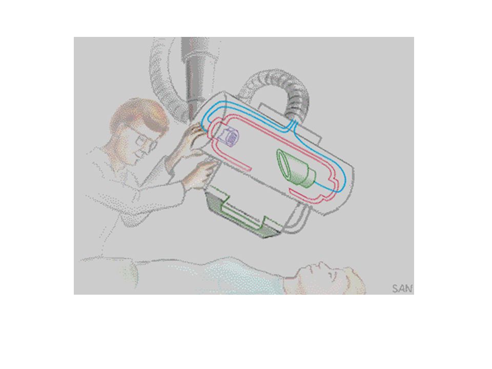
Puluj tube Phosphorescent tube acc. to Puluj, 1870.

19
Through his experiments into the nature of "cold light," Mr
Through his experiments into the nature of "cold light," Mr. Puliui invented an X-ray emitting device as early as The tubes of this invention became known as the "Pului lamp" and were mass-produced for a period. Mr. Puliui personally presented one of them to Mr. Roentgen. And it was Mr. Puliui, not Mr. Roentgen, who first demonstrated an X-ray photograph of a 13-year-old boy's broken arm and an X-ray photograph of his daughter's hand with a pin lying under it. A couple of years later, Mr. Roentgen was to publicly repeat the same experiments, but in doing so did not once credit Mr. Puliui's role in this discovery.

20
Wilhelm Conrad Röntgen
Wilhelm Conrad Röntgen. While working with a Crookes tube, a plate of Barium Platino-Cyanide (fluorescent crystals) on a table six feet away in his workroom glowed when he activated the tube. Even after covering the tube with black cardboard it kept glowing. He concluded that a new type of ray emitted from the tube, passed through the covering, and casted shadows of solid objects. The rays passes through most substances, including the soft tissues of the body, but left the bones and most metals visible. One of his earliest photographic plate from his experiments was a film of his wife, Bertha's hand with a ring, was produced on Friday, November 8, 1895
 on a table six feet away in his workroom glowed when he activated the tube. Even after covering the tube with black cardboard it kept glowing. He concluded that a new type of ray emitted from the tube, passed through the covering, and casted shadows of solid objects. The rays passes through most substances, including the soft tissues of the body, but left the bones and most metals visible. One of his earliest photographic plate from his experiments was a film of his wife, Bertha s hand with a ring, was produced on Friday, November 8,")
23
On New Kind of Rays

24
The news of Roentgen’s discovery spread quickly throughout the world
The news of Roentgen’s discovery spread quickly throughout the world. Scientists everywhere could duplicate his experiment because the cathode tube was very well known during this period. In early 1896, X-rays were being utilized clinically in the United States for such things as bone fractures and gun shot wounds. Internal structures of the body could be made visible without the necessity of surgery.

25
Roentgen discovered x-rays while experimenting with the Crookes tube
Roentgen discovered x-rays while experimenting with the Crookes tube. Crookes tube had almost a complete vaccum inside, in addition. It had a positive electrode (anode) which served as a target for electrons and the negative one (cathode) which served as a source of electrons. Unlike Coolidge tube it had a cold cathode. When high potential difference (kVp) was created between a cathode and anode, electrons rushed from a cathode and smashed in the anode. Upon collision with an anode or target they lost their kinetic energy. As a result of it, their loss of kinetic energy was transformed into the electromagnetic energy: x-rays and heat. An x-ray production process is a very inefficient process, in the midium range kVp (70 -80) kinetic energy of electrons is converted to more than 99% of heat and less than 1% of x-rays. In 1913 Dr. Coolidge invented an x-ray tube with a hot cathode. In that tube cathode was heated to liberate electrons necessary for the trip towards anode. This liberation process of electrons is called thermionic emission. The figure in the next slide demonstrates the x-ray production process in the Coolidge tube
 which served as a target for electrons and the negative one (cathode) which served as a source of electrons. Unlike Coolidge tube it had a cold cathode. When high potential difference (kVp) was created between a cathode and anode, electrons rushed from a cathode and smashed in the anode. Upon collision with an anode or target they lost their kinetic energy. As a result of it, their loss of kinetic energy was transformed into the electromagnetic energy: x-rays and heat. An x-ray production process is a very inefficient process, in the midium range kVp (70 -80) kinetic energy of electrons is converted to more than 99% of heat and less than 1% of x-rays. In 1913 Dr. Coolidge invented an x-ray tube with a hot cathode. In that tube cathode was heated to liberate electrons necessary for the trip towards anode. This liberation process of electrons is called thermionic emission. The figure in the next slide demonstrates the x-ray production process in the Coolidge tube.")
28
X-rays characteristics
Highly penetrating, invisible rays Electrically neutral Travel in straight lines. Travel with the speed of light in vaccum: 300, 000 km/sec or 186, 400 miles/sec. Ionize matter by removing orbital electrons Induce fluorescense in some substances. Fluorescent screen glow after being stricken with photons. Can't be focused by lenses nor by collimators.

31
light X-rays

34
kVp - kilovolt peak: Thousands of volts of electric potential applied accross cathode and anode. In a diagnostic radiology this potential ranges from kVp ( excluding mammography.) Anode is highly positively charged electrode and attracts the electrons. The higher the kVp, the faster the electrons travel from cathode to anode, as a result, the electromagnetic photons have higher energy. Consequently, kVp controls the energy ( quality) of an x-ray beam, not the speed of photons. Speed of photons is constant.
 Anode is highly positively charged electrode and attracts the electrons. The higher the kVp, the faster the electrons travel from cathode to anode, as a result, the electromagnetic photons have higher energy. Consequently, kVp controls the energy ( quality) of an x-ray beam, not the speed of photons. Speed of photons is constant..")
35
Low kVp

36
High kVp

37
mA mA - milliamperage: Number of electrons traveling from cathode to anode. mA describes x-ray tube current.

38
Low mA

39
High mA

40
CONVENTIONAL RADIOGRAPHY
PRODUCES STATIC IMAGES

41
FLUOROSCOPE WAS INVENTED BY THOMAS EDISON

42
Fluoroscopy is an imaging technique commonly used by physicians to obtain real-time moving images of the internal structures of a patient through the use of a fluoroscope. In its simplest form, a fluoroscope consists of an x-ray source and fluorescent screen between which a patient is placed. However, modern fluoroscopes couple the screen to an x-ray image intensifier and CCD video camera allowing the images to be recorded and played on a monitor.

43
FIRST X-RAY FATILITY IN THE US CLARENCE DALLY

44
OTHER INVENTIONS IN RADIOGRAPHY
INTENSIFYING SCREENS COLLIMATION FILTRATION DOUBLE EMULSION FILM

45
X-rays are highly penetrative rays that can ionize matter by removing electrons from the atoms. Are they harmful? Yes, they are. Nevertheless, radiography is considered a safe profession? Despite the fact that x-rays are harmful radiography is considered a safe profession. In order to understand this we must explain principles of the ALARA concept

46
"ALARA" is an acronym for "As Low As Reasonably Achievable"
"ALARA" is an acronym for "As Low As Reasonably Achievable". ALARA is a basic radiation protection concept or philosophy. It is an application of the "Linear No Threshold Hypothesis," which assumes that there is no "safe" dose of radiation. Under this assumption, the probability for harmful biological effects increases with increased radiation dose, no matter how small. Therefore, it is important to keep radiation doses to affected populations (for example, radiation workers, minors, visitors, students, members of the general public, etc.) as low as is reasonably achievable.
 as low as is reasonably achievable.")
47
ALARA Time Distance Shielding

48
Shielding

Similar presentations


 of Radioactivity>")
 and her.>")


 BPKIHS,Dharan.>")


>")


