Download presentation
Presentation is loading. Please wait.

1
Nose, Eyes, Ear, and Tongue
Ch 12: Special Senses Nose, Eyes, Ear, and Tongue

2
External Anatomy of the Eye
Eyelids: Protection, Lubrication Eyelashes: Protection Glands: Meibomian and Ciliary Meibomian: Oil glands, modified sebaceous glands on eyelids Ciliary: Modified sweat glands.

3
Third eye-lid Gathers dust and produces eye crispies. In other animals It can cover the Eye.

4
What is the Lacrimal apparatus and what does it do?
Lacrimal apparatus. Fig 12.5 Lacrimal glands: release tears Lacrimal ducts (eyelid) and canals (nose) Nasolacrimal ducts: empties into the nose Lacrimal Secretion: Tears have antibodies and lysozyme. Cleans, moistens. Why is it called a healthy cry? Lysozyme
 and canals (nose) Nasolacrimal ducts: empties into the nose. Lacrimal Secretion: Tears have antibodies and lysozyme. Cleans, moistens. Why is it called a healthy cry Lysozyme.")
5
What are the Muscles of the eye and how do they move it?
Lateral rectus: Moves eye laterally Medial rectus: Moves eye medially Superior rectus: Moves eye up Inferior rectus: Moves eye down S & I Obliques Fig 8.15, 197 Fig 12.7, pg 296 Practice using The Eye Muscles

6
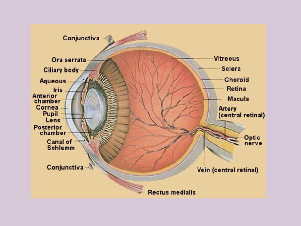
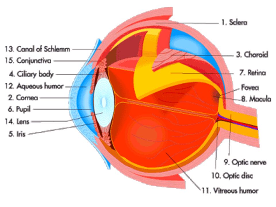
What are the internal structures of the eye? Pg. 295, Fig 12.6
Sclera: white of the eye Cornea: Clear Iris: colorful part of the eye Pupil: Opening of the Iris Lens and ciliary body: Ciliary body holds the lens in place. The lens focuses light on the back of the eye.

7
What fills the inner eye? Pg 298
Aqueous Humor: Circulates from the cornea to the Lens through the canal of Schlemm. Vitreous Humor: In the space behind the lens. Keeps the eyes shape, and keeps intraocular pressure stable. Floaters? Glaucoma?

9
What lines the back of the eye?
Retina: photoreceptor layer Rods: Black and white Cones: Color Choroid coat: Dark, vascularized layer. Absorbs light. *Non-humans are different Sclera: Thick, white covering

11
How do you see? Pg. 299 . How we See Details of Vision Adam

12
Light travels through the…
Cornea to the Aqueous humor to the Pupil to the lens. In the Lens light is bent so that it will hit the Retina of the eye. To get to the Fovea Centrals light must continue to pass through Vitreous humor. Light then hits the Retina where Rods and Cones interpret the image and sends it to the Optic nerve to the brain.

13
Where is your vision the best and the worst?
Worst vision: The blind spot. To find your blind spot go to pg 297 in the book. Blind spot is where the optic nerve takes the image to your brain. There are no rods or cones here. Your brain just fills in the gaps. Best vision: Fovea Centralis. Lateral to the blind spot, only contains cones. This is the spot of greatest visual acuity.

15
Why do cones see better than rods?
See Fig 12.8 Many Rods, one nerve = blurred edges. One Cone, one nerve = crisp lines. Which do you think causes Night Blindness? What type of cones do you have: Blue cones: see blue light Green cones: see green light Red cones: see red and green light.

16
Component colors are detected by cone cells in the retina
Component colors are detected by cone cells in the retina. All colors in the visible spectrum can be represented as a combination of red, green, and blue. In the retina, a full-color image is broken up into component colors by cone cells specialized to detect red light (long wavelength), green light (med. wavelength), blue light (short wavelength).
, green light (med. wavelength), blue light (short wavelength).")
17
What is colorblindness?
The lack of a type of cone, or all cones. Sex-linked gene. Men suffer from color blindness more often than females. Vision of the Colorblind

18
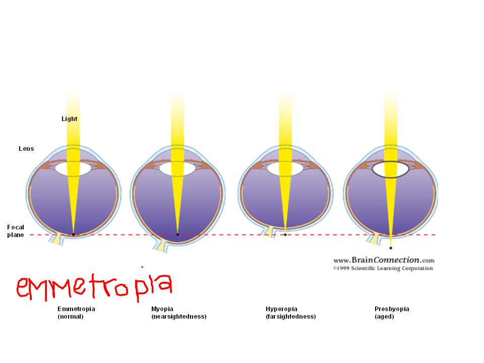
How is Lasik’s performed?
Steps to LASIK Surgery Steps to LASIK Surgery

19
Illusions More Illusions

20
Functions: Hearing and Equilibrium…………….
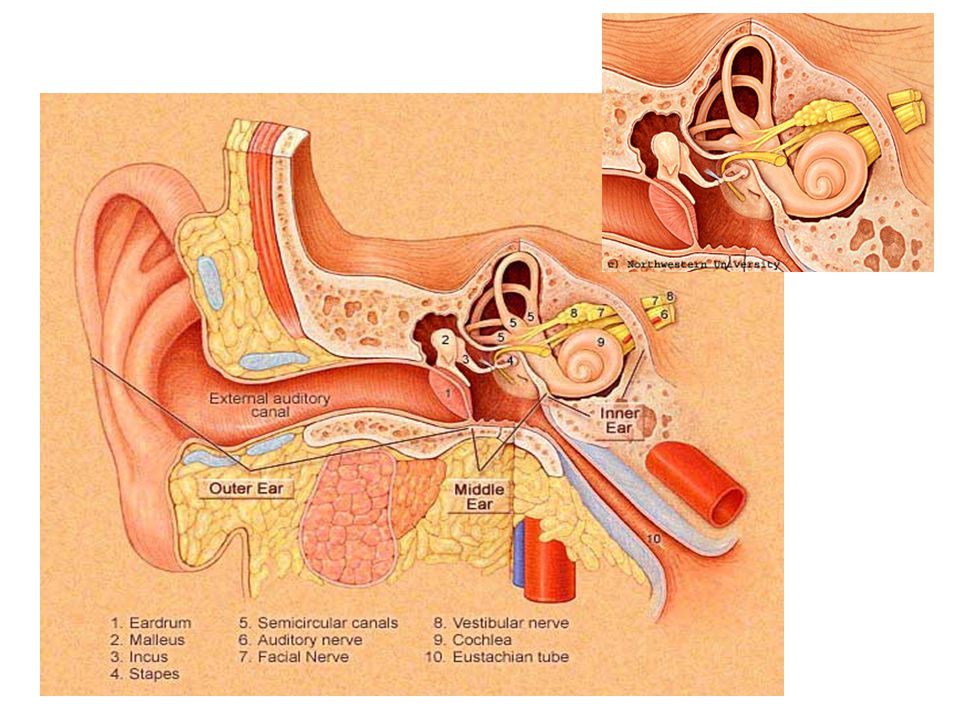
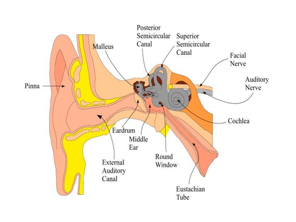
The Ear Pg. 302 & 303 Functions: Hearing and Equilibrium…………….

22
External Ear How the Ear hears. Auricle External Auditory Meatus-2.5 cm long leads to Middle ear

23
The Middle Ear Tympanic Cavity: air-filled space
Tympanic Membrane- ear drum Pressure changes cause it to vibrate, enhances the sound wave Auditory Ossicles: smallest bones in the body. Malleus, incus, and stapes. The Stapes vibrate at the oval window causing fluid in the inner ear to move, stimulating hearing receptors.

25
STAPES

26
INCUS

27
MALLEUS

28
Auditory tube mucous membranes connect directly with the middle ear linings. Thus, mucous membrane infections of the throat may spread through these tubes and cause a middle ear infection. Question: Why is it important to keep a babies head up when bottle feeding? How the Tubes work Xylitol gum and chewing it could help prevent ear infections.

29
Inner Ear. Pg. 304 Cochlea: hearing
Oval Window: the stapes vibrates this in order for your nerves to fire and for you to hear sound. Quick Review Sound on Cochlea Review of the Ear: In depth on Cochlea And Hearing

30
Inner Ear: pg. 306 - 307 Semicircular Canals: maintain equilibrium
The vestibular system

31
Hearing Illusions

32
The Nose. Fig 12.3 Smells using Olfactory Receptors
Sex and Smell Smells using Olfactory Receptors Yellow-brown mass located at the top of each nasal cavity Size of a postage stamp. Factoid: These swirl the Air so that Dust and germs Stick to the Mucus. How we smell

33
How do olfactory receptor cells smell?
Use Olfactory Hairs Must be covered in mucus Chemicals dissolve in mucus Trigger nerve Olfactory Nerve Connects to the Olfactory Bulb in Brain. Travels to temporal lobe for interpretation. Tied to the limbic system (emotional system) of the brain. Smells stimulate memories.
 of the brain. Smells stimulate memories.")
34
Interesting Nose facts
Olfactory neurons that are over stimulated shut off. (Can’t smell X after a while.) Anosmias: loss of smell due to head injuries, nasal cavity inflammation (cold, allergy, smoking) or age. Often caused by a zinc deficiency Olfactory auras: epileptics may have smell hallucinations before a seizure. Because dust in space does not settle Austronoauts sneeze about 100 times a day!
 Anosmias: loss of smell due to head injuries, nasal cavity inflammation (cold, allergy, smoking) or age. Often caused by a zinc deficiency. Olfactory auras: epileptics may have smell hallucinations before a seizure. Because dust in space does not settle Austronoauts sneeze about 100 times a day!")
35
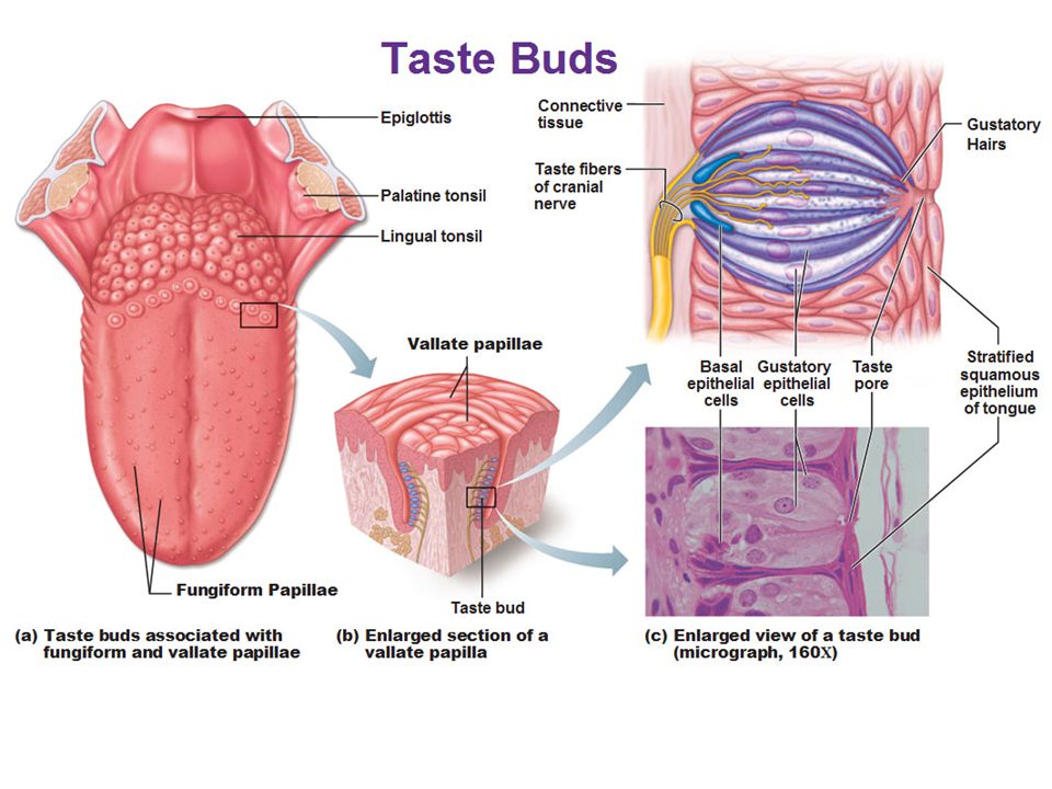
The Tongue. Fig. 12.4 . Factoid: You can detect and distinguish
between 10,000 odors, but only 5 tastes. .

37
How Do You Taste? Gustatory cells- respond to chemicals that are dissolved in saliva Taste buds- receptor sites for tastes. Most are on the tongue. Some are on the roof of the mouth and cheeks Papillae- on the sides of this structure is where taste buds are found How do you Taste?

38
Basic Taste Sensations
Sweet- sugar, OH- groups Sour- acidic, H+ Umami- meaty or savory Bitter- alkaloid bases, very few H+ bonds Salty- metal ions The science Of Picky Eaters

39
What effects taste? Temperature Smell Texture Genetics

Similar presentations


 Eyelids- shade and protect the eyes and provide lubrication Eyelids- shade and protect the eyes and.>")


 detects a physical or chemical change. 2. The physical or chemical change causes action potentials.>")


