Download presentation
Presentation is loading. Please wait.

1
Introduction to Parasitology & Lab Diagnosis of Parasitic Diseases
Dr. Sudheer Kher

2
Why study Parasitology?
Many of these parasites are causative agents of major public health problems of the world. Recent estimates of prevalence of parasites in the world are: Ascaris billion Hookworms billion Whipworms billion Filarial worms million Malaria million Schistosomes million Amebiasis million Taenia tapeworms million Clonorchis million Chagas’ Disease million These parasites cause untold suffering and death in the world today.

3
The burden of some major parasitic infections
Parasite Diseases No. people infected Deaths/yr Plasmodium malaria 273 million 1.12 million Soil transmitted helminths: Roundworm (Ascaris) Whipworm (Trichuris) Hookworm (Ancylostoma and Necator) Pnemonitis, intestinal obstruction Bloody diarrhoea, rectal prolapse Coughing, wheezing, abdominal pain and anaemia 2 billion 200,000 Schistosoma Renal tract and intestinal disease 200 million 15,000 Filariae Lymphatic filariasis and elephantiasis 120 million Not fatal but 40 million disfigured or incapacitated Trypanasoma cruzi Chagas disease (cardiovascular) 13 million 14,000 African trypanosomes African sleeping sickness 0.3 – 0.5 million 48,000 Leishamania Cutaneous, mucocutaneous and visceral leishmaniasis 12 million; 2 million new cases/yr 50,000
 Whipworm (Trichuris) Hookworm (Ancylostoma and Necator) Pnemonitis, intestinal obstruction. Bloody diarrhoea, rectal prolapse. Coughing, wheezing, abdominal pain and anaemia. 2 billion. 200,000. Schistosoma. Renal tract and intestinal disease. 200 million. 15,000. Filariae. Lymphatic filariasis and elephantiasis. 120 million. Not fatal but 40 million disfigured or incapacitated. Trypanasoma cruzi. Chagas disease (cardiovascular) 13 million. 14,000. African trypanosomes. African sleeping sickness. 0.3 – 0.5 million. 48,000. Leishamania. Cutaneous, mucocutaneous and visceral leishmaniasis. 12 million; 2 million new cases/yr. 50,000.")
7
Key definitions: What is ….?
Medical parasitology: “the study and medical implications of parasites that infect humans” A parasite: “a living organism that acquires some of its basic nutritional requirements through its intimate contact with another living organism”. Parasites may be simple unicellular protozoa or complex multicellular metazoa Eukaryote: a cell with a well-defined chromosome in a membrane-bound nucleus. All parasitic organisms are eukaryotes

8
Key definitions: What is ….?
Protozoa: unicellular organisms, e.g. Plasmodium (malaria) Metazoa: multicellular organisms, e.g. helminths (worms) and arthropods (ticks, lice) An endoparasite: “a parasite that lives within another living organism” – e.g. malaria, Giardia An ectoparasite: “a parasite that lives on the external surface of another living organism” – e.g. lice, ticks
 Metazoa: multicellular organisms, e.g. helminths (worms) and arthropods (ticks, lice) An endoparasite: a parasite that lives within another living organism – e.g. malaria, Giardia. An ectoparasite: a parasite that lives on the external surface of another living organism – e.g. lice, ticks.")
9
Key definitions: What is ….?
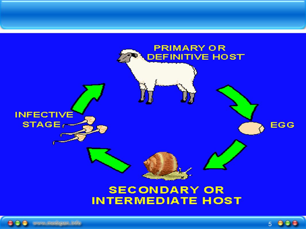
Host: “the organism in, or on, which the parasite lives and causes harm” Definitive host: “the organism in which the adult or sexually mature stage of the parasite lives” Intermediate host: “the organism in which the parasite lives during a period of its development only” Zoonosis: “a parasitic disease in which an animal is normally the host - but which also infects man” Vector: “a living carrier (e.g.an arthropod) that transports a pathogenic organism from an infected to a non-infected host”. A typical example is the female Anopheles mosquito that transmits malaria
 that transports a pathogenic organism from an infected to a non-infected host . A typical example is the female Anopheles mosquito that transmits malaria.")
10
Taxonomic classification of protozoa
Sub kingdom Phylum Sub-phylum Genus- examples Species- examples Protozoa Sarcomastig-ophora further divided into Sarcodina-- - move by pseudopodia Entamoeba E. histolytica Mastigophora move by flagella Giardia G. lamblia Apicomplexa no organelle of locomotion Plasmodium P. falciparum, P. vivax, P. malariae, P. ovale Ciliophora move by cillia Balantidium B. coli Microspora Spore-forming Enterocyto-zoa E. bienusi

11
Examples of important intestinal protozoa
Transmitted by the faecal-oral route and cause diarrhoea Giardia lamblia: world-wide distribution, lives in the small intestine and results in malabsorption Entamoeba histolytica: may invade the colon and cause bloody diarrhoea – amoebic dysentery. Also causes ameobic liver abscess. Cryptosporidium parvum: more prevalent in the immunocompromised Cyclospora cyatenensis - parasitises the small intestinal mucosa and may cause diarrhoea for several weeks Balantidium coli: a large motile ciliated parasite that lives in the colon of pigs, humans and rodents and can lead to colonic ulceration Enterocytozoon bienusi: a microsporidian that parasitises the small intestine. Also more common in the immunocompromised.

12
Examples of important systemic protozoa
Detected in the blood Plasmodium: the cause of malaria. There are 4 species that infect man: P. falciparum, P. vivax, P. ovale and P. malariae Toxoplasma gondi: transmitted by the ingestion of oocysts from cat faeces. Infection can lead to ocular problems and is also a cause of neonatal toxoplasmosis Leishmania: transmitted by sand flies, can lead to visceral, cutaneous and mucocutaneous leishmaniasis Trypanosoma: haemoflagellates which cause In Africa - sleeping sickness (transmitted by the Tsetse fly) In South America - Chagas disease (transmitted by the Reduviid bug) Typical lesion of cutaneous leishmaniasis Tsetse fly – the vector of African trypanosomiasis It has a painful bite!
 In South America - Chagas disease (transmitted by the Reduviid bug) Typical lesion of cutaneous leishmaniasis. Tsetse fly – the vector of African trypanosomiasis It has a painful bite!")
13
Taxonomic classification of helminths
Sub kingdom Phylum Class Genus – examples Metazoa Nematodes Round worms; appear round in cross section, they have body cavities, a straight alimentary canal and an anus Ascaris (roundworm) Trichuris (whipworm) Ancylostoma (hookworm) Necator (hookworm) Enterobius (pinworm or threadworm) Strongyloides Platyhelminthes Flat worms; dorsoventrally flattened, no body cavity and, if present, the alimentary canal is blind ending Cestodes Adult tapeworms are found in the intestine of their host They have a head (scolex) with sucking organs, a segmented body but no alimentary canal Each body segment is hermaphrodite Taenia (tapeworm) Trematodes Non-segmented, usually leaf-shaped, with two suckers but no distinct head They have an alimentary canal and are usually hermaphrodite and leaf shaped Schistosomes are the exception. They are thread-like, and have separate sexes Fasciolopsis (liver fluke) Schistosoma (not leaf shaped!)
 Trichuris (whipworm) Ancylostoma (hookworm) Necator (hookworm) Enterobius (pinworm or threadworm) Strongyloides. Platyhelminthes. Flat worms; dorsoventrally flattened, no body cavity and, if present, the alimentary canal is blind ending. Cestodes. Adult tapeworms are found in the intestine of their host. They have a head (scolex) with sucking organs, a segmented body but no alimentary canal. Each body segment is hermaphrodite. Taenia (tapeworm) Trematodes. Non-segmented, usually leaf-shaped, with two suckers but no distinct head. They have an alimentary canal and are usually hermaphrodite and leaf shaped. Schistosomes are the exception. They are thread-like, and have separate sexes. Fasciolopsis (liver fluke) Schistosoma (not leaf shaped!)")
14
Examples of important metazoa – intestinal nematodes
Trichuris (whipworm) A soil transmitted helminth prevalent in warm, humid conditions Can cause diarrhoea, rectal prolapse and anaemia in heavily-infected people Ancylostoma and Necator (hookworms) A major cause of anaemia in the tropics Strongyloides inhabits the small bowel infection more severe in immunospressed people (e.g. HIV/AIDS, malnutrition, intercurrent disease) Enterobius (pinworm or threadworm) prevalent in cold and temperate climates but rare in the tropics found mainly in children Ascaris (roundworm) Found world-wide in conditions of poor hygiene, transmitted by the faecal- oral route Adult worms lives in the small intestine Causes eosinophilia Heavy intestinal infections may occur with Ascaris. Adult worms can be several cms long.
 A soil transmitted helminth. prevalent in warm, humid conditions. Can cause diarrhoea, rectal prolapse and anaemia in heavily-infected people. Ancylostoma and Necator (hookworms) A major cause of anaemia in the tropics. Strongyloides. inhabits the small bowel. infection more severe in immunospressed people (e.g. HIV/AIDS, malnutrition, intercurrent disease) Enterobius (pinworm or threadworm) prevalent in cold and temperate climates but rare in the tropics. found mainly in children. Ascaris (roundworm) Found world-wide in conditions of poor hygiene, transmitted by the faecal- oral route. Adult worms lives in the small intestine. Causes eosinophilia. Heavy intestinal infections may occur with Ascaris. Adult worms can be several cms long.")
15
Examples of important metazoa –systemic nematodes
Filaria including: Onchocerca volvulus – Transmitted by the simulium black fly, this microfilarial parasite can cause visual impairment, blindness and severe itching of the skin in those infected Wuchereria bancrofti – The major causative agent of lymphatic filariasis Brugia malayi – Another microfilarial parasite that causes lymphatic filariasis Toxocara A world-wide infection of dogs and cats Human infection occurs when embryonated eggs are ingested from dog or cat faeces It is common in children and can cause visceral larva migrans (VLM)
")
16
Examples of important flatworms - cestodes
Intestinal - (“tapeworms”) Taenia saginata worldwide acquired by ingestion of contaminated, uncooked beef a common infection but causes minimal symptoms Taenia solium acquired by ingestion of contaminated, uncooked pork that contains cystercerci Less common, but causes cystercicosis – a systemic disease where cysticerci encyst in muscles and in the brain – may lead to epilepsy Systemic Echinococcus granulosus (dog tapeworm) and Echinicoccus multilocularis (rodent tapeworm) Hydatid disease occurs when the larval stages of these organisms are ingested The larvae may develop in the human host and cause space-occupying lesions in several organs, e.g. liver, brain
 Taenia saginata. worldwide. acquired by ingestion of contaminated, uncooked beef. a common infection but causes minimal symptoms. Taenia solium. acquired by ingestion of contaminated, uncooked pork that contains cystercerci. Less common, but causes cystercicosis – a systemic disease where cysticerci encyst in muscles and in the brain – may lead to epilepsy. Systemic. Echinococcus granulosus (dog tapeworm) and Echinicoccus multilocularis (rodent tapeworm) Hydatid disease occurs when the larval stages of these organisms are ingested. The larvae may develop in the human host and cause space-occupying lesions in several organs, e.g. liver, brain.")
17
Examples of important metazoa –trematodes (flukes)
Intestinal Fasciolopsis buski - A common parasite of humans and pigs in South- east Asia. This parasite is one of the largest trematodes to infect man (8cm in length) and lives in the upper intestine. Chronic infection leads to inflammation, ulceration and haemorrhage of the small intestine Fasciola hepatica (liver fluke)- Primarily, a parasite of sheep, humans become infected when they ingest metacercariae that have encysted on watercress. The adult trematode lives in the intra-hepatic bile ducts of the liver. “Fascioliasis” can lead to severe anaemia in humans Clonorchis sinensis (liver fluke)- Widespread in China, Japan, Korea and Taiwan, this parasite is acquired by ingestion of infective metacercariae in raw or pickled fish Paragonimus westermani ( lung fluke)- Widespread in the Far East and South east Asia, the parasite is acquired by ingestion of infective metacercariae in raw or pickled crustaceans Schistosoma haematobium, S. mansoni and S. japonicum – see below
 and lives in the upper intestine. Chronic infection leads to inflammation, ulceration and haemorrhage of the small intestine. Fasciola hepatica (liver fluke)- Primarily, a parasite of sheep, humans become infected when they ingest metacercariae that have encysted on watercress. The adult trematode lives in the intra-hepatic bile ducts of the liver. Fascioliasis can lead to severe anaemia in humans. Clonorchis sinensis (liver fluke)- Widespread in China, Japan, Korea and Taiwan, this parasite is acquired by ingestion of infective metacercariae in raw or pickled fish. Paragonimus westermani ( lung fluke)- Widespread in the Far East and South east Asia, the parasite is acquired by ingestion of infective metacercariae in raw or pickled crustaceans. Schistosoma haematobium, S. mansoni and S. japonicum – see below.")
18
Schistosomiasis (bilharzia)
")
19
Hookworm (1) Epidemiology
>1200m infections each year of which 100m are symptomatic It is due to 2 parasites both of which occur worldwide: Necator americanus - predominant species in sub-Saharan Africa, south Asia and the Pacific Ancylostoma duodenale –predominant in S. Europe, N. Africa, western Asia, northern China, Japan and the west coast of America Hookworm is a major cause of anaemia

20
Hookworm (2) Life cycle Adult worms live in the intestine and excrete eggs in the faeces In the absence of latrines, eggs contaminate soil and develop in warm, damp conditions eggs hatch and infective filariform larvae develop in about one week and remain infective in soil for many weeks filariform larvae penetrate the skin when a person walks barefoot in the soil larva migrate from the skin to the lungs via the lymphatic and blood systems larvae penetrate the capillary wall to enter the alveolus Larvae are propelled up the respiratory tree to the epiglottis where they are swallowed Develops to adult stage in upper intestine; adult worms are fully mature after about 5 weeks Eggs are excreted in the faeces Note: eating soil (pica) is a common practice. Ingested filariform larvae of A. duodenale can pass directly to the gut mucosa Egg of A. duodenale in faecal smear (size µm by µm) Filariform larvae
 is a common practice. Ingested filariform larvae of A. duodenale can pass directly to the gut mucosa. Egg of A. duodenale in faecal smear (size µm by µm) Filariform larvae.")
21
Hookworm (3) Pathology Hookworms move several times a day to different attachment sites in the upper intestinal mucosa to ingest blood They secrete an anticoagulant which causes the old attachment sites to continue to bleed Heavy hookworm infection results in chronic haemorrhage from the duodenal and jejunal mucosa The combination of constant blood loss due to hookworm infection and poor iron intake in the diet results in iron deficiency anaemia A. duodenale ingests 4-5 times more blood each day than N. americanus In a child, the continued daily loss of 10ml of blood can lead to severe anaemia Adult male and female worms of A. duodenale

22
Hookworm (4) Symptoms and signs Minor
Often itchy papules are found at the site where the larva penetrated the skin There may be cough and wheezing as the larva migrates through the lungs Major Hookworm anaemia Tiredness, aches and pains Pallor Breathlessness Oedema Diagnosis Microscopic examination of faecal smears to demonstrate significant numbers of hook worm eggs Measure Hb, serum ferritin, iron Exclude other causes of anaemia Treatment Mebendazole (cheap) – 100mg, twice daily for 3 days Mebendazole is contraindicated in pregnancy – use Bephenium hydroxynaphthoate “alcopar” For anaemia: ferrous sulphate mg three times a day for 3 months (adult regimen) Prevention and control Health education and improve sanitation facilities – install pit latrines Encourage use of protective footwear Discourage soil eating (pica) Mass drug treatment of communities Iron supplementation in areas of low iron intake
 – 100mg, twice daily for 3 days. Mebendazole is contraindicated in pregnancy – use Bephenium hydroxynaphthoate alcopar For anaemia: ferrous sulphate mg three times a day for 3 months (adult regimen) Prevention and control. Health education and improve sanitation facilities – install pit latrines. Encourage use of protective footwear. Discourage soil eating (pica) Mass drug treatment of communities. Iron supplementation in areas of low iron intake.")
23
Lymphatic filariasis (1)
Epidemiology 120m people infected in >80 countries in Africa, Asia, the Pacific islands and South and Central America 40m of those infected are disfigured or severely incapacitated 95% cases due to Wuchereria bancrofti, other species include Brugia malayi and Brugia timori A female Anopheles mosquito taking a blood meal

24
Lymphatic filariasis (2)
Life cycle Wuchereria bancrofti is mainly transmitted by Culex mosquitoes in India Anopheline mosquitoes in Africa B. malayi and B. timori are transmitted mainly by Mansonia mosquitoes Larval forms of the parasite (microfilariae) are taken up by a female mosquito when it takes a blood meal from a human infected with adult worms The microfilariae develop inside the mosquito When the mosquito takes another blood meal the infective filariform larvae enter the bite wound Filariform larvae migrate to the lymphatics and lymph glands Larvae develop into sexually mature adult worms over 3-12 months depending on the species of filarial worm Microfilaria of B. malayi in thick blood film (H&E stain; source: CDC) Adult worms of B. malayi in section in a lymph node (source: Univ South Carolina)
 are taken up by a female mosquito when it takes a blood meal from a human infected with adult worms. The microfilariae develop inside the mosquito. When the mosquito takes another blood meal the infective filariform larvae enter the bite wound. Filariform larvae migrate to the lymphatics and lymph glands. Larvae develop into sexually mature adult worms over 3-12 months depending on the species of filarial worm. Microfilaria of B. malayi in thick blood film (H&E stain; source: CDC) Adult worms of B. malayi in section in a lymph node (source: Univ South Carolina)")
25
Lymphatic filariasis (3)
Pathology Adult worms live in the afferent lymphatic vessels and cause severe disruption to the lymphatic system Scrotal damage and massive swelling may occur when adult Wuchereria bancrofti lodge in the lymphatics of the spermatic cord Late stage disease is typified by elephantiasis – painful and disfiguring swelling of the limbs Trauma and secondary bacterial infection of affected tissues is common Elephantiasis of the leg (source: WHO/TDR/Crump)
")
26
Lymphatic filariasis (4)
Symptoms and signs – 3 stages 1. Asymptomatic stage There is internal damage to the lymphatics and kidneys 2. Acute stage – Filarial lymphangitis Characterised by bouts of fever heat, redness, pain, swelling and tenderness of the lymph nodes and ducts 3. Chronic stage Usually results in elephantiasis as a result of chronic lymphoedema There is a massive overgrowth of tissue resulting in severe deformities The legs are often affected and result in inability to walk The scrotum is often affected in men and the breasts and vulva in women Elderly male with massive hydrocoele, and elephantiasis of the leg. Also has nodules in the groin due to onchocerciasis (source: WHO/TDR/Crump)
")
27
Lymphatic filariasis (5)
Diagnosis Microscopic examination of Giemsa stained thick blood films for the presence of microfilariae W. bancrofti shows marked nocturnal periodicity, so it’s best to collect blood samples between 10pm and 1 am Serology Treatment Diethylcarbamazine (DEC) rapidly kills microfilariae and will kill adult worms if given in full dosage over 3 weeks Release of antigens from dying microfilaria causes allergic-type reactions – add an antihistamine and aspirin to treatment regimen Other treatment options are ivermectin combination of DEC and albendazole Prevention and control Rapid diagnosis and treatment of infected individuals Mass drug administration to at risk communities Vector control: eliminate mosquito breeding sites through improved sanitation and enviromental management Personal protection against mosquito bites by insecticides, bednets and repellants
 rapidly kills microfilariae and will kill adult worms if given in full dosage over 3 weeks. Release of antigens from dying microfilaria causes allergic-type reactions – add an antihistamine and aspirin to treatment regimen. Other treatment options are. ivermectin. combination of DEC and albendazole. Prevention and control. Rapid diagnosis and treatment of infected individuals. Mass drug administration to at risk communities. Vector control: eliminate mosquito breeding sites through improved sanitation and enviromental management. Personal protection against mosquito bites by insecticides, bednets and repellants.")
28
Laboratory Diagnosis of Parasitic Infections
Purpose – Confirmation of clinical suspicion Identification of unsuspected infection Methods same as used in Bacteriology & Virology but significance of different methods varies. Isolation least important, morphological identification very important. Serology relatively less important

29
Morphological identification
Examination of faeces – Gross Microscopy Saline mount Iodine Mount Thick smears – not commonly used Permanent stained smears Iron hematoxylene Whearley’s trichrome stain Concentration methods Floatation techniques Sedimentation techniques

30
Morphological identification
Examination of Blood Thin Smear Thick smear Wet mount for microfilaria Stains used

31
Cultivation of parasites
Culture methods – Amoeba Leishmania & Trypanosoma Malarial parasite Animal inoculation – Not practical Xenodiagnosis – Vectors infected experimentally Immunological diagnosis

32
Immunological diagnosis
Serology – All tests available IHA ELISA CIEP IF CFT More useful in Amoebiasis Leishmaniasis Malaria Toxoplasmosis Trichinosis Filariasis Echinococcosis Skin Tests – Specificity low, cross reactions common Casoni’s test Leishmanin test

33
Sources of information
The Special Programme for Research and Training in Tropical Diseases (TDR UNICEF, UNDP, World Bank, WHO) website: ww.who.int/tdr/media/image.html University of South Carolina School of Medicine: Lecture notes on Tropical Medicine, Dion R Bell, Fourth edition, 1996, Blackwell Science. Parasites and human disease, W. Crewe and D.R.W. Haddock, 1985, First edition, Edward Arnold
 website: ww.who.int/tdr/media/image.html. University of South Carolina School of Medicine: Lecture notes on Tropical Medicine, Dion R Bell, Fourth edition, 1996, Blackwell Science. Parasites and human disease, W. Crewe and D.R.W. Haddock, 1985, First edition, Edward Arnold.")
Similar presentations


 Drs. Babcock and Hopkins Spring 2009>")


 : Protozoa: 1- Protozoa are unicellular (eukaryotic) or acellular organisms. 2- Protozoan is measured in microns;>")


