Download presentation
Presentation is loading. Please wait.

2
*

6
Sag PD Cor T1

7
*

8
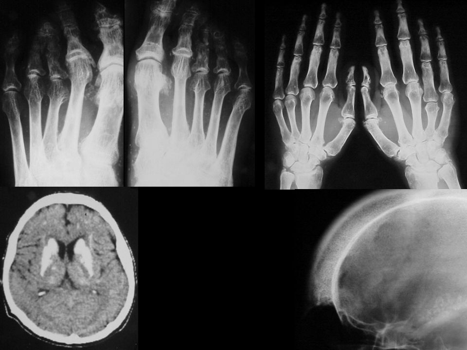
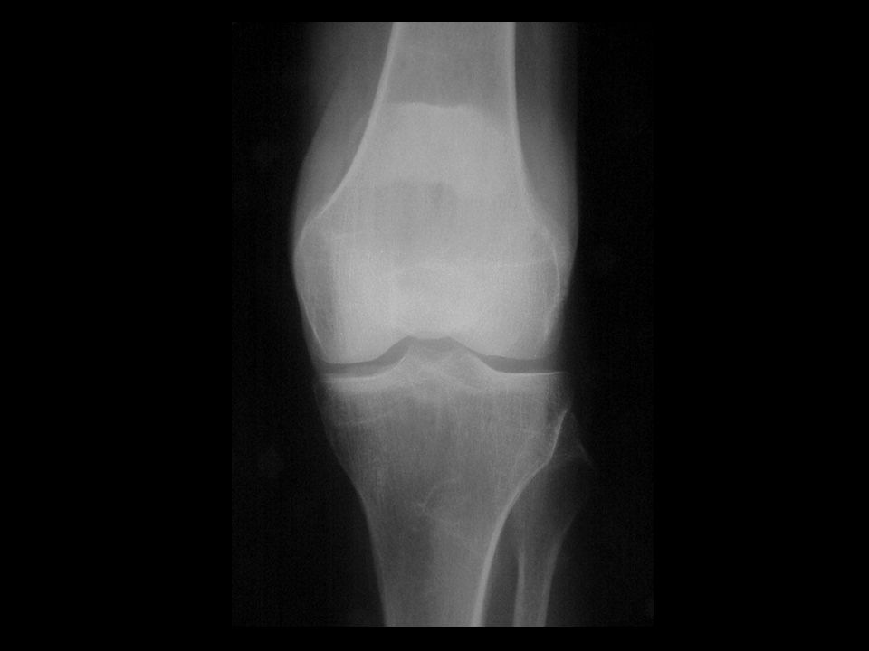
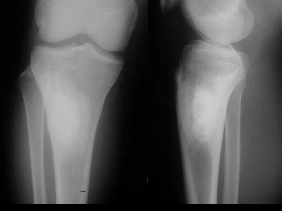
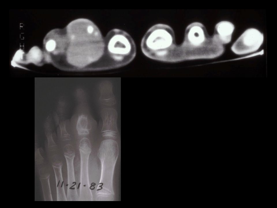
47F 5Y increasing stiffness and decreased ROM. Initial presentation

12
* Ankylosing Spondylitis Dagger sign
Bilateral hip inflammatory arthritis *

13
Ankylosing Spondylitis
Young white men When fuse posteriorly, may spare anterior Scalloping of Cx spine Dagger + Bamboo = Tram tracks

14
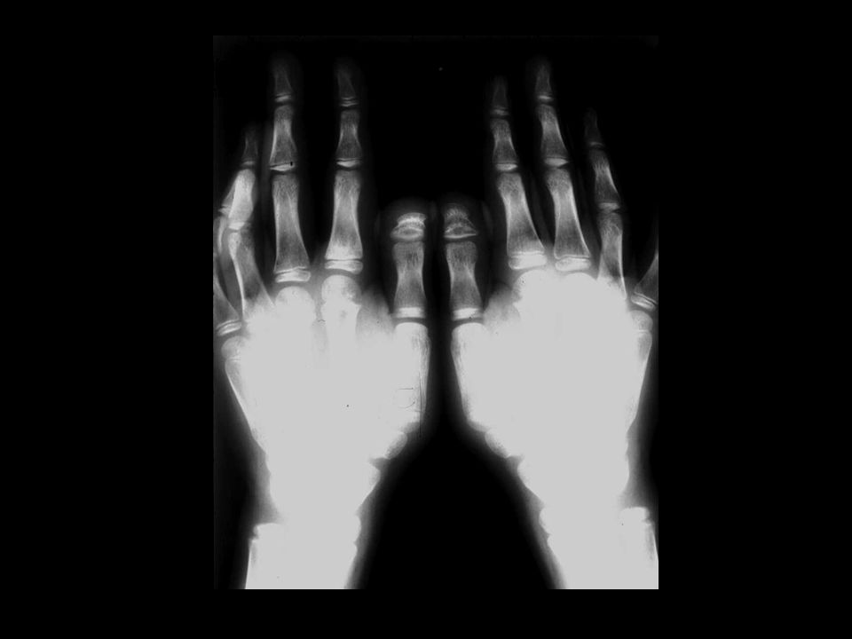
19M increasing mass on hand, refused treatment
3

17
Osteosarcoma *

18
Osteosarcoma Most common primary malignant bone tumor in young adults and children Second to MM in older Osteoid immature bone Bimodal Close to knee, away from elbow Young-cylindrical, Old-flat

19
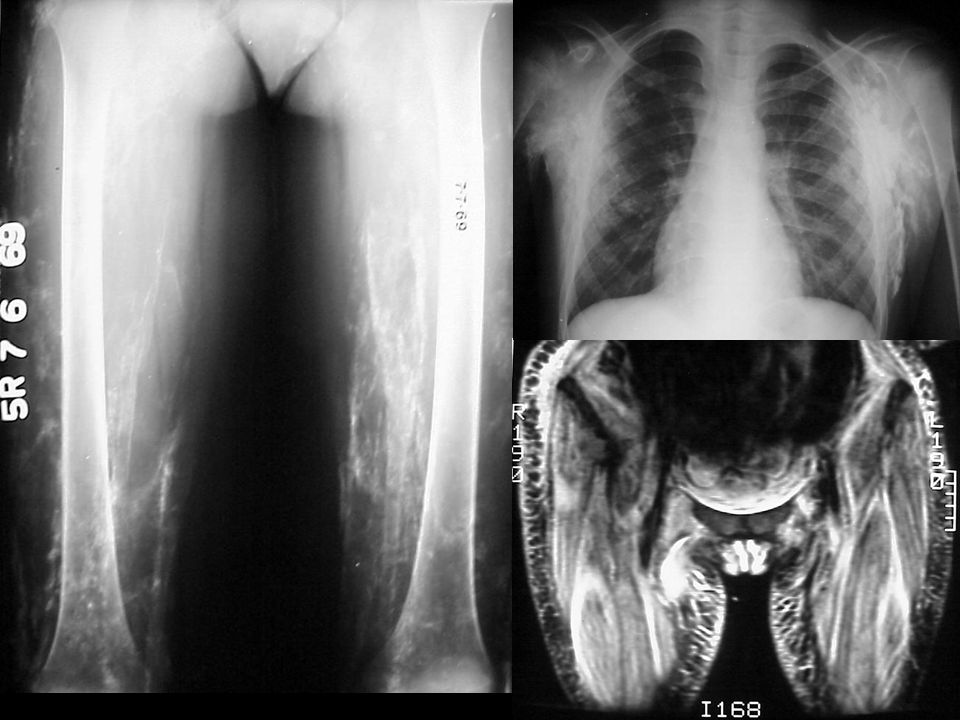
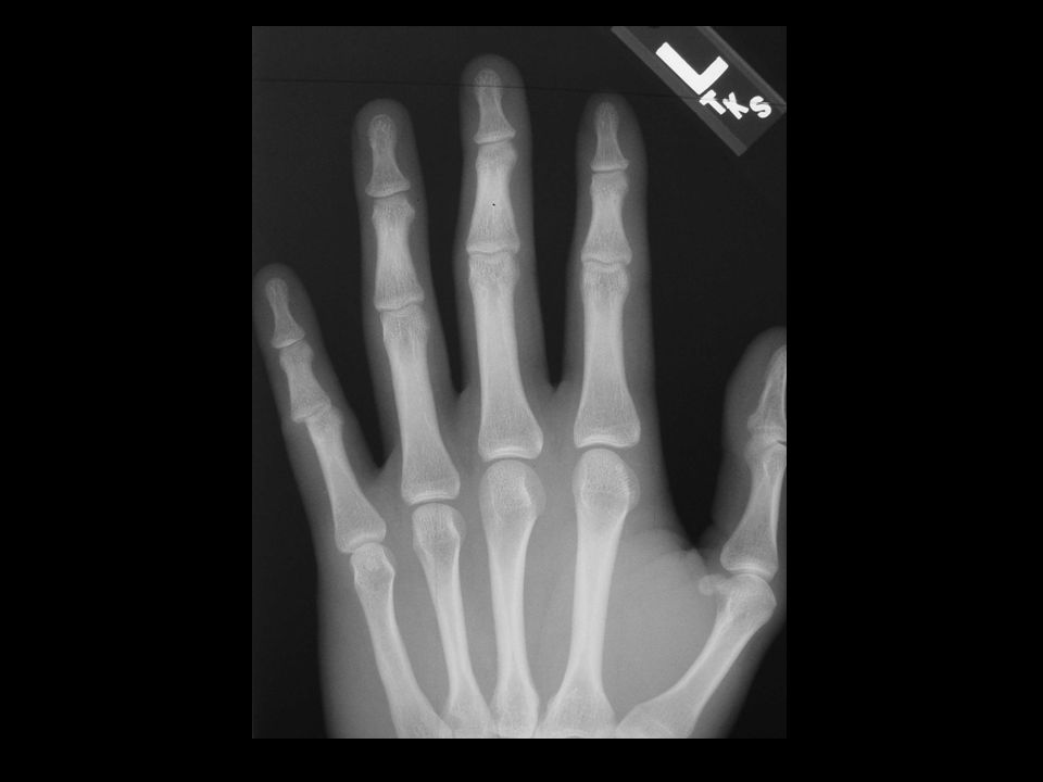
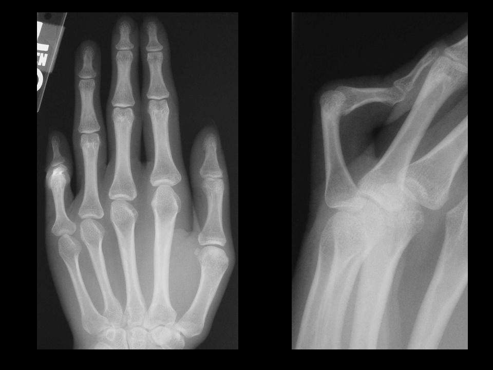
25M with wrist pain 3

22
Osteopoikilosis *

23
Osteopoikilosis Juxtaarticular bone islands ?AD, M>F, asymptomatic
Ovoid 2-10mm Benign sclerosing bone dysplasia

24
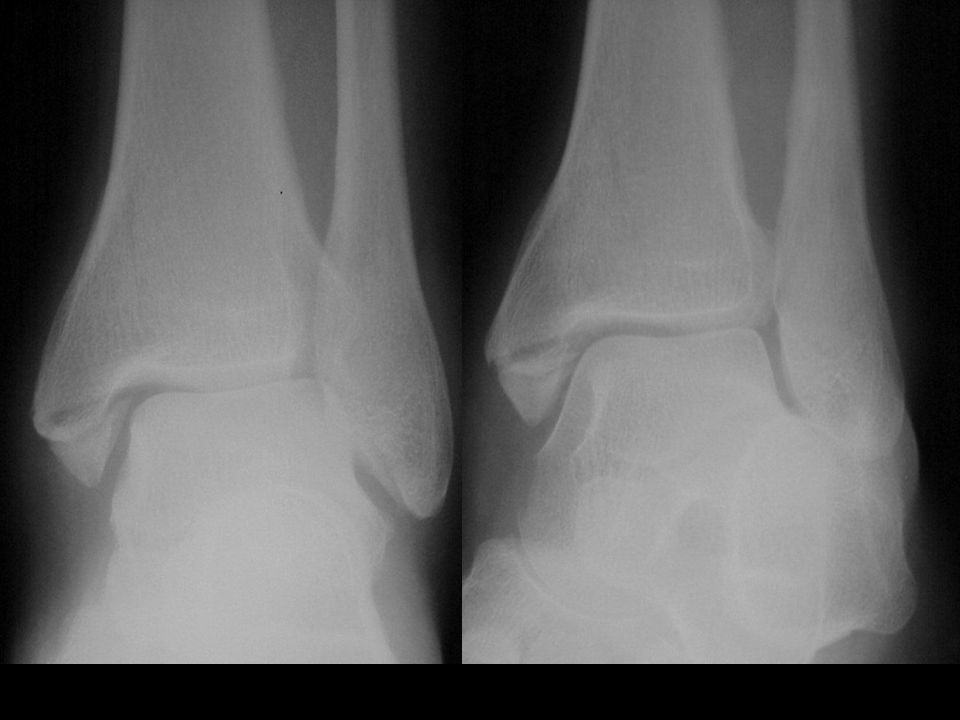
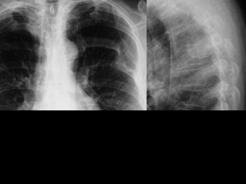
95 M pain R hip 4

25
Cor T1

28
Cor STIR Pagets disease Pathological Fx Asterix enhancement *

29
Pagets disease O. deformans 3% >40, northern latitudes, >M
Lytic, vascular fibrous connective tissue Inactive, decreased turnover with sclerosis Mixed, common, both together

30
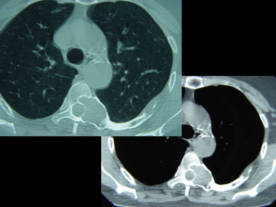
40M painful knee 4

31
Cor T1

32
Sag PD B thalassemia intermedia *

33
B thalassemia Major-Homozygous-Cooley anaemia lack of B
Italian and Greek Skull, hair on end, widened diploic Porosis, thin cortex, Erlenmyer flask Arthropathy

34
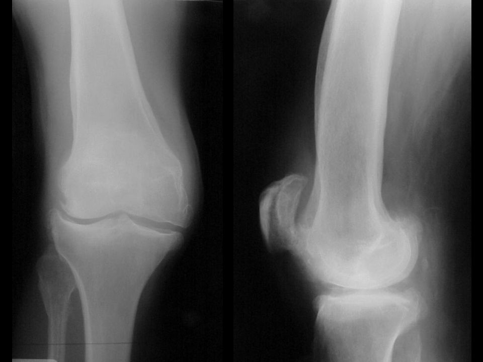
23M knee pain 1

35
Distal MCL injury *

36
Distal MCL injury Ossification points to joint
MHE points away from joint

38
Sag T1 Sag PDFS

39
Sag T1 Sag PDFS

43
C5

44
50M heel pain 2

45
Sag T1

46
Sag PDFS *

47
Involuting lipoma Any age, equal sex
Calcaneus>Femur>Tibia>Fibula Can have central dystrophic calcification

48
13F painful foot No trauma

49
Sag T1

50
Sag PDFS *

52
57M L hip pain 3

55
Sandwich vertebrae Osteopetrosis *

56
Osteopetrosis Marble bone, Defective osteoclasts
AR, Infantile, Systemic, Leukaemia AD, Adult, Fxs, anaemia, CN palsy Sclerotic, peri and endosteal Erlenmyer flask Bone in bone, sandwich vertebrae Calvaria and mandible spared

57
Variable patients Same condition

63
*

64
Pseudohypoparathyroidism
X-linked, renal and skeletal resistance to PTH Short, retarded, Decreased Ca, normal/increased PTH Brachydactyly 1,4,5 MC Ca basal ganglia, skin, SubQ

65
Variable patients Same condition
8

69
Dermatomyositis Damaged chondroitin sulfate
Atrophy, oedema, necrosis of muscle 30-60, F>M Calcification extremities and girdles Pointing of tufts Ass. Malignancy, lung, kidney, ovary, breast

70
35M with recent trauma Flex / Ext

71
C5

72
C6 Fx subluxation C5 C6 *

73
Signs of instability Spinous process fanning Widening of disk space
Horizontal displacement >3.5mm Angulation >11 degrees Disruption of facets Multiple fractures at one level

75
*

76
*

77
*

78
*

79
*

80
*

81
10M painful neck

83
* Transient disc calcification of children

84
Transient disc calcification of children
Painful Pain lasts weeks Calcification lasts months Adjacent vertebrae flattened

85
28F pain in lumbar region

86
* Anterior limbus with instability

87
Limbus Vertebrae More often anterior and superior
More significant posterior Disc herniates through ring apophysis Commonest lower lumbar

88
37F acute torticollis, stiffness and shortness of breath

89
Fibrodysplasia ossificans progressiva
*

90
Fibrodysplasia ossificans progressiva
MOP / Stone man Rare, AD, sporadic Presents in childhood Stiffness, Heterotopic ossification Malformed fingers and toes Bone morphogenic protein (BMP) signaling pathway problem
 signaling pathway problem.")
91
48M joint pains and dark pigmentation on ears

92
Alkaptonuria *

93
Alkaptonuria / Ochronosis
Absence of homogentisic acid oxidase Pigmentation Arthropathy Osteoporotic with dense disc calcification Larger joints show DJD

94
6mM Calcareous nodules

95
Idiopathic calcinosis
universalis *

96
Idiopathic calcinosis universalis
Rare, unknown cause Infants - subcutaneous Children - spreads to muscles Calcium phosphate and carbonate Serum calcium and phosphorous normal DDx - DMS, HPT, Calcium gluconate

97
28BF mass in shoulder

98
Idiopathic Tumoral Calcinosis
*

99
Idiopathic Tumoral Calcinosis
Periarticular calcified masses Shoulder, hip, elbow B>W, M=F Recur if resected Elevated phosphate, normal calcium Renal tubular phosphate resorption

101
*

102
Ax PDFS Ax T1 *

103
Cor T2 Cor T1 *

104
*

105
*

106
Sag T1 *

108
81M shoulder pain

109
* Chondrocalcinosis

110
Chondrocalcinosis CPPD Hyperparathyroidism Hemochromatosis Acromegaly
Gout Wilsons disease

111
45F swelling of arm with numbness of 2nd and 3rd fingers

112
* Ax PDFS Ax T1 Fibrolipomatous hamartoma of the median nerve
with macrodystrophia lipomatosis *

113
Fibrolipomatous hamartoma of the median nerve with macrodystrophia lipomatosis
Nerve territory directed macrodactyly Localised form of gigantism Median or Plantar nerves Possible relation with neurofibromatosis

114
35M 8w post injury

115
Cor T2 Cor T1 Post traumatic myositis ossificans *

116
Post traumatic myositsis ossificans
4/52 Faint peripheral Ca Periosteal reaction 8/52 Circumscribed cortex Central lacy pattern 5/12 Maturity >6/12 Regression Separate from bone 1 year Usually disappears Periosteal reaction remains

117
25M Blocker

118
* Myositis ossificans

119
Sport related myositis ossificans
Single direct blow Repeated minor trauma Adductor longus-Rider’s bone Brachialis-Fencer’s bone Soleus-Dancer’s bone Blocker’s arm

121
48M Alcohol ++

122
*

123
Grading of AVN Steinberg modification of Arlet/Ficat
0 - Abnormal MRI no symptoms 1 - Abnormal MRI, pain 2 - Mixed sclerosis and lucency on x-ray 3 - Subchondral collapse 4 - Marked collapse 5 - Secondary acetabular OA

124
Causes of AVN Trauma Steroids Alcohol Pancreatitis Protease inhibitors
Gauchers Sickle cell Caisson Perthes / Idiopathic

125
34M Developing lump anterior to knee

127
Ax PDFS Ax T1

128
* Sag T1

129
Soft tissue chondroma Rare 20-40Y Hands and feet
Well demarcated and lobulated Curvilinear, ringlike or nodular calcification High signal T2

131
PDFS *

133
*

134
*

135
*

136
*

137
35M Knee injury

138
PDFS *

139
ACL/MCL Empty lateral gutter

140
86M Stiffness and Locking

142
Multiple bodies in Popliteal recess
Primary V’s secondary osteochondromatosis Multiple similar size Origin

143
62M Fullness in suprapatella region

144
*

145
Body growing in joint Laminated Slow growing

146
30M Outdoors man

147
*

148
Snake bite Venom not infection Due to proteases

149
36M Prior trauma

150
*

151
Florid reactive periostitis
BPOP Bizarre parosteal osteochondromatous proliferation Manifestation of PTMO in hands Periosteal proliferation > ST ossification

152
50M Trauma Whiplash injury
1

153
*

154
Extension tear drop Fx Small fragment Usually more superior Cx spine

157
* 1

158
*

159
2W earlier 2W later

160
*

161
*

163
36M Trauma 3Y ago MVA Now myelopathy

166
*

167
Chronic non-union of C2 Fx
Type 1 steep oblique Due to alar ligament Type 2 neck of odontoid process Prone to non-union Type 3 extends into body Often heal with conservative Rx

168
42M Fall 1

169
* 1

170
Anterior shoulder dislocation
Hill Sach’s lesion (Hatchett ) Stryker view Bony or soft tissue Bankart Westpoint view Posterior dislocation Trough Fx Bennet’s lesion Bony
 Stryker view. Bony or soft tissue Bankart. Westpoint view. Posterior dislocation. Trough Fx. Bennet’s lesion. Bony.")
171
21M Injury weeks ago Recent surgery

172
*

173
Volkmann’s ischaemic contracture
Soft tissue contractures Volkmann’s Burns Neurologic conditions RhA, SLE Arthrogryposis multiplex congenita

174
40M Known medical condition Recent trauma
2

175
2W earlier 2W later

176
* 2W later

177
Hemophilic pseudotumor
Uncommon manifestation of Hemophilia Femur > Pelvis > Tibia > Small bones of hands and feet Intraosseous or subperiosteal Lytic, expansile, can look aggressive, ST mass

178
75F Lifelong limp 1

179
*

180
DDH Adults

181
DDH Infants Acetabular angle Lateral shift Superior shift Shenton’s
Perkin’s Hilgenreiner’s Center Edge

182
40M Waterskier 1

183
*

185
Old ischial avulsion Avulse bone < 25Y
Waterskier, Hurdler, Sprinter

188
*

190
*

194
54F Fall 2

196
*

197
Transverse Patella FX Direct or indirect Transverse 70%, indirect
Longitudinal, stellate or comminuted Bipartite - superolateral Dorsal defect - superolateral Direct

198
63F Longstanding decrease ROM
1

199
*

200
Chronic anterior shoulder dislocation, with neoglenoid
Failure to diagnose May have increased ROM

201
19F Slowly growing (1Y) lump on thigh
3

204
*

205
Alveolar Soft Part Sarcoma
Malignant granular cell myoblastoma Young adult females Thigh muscles Slow growth, calcifcation, invade bone Metastasizes late Vascular, may have flow voids Path - similar to paraganglioma

206
19F MVA 1

207
*

208
Odontoid Fx Type 1 - Steep oblique Type 2 - Neck
Sometimes tip Fx also called type 1 Type 2 - Neck Prone to non-union Type 3 - Involves body Usually heal conservatively

209
65M Neck pain Myelopathy 3

212
* *

213
CPPD arthropathy Deposited in transverse ligament Associated
Tumor like masses may compress cord Atlanto axial subluxation Spontaneous odontoid Fx

214
35M Deformity

216
Maffucci syndrome Multiple enchondromas ST Hemangiomas
Malignant potential close to 100% Olliers enchondromatosis 25-30% Developmental, not hereditary Growth deformities

225
12M Deformity

227
Noonans Short Metacarpal Idiopathic Post trauma
Iatrogenic, Fx, Growth plate inj, Thermal, Electrical Turners, 4th +/- 3rd or 5th Pseudo- and pseudopseudohypoparathyroidism 4th and 5th

228
14M Deformity

230
Carpal osteolysis Onset childhood Carpals, Tarsals, elbows
Associated nephropathy

231
29F FOOSH

233
Scaphoid Fx on lateral view
Many scaphoid fractures are best seen on lateral

234
68M Wrist instability

236
VISI Suggests lunotriquetral ligament tear
DISI- scapholunate ligament tear Angle between scaphoid and lunate < 30 Pie shaped lunate

237
33F No history of trauma

239
Keinbocks Ulna minus Trauma Osteonecrosis

240
45F Hand pain

242
Acroosteolysis Tuft CVD- Scleroderma, CREST, Raynauds Psoriasis
Neuropathic DM, Leprosy, Myelomeningocele, Syrinx, Cong indifference to pain (Leesch Nyan) Trauma Thermal, Burns, frostbite electrical Hyperparathyroidism Epidermolysis bullosa Porphyria, Subungal exostosis, Snake and scorpion venom Phenytoin toxicity in infants
 Trauma. Thermal, Burns, frostbite electrical. Hyperparathyroidism. Epidermolysis bullosa. Porphyria, Subungal exostosis, Snake and scorpion venom. Phenytoin toxicity in infants.")
243
40M Knife injury

245
Flexor tendon laceration Displaced sesamoid
Sesamoid useful marker of tendon

249
*

250
*

251
*

252
*

254
20M Hurt hand catching ball

256
Dislocations Need 2 views for trauma

257
22F Deformity

259
Boutonnierre Rupture of middle slip of extensor tendon as it passes over PIPJ Lateral slips migrate volarly Occasionally avulsion Needs early Dx

260
76F RhA

261
*

262
Cranial Settling Atlantoaxial settling Erosion of lateral masses
Different from basilar invagination

263
57M Bilateral shoulder pain

264
*

265
DDx Unilateral Bilateral Amyloid, TB Crystal Occupational OA
Syrinx - neuropathic Previous inflammatory arthritis Clavicles normal Hemophilia

266
24M FOOSH

267
*

268
Trans scaphoid/triquetrum perilunate Fx dislocation
Pie shaped lunate

269
31M Blow to flexed thumb

270
*

271
Rolando Fx Axial blow More difficult to anatomically reduce

273
March 01

276
*

277
*

278
*

280
57M Right hip pain

281
March 01

282
May 01

283
Dec 02 *

284
Hepatic metastases Rare to bone Similar to other hypervascular mets

285
50M MVA

288
Anterior hip dislocation
5% of hip dislocations Can have associated impaction injuries Leg externally rotated

289
32M Bilateral chronic hip pain

291
*

292
Perthes White boys 4-7y Younger onset – better outcome
DDx for bilateral MED, Morquios, SCD, Gauchers, Hypothyroid, CDP, Warfarin embryopathy

293
17F Mechanical symptoms

295
*

296
Osteochondroma Point away from joint Cartilage cap is hyaline
Cap thickness > 1cm concerning Pain important to dx malignancy

297
75M Knee locking

299
*

300
Primary synovial osteochondromatosis
Metaplasia of synovium May not be visible on X-ray Primary similar size Synovial hemangiomas have lucent centers

301
55M 1Y post trauma

302
*

303
Dystrophic calcification in Quadriceps tear
Metastatic Dystrophic Tumoral

309
Left Right *

312
46M Previous trauma Chronic bowel problems

315
*

316
Hypertrophic Osteoarthropathy
Pulmonary CA bronchus, Lymphoma, Abscess, Bronchiectasis, Metastases Pleural LFTP (highest association), Mesothelioma Cardiovascular CCHD GI UC, Crohns, Dysentry, Lymphoma, Whipples, Coeliac, Cirrhosis, Nasopharnygeal CA, Juvenile polyposis
, Mesothelioma. Cardiovascular. CCHD. GI. UC, Crohns, Dysentry, Lymphoma, Whipples, Coeliac, Cirrhosis, Nasopharnygeal CA, Juvenile polyposis.")
317
16M Slowly increasing pain in tibia

320
T1FSGd *

321
Osteosarcoma Conventional Telangiectatic Parosteal Periosteal
Multicentric Soft tissue

322
14M Pain with running

324
1m later *

325
Stress Fracture Fatigue Insufficiency Pathologic

326
54M Twisting injury

329
*

330
Maisonneuve Fx Transverse fracture of medial malleolus without distal fibula Fx, ask for proximal fibula

331
31F Arthritis

332
Left

333
Left Right *

334
RhA noeostosis Reiters is more plantar and less symmetric

335
53M Pain lower back

337
Ankylosing Spondylitis
Enthesopathy

345
*

347
42F Foot stiffness

349
Compartment syndrome ossification
Extensive sheet like ossification Dystrophic

350
24F Fall

352
Fracture blisters DDx pseudoaneurysm

353
8M Swelling of toe

355
Digital fibroma Recurring digital fibroma of infancy Can become large
Painless Fingers and toes DDx Enchondroma, Epidermoid inclusion, Digital fibroma, Subungal lesions, Glomus

358
Melorrheostosis A benign sclerosing bone dysplasia Osteopathia striata
Osteopoikilosis Dripping candle wax Sclerotomes

359
43M Trauma

361
Osteoma (Ivory) Gardeners syndrome
Adenomatous polyps, Dental lesions, ST tumors, osteomas

362
67F Lump

365
Fibrous Dysplasia Common Hamartomatous fibro-osseous metaplasia
70% monoostotic Polyostotic tends to be unilateral Usually expansile Shepherds crook, ground glass Any bone, but spine unusual

366
33M Tackled at rugby

368
Anterior dislocation Hill sachs Bony Bankart
Stryker for Hill Sachs Westpoint for bony Bankart Can occur after one dislocation

369
56F Lump and pain

371
GCT Multinucleated giant cells in fibroid stroma
Knee, distal radius, proximal humerus Lytic, subarticular, narrow zone of transition without sclerosis Can look aggressive After epiphyseal fusion

372
42M Mechanical symptoms

374
*

375
Sessile osteochondroma
Anterior at knee Also have mechanical symptoms

Similar presentations


.>")
>")


>")

