Download presentation
Presentation is loading. Please wait.

1
Bio& 242, Human A&P 2: Unit 1/Lecture 1

2
Overview of GI tract Functions
Mouth---bite, chew, swallow Pharynx and esophagus----transport Stomach----mechanical digestion; absorption of water & alcohol; chemical digestion Small intestine--chemical digestion & absorption Large intestine----absorb electrolytes & vitamins (B and K) Rectum and anus---defecation
 Rectum and anus---defecation.")
3
Digestion includes 7 basic processes.
Ingestion: taking food into the mouth (eating). Secretion: release by cells within the walls of the GI tract and accessory organs of water, acid, buffers, and/or enzymes into the lumen of the tract. Mixing and propulsion (Peristalsis): result from the alternating contraction and relaxation of the smooth muscles within the walls of the GI tract. Mechanical digestion: consists of movements of the GI tract that aid chemical digestion. Chemical digestion: is a series of catabolic (hydrolysis) reactions that break down large carbohydrate, lipid, and protein food molecules into smaller molecules that are usable by body cells.
. Secretion: release by cells within the walls of the GI tract and accessory organs of water, acid, buffers, and/or enzymes into the lumen of the tract. Mixing and propulsion (Peristalsis): result from the alternating contraction and relaxation of the smooth muscles within the walls of the GI tract. Mechanical digestion: consists of movements of the GI tract that aid chemical digestion. Chemical digestion: is a series of catabolic (hydrolysis) reactions that break down large carbohydrate, lipid, and protein food molecules into smaller molecules that are usable by body cells.")
4
Digestion includes 7 basic processes.
Absorption: passage of end products of digestion from the GI tract into blood or lymph for distribution to cells. Defecation: emptying of the rectum, eliminating indigestible substances from the GI tract.

5
Mouth and it’s associated glands
The mouth (oral or buccal cavity): is formed by the cheeks, hard and soft palate, lips, and tongue. The vestibule: is bounded externally by the cheeks and lips and internally by the gums and teeth. The oral cavity proper: is a space that extends from the gums and teeth to the fauces, the opening between the oral cavity and the pharynx or throat.
: is formed by the cheeks, hard and soft palate, lips, and tongue. The vestibule: is bounded externally by the cheeks and lips and internally by the gums and teeth. The oral cavity proper: is a space that extends from the gums and teeth to the fauces, the opening between the oral cavity and the pharynx or throat.")
6
Mouth and it’s associated glands
Lips and cheeks: contains buccinator muscle that keeps food between upper & lower teeth Vestibule: area between lips and teeth Oral cavity proper: roof = hard, soft palate and uvula floor = the tongue Sides = teeth and gums Anterior = teeth and gums

7
Mouth and it’s associated glands
Mouth: Ingestion Teeth: Mastication = Mechanical digestion Salivary Glands: Parotid; Stenson's duct Submandibular; Wharton's duct Sublingual; Rivinus' duct Mumps is an inflammation and enlargement of the parotid salivary glands caused by infection with the mumps virus (myxovirus). Salivation is entirely under autonomic nervous control via the parasympathetic
. Salivation is entirely under autonomic nervous control via the parasympathetic.")
8
Mouth continued Salivary Glands:
Water: Moistens Food and dissolves food for tasting Mucus: Lubricates and binds food into bolus Amylase: Starts break- down of Starch and Glycogen Bicarbonate (HCO3): Buffering action neutralizes acidic food in the mouth Enzymes: (lysozymes) helps destroy bacteria in the mouth
: Buffering action neutralizes acidic food in the mouth. Enzymes: (lysozymes) helps destroy bacteria in the mouth.")
9
Structure and Function of the Tongue
Tongue: forms the floor of the oral cavity. It is composed of skeletal muscle covered with mucous membrane. Extrinsic and intrinsic muscles: permit the tongue to be moved to participate in food manipulation for chewing and swallowing and in speech. Extrinsic muscles: move the tongue, learned in the muscle unit. Intrinsic Muscles: change the shape of the tongue, some people have a gene mutation the limits the use of these muscles Lingual frenulum: fold of mucous membrane that attaches to the midline of the undersurface of the tongue. Children born with to pronounced frenulum are tongue tied Papillae: cover upper surface and sides of the tongue. lingual lipase: Enzyme produced by glands on the dorsum of the tongue. initiates digestion of triglycerides.

10
Structure and Function of the Tongue
Muscles of tongue are attached to hyoid, mandible, hard palate and styloid process Papillae: elevations that create a rough surface for manipulating food and providing sensations Filiform: temperature, touch, and pressure sensations Fungiform: taste buds Foliate: taste buds Circumvallate: taste buds

11
Structure and Function of the Tongue

12
Structure and Function of the Teeth
Teeth: project into the mouth and are adapted for mechanical digestion Tooth principal portions: crown, root, and neck. Tooth composition: dentin, a calcified connective tissue that gives the tooth its basic shape and rigidity enamel, covers dentin and protects the tooth from the wear of chewing. cementum, covers dentin of the root and is a bone-like substance, periodontal ligaments, attaches root to the alveolar process pulp cavity: space in the crown and the root canals in the root.

13
Composition of Teeth Enamel hardest substance in body
calcium phosphate or carbonate Dentin calcified connective tissue Cementum bone-like periodontal ligament penetrates it

14
Dentition There are two sets of teeth or dentition:
deciduous (primary), milk teeth, or baby teeth permanent (secondary) Adult or permanent teeth Primary or baby teeth 20 teeth that start erupting at 6 months 1 new pair of teeth per month Consisting of incisors, canines, and molars Permanent teeth 32 teeth that erupt between 6 and 12 years of age replacing primary teeth Consisting of incisors, canines, premolars and molars
, milk teeth, or baby teeth permanent (secondary) Adult or permanent teeth. Primary or baby teeth. 20 teeth that start erupting at 6 months. 1 new pair of teeth per month. Consisting of incisors, canines, and molars. Permanent teeth. 32 teeth that erupt between 6 and 12 years of age replacing primary teeth. Consisting of incisors, canines, premolars and molars.")
15
Dentition Differing structures of teeth indicate function
Incisors: biting teeth 4 upper and 4 lower Canines or “cuspids”: tearing teeth 2 upper and 2 lower Premolars or “bicuspids”: crushing and grinding teeth lacking in primary teeth Molars or “tricuspids”: crushing and grinding teeth 3 upper and 3 lower 3rd molars are “wisdom” teeth

16
PHARYNX Pharynx: funnel-shaped tube that extends from the internal nares to the esophagus posteriorly and the larynx anteriorly. Moves bolus of food It is composed of skeletal muscle and lined by mucous membrane. Divide into three regions Nasopharynx: functions in respiration only, oropharynx: Functions in both respiration and digestion. Located between the naso and laryngopharynx and posterior to oral cavity. laryngopharynx Functions in both respiration and digestion. Located inferior to oropharynx. Two Arches Platatoglossal: anterior most arch Platatopharyngeal: posterior most arch Platatine tonsils sit between these arches.

18
Deglutition or swallowing
starts when bolus is pushed into the oropharynx sensory nerves send signals to deglutition center in brainstem soft palate is lifted to close nasopharynx larynx is lifted as epiglottis is bent to cover glottis

19
Stages of Deglutition Voluntary Stage: a. Buccal or Oral activity
formation and movement of the bolus

20
Stages of Deglutition Pharyngeal Stage:
involuntary Soft-palate is pulled upward closing off the nasopharynx Palatoglossal and palatopharyngeal arches are pulled medially forming a sagittal slit with the fauces.

21
Stages of Deglutition Pharyngeal Stage: Vocal cords close
Epiglottis swings backward over larynx and larynx is pulled upward to close off the opening of the larynx Upper esophageal sphincter relaxes to that bolus can enter the esophagus.

22
Stages of Deglutition Esophageal Stage: involuntary
Esophagus: Moves bolus from pharynx to the stomach Peristalsis pushes the bolus downward through the esophagus. Lower esophageal sphincter relaxes and the bolus enters the stomach

23
GI Tract Structures: Stomach

24
GI Tract Functions: Stomach
Stomach: Stores, mixes, and dissolves food, converts food into chyme, regulates emptying of chyme into small intestines

25
GI Tract Functions: Stomach
Stomach continued: Mucus: lubricates and protects the stomach mucosa Hydrochloric acid: dissolves food particles and kills most microorganisms Pepsinogen: inactive form of the enzyme pepsin. It is activated by HCl. Splits apart peptide bonds to begin the digestion of proteins

26
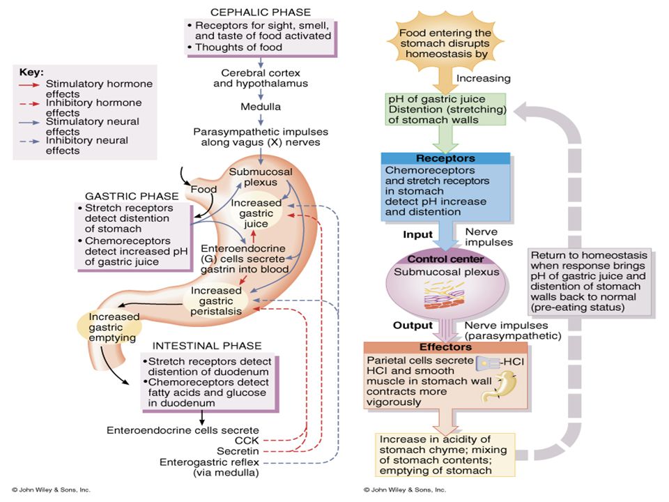
GI Tract Functions: Stomach
Stomach continued: 4. intrinsic factor: required for the absorption of vitamin B12 5. Gastrin: hormone that controls HCl release along with other activities to be discussed later

27
GI tract functions: Small Intestine
Segmentation: mixes chyme with digestive juices (from small intestine, pancreas, and liver) and brings chyme into contact with the mucosa for absorption, peristalsis propels chyme toward the large intestine.
 and brings chyme into contact with the mucosa for absorption, peristalsis propels chyme toward the large intestine.")
28
GI tract functions: Small Intestine

29
GI tract functions: Small Intestine

30
GI tract functions: Small Intestine

31
GI tract functions: Small Intestine
Duodenum: shortest region, about 25cm. Continues the digestion of carbohydrates, proteins, and lipids. Begins the digestion of nucleic acids. Receives the digestive fluids from the pancreas and liver via the hepatopancreatic ampulla.

32
GI Tract Functions : Small Intestine
Jejunum: Middle region of the small intestine, about 1 m (3 to 4 ft) (8 ft in a cadaver due to smooth muscle relaxation) Completes the digestion of carbohydrates, proteins, lipids, and nucleic acids Ileum: final and longest region of the small intestine, about 2m (6 ft). (12 feet in a cadaver) Involved in absorption of about 90% of the nutrients produced by digestion.
 (8 ft in a cadaver due to smooth muscle relaxation) Completes the digestion of carbohydrates, proteins, lipids, and nucleic acids. Ileum: final and longest region of the small intestine, about 2m (6 ft). (12 feet in a cadaver) Involved in absorption of about 90% of the nutrients produced by digestion.")
33
Functions of the Small Intestinal Mucosa
Mucus: lubricates chyme and protects mucosa Assorted enzymes: a. pancreatic enzymes b. brush-border enzymes (details later) Secretin: stimulates pancreatic bicarbonate secretion. Cholecystokinin: stimulates gallbladder contraction, pancreatic enzyme secretion, and stomach emptying
 Secretin: stimulates pancreatic bicarbonate. secretion. Cholecystokinin: stimulates gallbladder contraction, pancreatic. enzyme secretion, and. stomach emptying.")
34
Functions of the Small Intestinal Mucosa
4. Gastric-inhibitory peptide: inhibits stomach acid secretion and motility

36
Overview of fluid intake and secretion compared to fluid absorption by the digestive tract

Similar presentations


>")


