Download presentation
Presentation is loading. Please wait.

1
Nerve & Muscle Physiology
Jeff Ericksen, MD VCU Health Systems PM&R

2
Topics * Relevant anatomy Cell functions for signal transmission
Transport, resting potential, action potential generation & propagation Neuromuscular transmission Muscle transduction Volume Conductor theory Relevant anatomy for nerve conduction and muscle contraction will be covered. We will look at how the cell membrane functions as a signal generator and conductor. Conversion or transduction of the peripheral nerve signal into a mechanical event will also be discussed as well as how these events are recorded in the electrodiagnosis laboratory.

3
Acknowledgements Electrodiagnostic Medicine by Daniel Dumitru, MD
Chapter 1: Nerve and Muscle Anatomy and Physiology Superb text covering all aspects of EMG/NCS

5
Cell membrane Necessary for life as we know it Border role for cell
Separates intracellular from extracellular milleau Allows ion and protein concentration gradients to exist Creates electric charge gradients

6
Cell membrane Provides structure for cell
Modulates cell interaction with environment Mechanical, hormone-receptor Controls material flow into/out of cell Nutrition/waste management

7
3 Key Membrane Components
Lipids 45-49% phospholipids, cholesterol & glycolipids = amphipathic molecules Polar = hydrophilic vs. nonpolar = hydrophobic Proteins 45-49% Carbohydrates 2-10%

9
Lipid characteristics
Membrane phospholipids have polar head group with 2 nonpolar tails In water - nonpolar tail groups form an inside excluding water 2 arrangements possible Micelle = tails inside, heads face out Bilayer

11
Lipid bilayer or fluid mosaic model
Phospholipid sheet with tails aligned in center, heads facing out for a head-tail-head sandwich No H2O at center, 75 Angstroms Model as 2-D liquid with 2 degrees of freedom of motion for lipid Long axis rotation Lateral diffusion Center of the bilayer is free of water Width is about 75 angstroms 3rd degree of mobility is for lipid to flip flop from one side to another in bilayer Cholesterol molecules are woven into bilayer, possibly for stability and reduces permeability to small molecules. Glycolipids exist on extacellular outer surface only, bound to carbohydrates, function unknown.

15
Proteins in membrane provide cell functions
2 membrane protein types Transmembrane = integral - across whole layer, amphipathic Hydrophobic midportion acts with lipid layer tails Hydrophilic section faces intra/extra environment Peripheral proteins - inside or outside of bilayer

16
Proteins

18
Membrane transport Lipid soluble molecules cross readily but large water soluble molecules need transport across bilayer Transport proteins - specific for ion or molecule to cross Channel proteins - span bilayer, large center, allow ion/molecule passage based on size Carrier proteins - binding with specific material, conformational change then crossing membrane

19
Membrane transport Diffusion Active transport
Driven by kinetic energy of random motion Thru lipids or proteins Follows concentration gradient Active transport Needs energy source Fights concentration or energy gradient

20
Simple vs. Facilitated diffusion
Crosses membrane bilayer or channel without binding Increases with kinetic energy + lipid solubility + concentration gradient Protein channels specific for ions, often gated by cell functions Facilitated Transmemb proteins Needs protein binding, conformational change Speed of transport limited by conformational change Simple diffusion speed is dependent on kinetic energy and strength of concentration or charge gradient. Protein channel control with ligand or voltage gating. Ligand gating = binding molecule induces conformational change to allow transport. Voltage gating = specific voltage change induces conformational change.

21
Membrane transport Carrier proteins Energy Diffusion Active transport
Channel protein Energy Simple diffusion Facilitated diffusion Diffusion Active transport

22
Active transport Acting on semi-permeable membrane allows the cell to maintain a high intracellular concentration vs. extracellular fluid Requires active process as diffusion would eventually equilibrate concentrations across membrane

23
Active transport Transmembrane carrier protein uses ATP energy to pump ions against concentration gradient to develop transmembrane resting potential

24
Resting membrane potential
Excitable cells can generate and conduct action potentials over distances Intracellular space carries potential difference of mV, inside with negative charge excess relative to outside

25
Resting membrane potential created by semi-permeable membrane and ions
Extracellular 440 20 560 Intracellular Na 50 K 400 Cl 52

27
Nernst used thermodynamics in 1888 to determine work done by membrane
Work to move ion against concentration gradient is opposite to work to move against electrochemical gradient Can calculate contributions from different ions K = -75 mV, Na = +55 mV

28
Nomenclature Polarized membrane: Intracellular potential is negative relative to extracellular space Depolarization = less polarization of the membrane -80mV -> +20mV Hyperpolarization = more polarization of membrane -80mV -> -100mV

29
Na influx with K efflux Na driven by negative charge excess inside + concentration gradient K driven by concentration gradient If continued, would lose resting potential

30
Na - K ATP dependent pump
Plasma membrane structure uses active transport 2 K in for 3 Na out actively Thus 3 Na must diffuse in for 2 K out

32
Membrane potential from Goldman-Hodgkin-Katz equation
Resting potential mostly from K contributions If sudden Na permeability change, potential approaches Nernst Na potential rapidly Action potential!

33
Voltage dependent ion channels
Ion flow across through membrane channels is initiated by membrane potential changes If potential exceeds a threshold, rapid increase in Na permeability followed by later K permeability increase

34
Voltage dependent ion channels
Extracellular Na activation gate with intracellular inactivation gate and slow K activation gait Conformational changes due to membrane potential changes influence ion permeability

35
Voltage gated channels

36
Channels and voltage influence
If resting potential depolarized by mV, then activation gate opened with 5000x increase in Na permeability followed by inactivation gate closure 1 msec later Slow K activation gate opens when Na inactivation gate closes to restore charge distribution, slight hyperpolarization

39
Refractory periods Absolute = state when activation gait cannot be reopened with a strong depolarization current, the membrane potential is relatively more positive Relative = state when activation gait can be reopened by strong depolarizing current as membrane potential returns to equilibrium state

40
Action potential timing

43
Action potential propagation
Na + charge influx spreads longtiduinally down path of least resistance to induce depolarization in adjacent membrane, some transmembrane spread As + charge builds up, attracts intracellular - charges and they are neutralized by new ICF + charges

44
AP propagation Less electrochemical hold of ECF + charges which migrate and allow depolarization of membrane further Process is repeated down axon until end is reached AP is identical to AP from upstream nerve area, all or none event

47
Nerve membrane modeling
Capacitor = charge storage device, separate poles separated by a nonconducting material or dielectric Hydrophobic center to lipid bilayer is good dielectric, allows membrane to function well as a capacitor

48
Nerve membrane modeling
Resistor = direct path to current flow but with some impedance Nerve axon has both transmembrane resistance as well as longitudinal resistance

49
Current spread Membrane capacitor model suggests transmembrane resistance is high, hence current flows more longitudinally vs. transmembrane capacitance flow or ionic channel resistance flow

50
Slow process Longitudinal AP spread requires sequential depol. to threshold, membrane capacitor discharge and then alteration of proteins to turn on Na activation channels. This process can be slow. Hence unmyelinated nerve conducts slowly = m/sec.

51
Need velocity to interact with environment!
longitudinal resistance will speed diameter will resistance Eliminate need to fire all surrounding tissue will velocity of conduction Insulate nerve to prevent leakage, spread out the gated Na channels Myelin & Nodes of Ranvier

52
Myelin All peripheral nerve axons surrounded by plasma membrane of a Schwann cell Single layer of membrane = unmyelinated nerve, multiple layers = myelinated nerve Gap between Schwann cell covers = node of Ranvier

53
Myelinated axons Outer myelin sheath + axon plasma membrane = axolemma covering axoplasm Schwann cell membrane has lipid sphingomyelin, highly insulating No Na channels under myelin, only at nodes. K channels under myelin in perinodal area

56
Current conduction with myelin insulation
AP at node, Na charge influx and current spreads longitudinally down axon Minimal leak between nodes, reduced by 5000 vs. unmyelinated nerve Charge separation, reduced protein leak channels & increased membrane resistance account for this

57
Current conduction Circuit is closed by efflux of ionic current at node Na ions accumulate beneath node, reduces electrochemical pull on ECF Na above node, they migrate back to upstream node to close loop Above tends to increase + charge inside membrane or depolarize to give AP

58
AP generation at node Nodes contain high # Na channels which open with depolarization Na influx starts process again

60
Myelin effects Conduction velocity increases
Current and action potential jumps from node to node = saltatory conduction Optimal internodal length is 100x axon diameter Optimal myelin/axon ratio is 60/40

62
Neuromuscular junction, transducing the electrical signal to mechanical force

63
Multiple branches from large motor axons

64
What happens if varying myelin and diameter in branches?

65
NMJ anatomy Presynaptic Postsynaptic Terminal axon sprout
Mitochodria Synaptic vesicles = ACH Presynaptic membrane Postsynaptic Motor endplate Single muscle fiber Mitochondria Ribosomes Pinocytotic vesicles Postsynaptic membrane ACH receptors

68
NMJ Electrochemical conduction
Considerable slowing in smaller diam less myelinated branches AP depolarizes terminal axon, Na conductance increases Calcium conductance also dramatically increased Influx Ca++ in terminal axon Possibly facilitates fusion of ACH vesicles with presynaptic membrane

69
Electrochemical conduction….
Vesicular fusion with presynaptic membrane Open to synaptic cleft, release quantum of ACH 100 vesicles per AP in mammals, 10k ACH per vesicle Ca++ stays in terminal axon 200 ms, keeps axon readily excitable for repeat stimulation

70
ACH release Rapid diffusion across cleft in .5 msec timing, bind receptors Large transmembrane proteins with ACH site and ion channel Ligand activated vs. voltage activated ACH binding induces conformational change in ion channel 1 ms opening of cation specific channel = Na, K, Ca, repels anions with charge

71
Postsynaptic ion channel opening with ACH binding
Predominant influx is Na, K blocked by electrochem gradient, Ca concentration gradient not that large Na influx locally depolarizes muscle membrane= endplate potential reversal which is not propagated = EPP Single packet of ACH from vesicle gives MEPP

72
Muscle action potential
Generated if sufficient ACH released to cause postsynaptic membrane to reach threshold, muscle membrane depolarized and propagated impulse follows Muscle AP travels along muscle membrane = sarcolemma Similar to nerve, increased Na permeability in + feedback loop

73
T-tubules Small volume favors K accumulation during repolarization after AP, tends to make membrane easy to depolarize again Penetrate into muscle to spread AP into fiber High surface area of T-tubules increases capacitance qualities and slows conduction in muscle

74
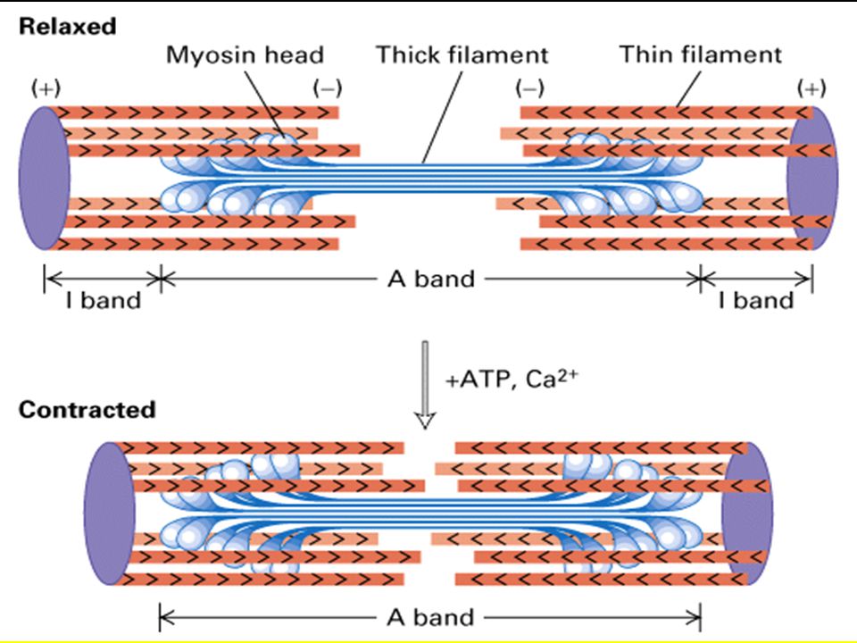
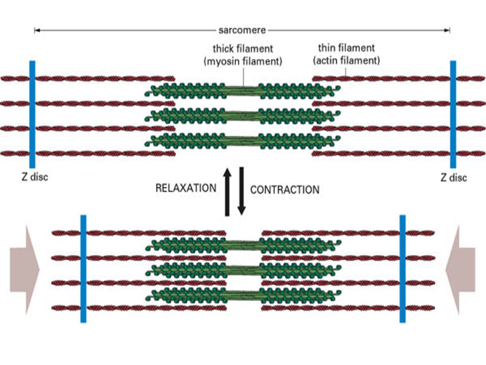
Excitation-Contraction
AP in T-tubule induces Ca++ release in SR terminal cisternae, exposure for 1/30 sec, then reuptake via pump Ca++ bind to troponin C, induces conformational change of troponin complex and influences tropomyosin to actin relationship - mechanical force

80
The End!

Similar presentations


.>")


 Nervous system functions Structure of a neuron Sensory, motor, inter- neurons Membrane potential Sodium.>")


