Download presentation
Presentation is loading. Please wait.

1
Tuberculosis

2
A Small Disclaimer… This presentation will NOT teach you everything there is to know about tuberculosis

3
Image credit: National Library of Medicine
History of TB (1) TB has affected humans for millennia Historically known by a variety of names, including: Consumption Wasting disease White plague TB was a death sentence for many Vintage image circa 1919 Image credit: National Library of Medicine 3 3
 TB has affected humans for millennia. Historically known by a variety of names, including: Consumption. Wasting disease. White plague. TB was a death sentence for many. Vintage image circa Image credit: National Library of Medicine")
4
History of TB (2) Scientific Discoveries in 1800s
Until mid-1800s, many believed TB was hereditary 1865 Jean Antoine-Villemin proved TB was contagious 1882 Robert Koch discovered M. tuberculosis, the bacterium that causes TB Mycobacterium tuberculosis Image credit: Janice Haney Carr 4 4

5
History of TB (3) Sanatoriums
Before TB antibiotics, many patients were sent to sanatoriums Patients followed a regimen of bed rest, open air, and sunshine TB patients who could not afford sanatoriums often died at home Sanatorium patients resting outside 5 5

6
Breakthrough in the Fight Against TB (1)
Drugs that could kill TB bacteria were discovered in 1940s and 1950s Streptomycin (SM) discovered in 1943 Isoniazid (INH) and p-aminosalicylic acid (PAS) discovered between 1943 and 1952 6 6
 discovered in Isoniazid (INH) and. p-aminosalicylic acid (PAS) discovered between 1943 and")
7
most common and important agent of human disease is M. tuberculosis.
M. bovis (characteristically resistant to pyrazinamide, once an important cause, currently the cause of a small percentage of cases worldwide) M. microti : less virulent and rarely encountered M. pinnipedii (seals and sea lions in the southern hemisphere and recently isolated from humans ) M. canettii (a rare isolate from East African cases that produces unusual smooth colonies on solid media)
 M. microti : less virulent and rarely encountered. M. pinnipedii (seals and sea lions in the southern hemisphere and recently isolated from humans ) M. canettii (a rare isolate from East African cases that produces unusual smooth colonies on solid media)")
8
neutral on Gram's staining.
M. tuberculosis is a rod-shaped, non-spore-forming, thin aerobic bacterium measuring 0.5μm by 3 μm neutral on Gram's staining. once stained, the bacilli cannot be decolorized by acid alcohol due mainly to the organism’s high content of mycolic acids, long-chain cross-linked fatty acids, other cell-wall lipids

9
cell wall, lipids are linked to underlying arabinogalactan and peptidoglycan. This structure confers very low permeability of the cell wall, thus reducing the effectiveness of most antibiotics cell wall, lipoarabinomannan, is involved in the pathogen-host interaction and facilitates the survival of M. tuberculosis within macrophages

10
Microorganisms other than mycobacteria that display some acid fastness include species of Nocardia and Rhodococcus, Legionella micdadei, and the protozoa Isospora and Cryptosporidium

12
TB Transmission (3) TB is spread person to person through the air via droplet nuclei M. tuberculosis may be expelled when an infectious person: Coughs Sneezes Speaks Sings Transmission occurs when another person inhales droplet nuclei 12 12

13
Dots in air represent droplet nuclei containing
TB Transmission (4) Dots in air represent droplet nuclei containing M. tuberculosis 13
 Dots in air represent droplet nuclei containing. M. tuberculosis. 13.")
14
SOURCE CASE Only patients with pulmonary tuberculosis can be regarded as infectious. (placenta, skin, milk) Smear positive Cavitary pulmonary disease Endobronchial or laryngeal tuberculosis Coughing(wearing mask with patient) Use of chemotherapy (Isoniazide) persons with both HIV infection and tuberculosis are less likely to have cavitations, they may be less infectious than persons without HIV co-infection.
 Use of chemotherapy (Isoniazide) persons with both HIV infection and tuberculosis are less likely to have cavitations, they may be less infectious than persons without HIV co-infection.")
15
A single bacillus in a tiny droplet nucleus is more hazardous than several bacilli in larger airborne particles Coughing is the most effective mechanism for generating aerosols that create droplet nuclei. Thin, watery secretions are more easily fragmented into small respirable droplets than is more viscous mucus.

16
Masks worn by persons exposed to an infectious source are less effective than are masks worn by patients, because most airborne droplet nuclei are much smaller than their parent droplets.

17
prevalence of PPD + among young contacts of patients with newly discovered tuberculosis increased as the radiographic extent of involvement increased contacts of patients who have organisms + in sputum smears have a much higher prevalence of infection patients who had positive sputum smears but who were receiving antituberculosis drugs were much less infectious for guinea pigs than were untreated patients.

18
under standard conditions of temperature and humidity indoors : survived
60% to 71% for 3 hours, 48% to 56% for 6 hours, 28% to 32% for 9 hours. exposure to ultraviolet radiation kills tubercle bacilli

19
close contacts (generally household) 30% are infected, rate of tuberculosis is in the range of 15 per 1000 nonclose contacts (generally out-of-household) 15% are infected, 3 per 1000 nonclose contacts develop tuberculosis Because the risk of tuberculosis is higher among close contacts, they should also be considered high-priority candidates for INH preventive therapy.
 15% are infected, 3 per 1000 nonclose contacts develop tuberculosis. Because the risk of tuberculosis is higher among close contacts, they should also be considered high-priority candidates for INH preventive therapy.")
20
2- nonimmunologic defenses ,
risk of developing disease after being infected depends on endogenous factors, such as the individual's: innate 1- immunologic 2- nonimmunologic defenses , level of function of cell-mediated immunity (CMI)
")
21
FROM INFECTION TO DISEASE
PRIMARY TUBERCULOSIS -common among children (up to 4yr) -may be severe and disseminated -usually not transmissible -within two years after infection SECONDARY TUBERCULOSIS -often infectious late adolescent and early adulthood -women(25-34yr)
 -may be severe and disseminated -usually not transmissible -within two years after infection. SECONDARY TUBERCULOSIS -often infectious -late adolescent and early adulthood -women(25-34yr)")
22
risk is much higher among HIV-infected persons
one-third of cases of active tuberculosis in some inner-city communities were due to recent transmission rather than to reactivation of latent infection.

23
Risk Factors for Active Tuberculosis
Recent infection (<1 year) Fibrotic lesions (spontaneously healed) Intravenous drug use Gastrectomy Jejunoileal bypass Posttransplantation period (renal, cardiac) Malnutrition and severe underweight Silicosis Lymphoma Leukemia Other malignancy HIV Hemophilia CRF Diabetes(insulin dependent) Immunosuppresion
 Fibrotic lesions (spontaneously healed) Intravenous drug use. Gastrectomy. Jejunoileal bypass. Posttransplantation period (renal, cardiac) Malnutrition and severe underweight. Silicosis. Lymphoma. Leukemia. Other malignancy. HIV. Hemophilia. CRF. Diabetes(insulin dependent) Immunosuppresion.")
24
Infection and Macrophage Invasion
usually <10% of droplet nuclei reach the alveoli Invasion of macrophages results largely from binding of the bacterial cell wall with a variety of macrophage cell-surface molecules Phagocytosis is enhanced by complement activation leading to opsonization of bacilli

25
LAM inhibits the intracellular increase of Ca2+
LAM inhibits the intracellular increase of Ca2+. Thus the Ca2+/calmodulin pathway (leading to phagosome-lysosome fusion) is impaired, and the bacilli may survive within the phagosomes replication begins and the macrophage eventually ruptures and releases its bacillary contents
 is impaired, and the bacilli may survive within the phagosomes. replication begins and the macrophage eventually ruptures and releases its bacillary contents.")
26
Virulance of the bacillus
lipid rich cell wall glycolipid capsule genes confer virulance(katG,rpoV) number of invading bacilli katG gene encodes for catalase/peroxidase enzymes that protect against oxidative stress rpoV is the main sigma factor initiating transcription of several genes
 number of invading bacilli. katG gene encodes for catalase/peroxidase enzymes that protect against oxidative stress. rpoV is the main sigma factor initiating transcription of several genes.")
27
PATHOGENESIS Host response tissue damage response(DTH) delayed-type hypersensitivity (DTH) reaction to various bacillary antigens; it destroys unactivated macrophages that contain multiplying bacilli but also causes caseous necrosis of the involved tissues -macrophage activating response (T-cell mediated phenomenon) activation of macrophages that are capable of killing and digesting tubercle bacilli
 delayed-type hypersensitivity (DTH) reaction to various bacillary antigens; it destroys unactivated macrophages that contain multiplying bacilli but also causes caseous necrosis of the involved tissues. -macrophage activating response (T-cell mediated phenomenon) activation of macrophages that are capable of killing and digesting tubercle bacilli.")
28
Granuloma Formation development of specific immunity and the accumulation of large numbers of activated lymphocytes and macrophages that evolve toward epithelioid and giant cell morphologies the tissue-damaging response can limit mycobacterial growth within macrophages growth is inhibited within this necrotic environment by low oxygen tension and low pH some lesions may heal by fibrosis, with subsequent calcification, whereas inflammation and necrosis occur in other lesions.

29
The Delayed-Type Hypersensitivity Reaction
minority of cases, the macrophage-activating response is weak mycobacterial growth can be inhibited only by intensified DTH reactions, which lead to lung tissue destruction. lesions enlarge and the surrounding tissue is progressively damaged cavities are formed liquefied caseous material, containing large numbers of bacilli, is drained through bronchi.

30
In young children with poor natural immunity, hematogenous dissemination may result in fatal miliary tuberculosis or tuberculous meningitis early stages of infection, bacilli are transported by macrophages to regional lymph nodes gain access to the bloodstream and disseminate throughout the body.

31
CELL MEDIATED IMMUNITY
T lymphocyte,mainly Th1(CD4+) Local macrophage IL1- IL6 –TNFα- IL2- IFNγ Active cell aggregate around lesion and effectively neutralize tubercle bacilli Caseous necrosis Viable bacilli may remain dormant Healed lesion may undergo calcification (Ranke complex)
 Local macrophage. IL1- IL6 –TNFα- IL2- IFNγ. Active cell aggregate around lesion and effectively neutralize tubercle bacilli. Caseous necrosis. Viable bacilli may remain dormant. Healed lesion may undergo calcification (Ranke complex)")
32
TST, which is used primarily for the detection of M
TST, which is used primarily for the detection of M. tuberculosis infection in persons without symptoms related mainly to previously sensitized CD4+ T lymphocytes DTH is associated with protective immunity , it by no means guarantees protection against reactivation previous latent or active tuberculosis may not confer fully protective immunity

33
Interferon-γ Release Assays
Compared with the tuberculin skin test, the INF-γ release assays have the advantage of : being accomplished with one patient visit, being more specific in the presence of BCG vaccination infection with nontuberculous mycobacteria, having less reader variability, not recalling waned immunity

34
POSSIBLE OUTCOMES Immediate clearance of the organism
Chronic or latent infection Primary disease Reactivation

35
Condition associated with reactivation
HIV infection End stage renal disease Diabetes mellitus Corticosteroid use Diminution in CMI associated with old age Reactivation TB tends to be localized

36
Clinical manifeststion
Extra pulmonary tuberculosis Tuberculous lymphadenitis Pleural tuberculosis Upper airway tuberculosis Genitourinary tuberculosis Skeletal TB , Meningitis & tuberculoma Gastrointestinal , Pericardial Miliary or disseminated TB Eye TB Pulmonary tuberculosis -primary Secondary

37
Bacilli may reach any part of the body, but common sites include:
Sites of TB Disease (1) Bacilli may reach any part of the body, but common sites include: 37 37
 Bacilli may reach any part of the body, but common sites include:")
38
Sites of TB Disease (2) Location Frequency Pulmonary TB
Lungs Most TB cases are pulmonary Extrapulmonary TB Places other than lungs such as: Larynx Lymph nodes Pleura Brain Kidneys Bones and joints Found more often in: HIV-infected or other immunosuppressed persons Young children Miliary TB Carried to all parts of body, through bloodstream Rare 38 38

39
Module 1 – Transmission and Pathogenesis of Tuberculosis
TB Pathogenesis (4) Droplet nuclei containing tubercle bacilli are inhaled, enter the lungs, and travel to small air sacs (alveoli) Module 1 – Transmission and Pathogenesis of Tuberculosis
 Droplet nuclei containing tubercle bacilli are inhaled, enter the lungs, and travel to small air sacs (alveoli) Module 1 – Transmission and Pathogenesis of Tuberculosis.")
40
TB Pathogenesis (5) Tubercle bacilli multiply in alveoli, where
infection begins Module 1 – Transmission and Pathogenesis of Tuberculosis

41
Module 1 – Transmission and Pathogenesis of Tuberculosis
TB Pathogenesis (6) A small number of tubercle bacilli enter bloodstream and spread throughout body Module 1 – Transmission and Pathogenesis of Tuberculosis
 A small number of tubercle bacilli enter bloodstream and spread throughout body. Module 1 – Transmission and Pathogenesis of Tuberculosis.")
42
TB Pathogenesis (7) LTBI
Within 2 to 8 weeks the immune system produces special immune cells called macrophages that surround the tubercle bacilli These cells form a barrier shell that keeps the bacilli contained and under control (LTBI) Module 1 – Transmission and Pathogenesis of Tuberculosis
 Module 1 – Transmission and Pathogenesis of Tuberculosis.")
43
TB Pathogenesis (8) TB Disease
If the immune system CANNOT keep tubercle bacilli under control, bacilli begin to multiply rapidly and cause TB disease This process can occur in different places in the body Module 1 – Transmission and Pathogenesis of Tuberculosis

44
Latent TB Infection (LTBI) TB Disease (in the lungs)
LTBI vs. TB Disease Latent TB Infection (LTBI) TB Disease (in the lungs) Inactive, contained tubercle bacilli in the body Active, multiplying tubercle bacilli in the body TST or blood test results usually positive Chest x-ray usually normal Chest x-ray usually abnormal Sputum smears and cultures negative Sputum smears and cultures may be positive No symptoms Symptoms such as cough, fever, weight loss Not infectious Often infectious before treatment Not a case of TB A case of TB Module 1 – Transmission and Pathogenesis of Tuberculosis
 TB Disease (in the lungs) Inactive, contained tubercle bacilli in the body. Active, multiplying tubercle bacilli in the body. TST or blood test results usually positive. Chest x-ray usually normal. Chest x-ray usually abnormal. Sputum smears and cultures negative. Sputum smears and cultures may be positive. No symptoms. Symptoms such as cough, fever, weight loss. Not infectious. Often infectious before treatment. Not a case of TB. A case of TB. Module 1 – Transmission and Pathogenesis of Tuberculosis.")
45
Signs and Symptoms of TB
Cough for more than three weeks Coughing up blood Unexplained weight loss Night sweats Fever Feeling tired Not everyone with TB looks really sick…

46
Systemic effcts Fever Malaise Weight loss pancytopenia Leukocytosis
Anemia Leukopenia hyponatremia

47
Primary pulmonary tuberculosis
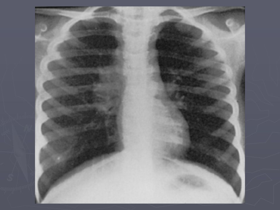
Often seen in children Frequently localized to middle and lower lung zone Paratracheal or hilar lymphadenopathy Progressive primary tuberculosis (cavation) Pleural effusion, collapse lung, Bronchectasis Dissemination ,may develop milliary tuberculosis and meningitis Ghon lesion(healed lesion)
 Pleural effusion, collapse lung, Bronchectasis. Dissemination ,may develop milliary tuberculosis and meningitis. Ghon lesion(healed lesion)")
48
Secondary tuberculosis
Symptom: productive cough, hemoptysis, dyspnea, fever, night sweat Signs: not specific, dullness with decreased fermitus (pleural effusion), rales, sign of consolidation, amphoric sound,clubbing
, rales, sign of consolidation, amphoric sound,clubbing.")
49
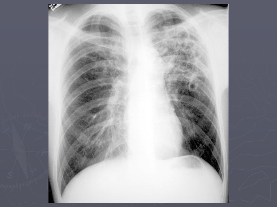
Chest.x.ray secondary tuberculosis
Apical and post. segments of the upper lobes(80-90%) Superior segments of lower lobes Infiltration and cavitary lesion Nodules and branching linear densities (tree in bud) Pleural effusion Solitary nodule(atypical)
 Superior segments of lower lobes. Infiltration and cavitary lesion. Nodules and branching linear densities (tree in bud) Pleural effusion. Solitary nodule(atypical)")
53
Other pulmonary presentation
Endobronchial tuberculosis ARDS Tuberculous pneumonitis (lower lobe) Lower lobe TB(elderly,diabetes,renal and hepatic dis. ,corticosteroid) Tuberculoma (rounded mass lesion)
 Lower lobe TB(elderly,diabetes,renal and hepatic dis. ,corticosteroid) Tuberculoma (rounded mass lesion)")
54
Complication of pulmonary TB
Massive hemoptysis (rare) Spontaneous pneumothorax Empyema Bronchectasis Extensive pulmonary destruction Tuberculosis of the upper airway(chronic productive cough, hoarseness,dysphagia)
 Spontaneous pneumothorax. Empyema. Bronchectasis. Extensive pulmonary destruction. Tuberculosis of the upper airway(chronic productive cough, hoarseness,dysphagia)")
55
Pleural tuberculosis Hypersensitivity reaction to mycobacterial antigens in pleural space Rupture or leakage from subpleural focus of disease Ph.E:dullness to percussion and absence of breath sound

56
Pleural fluid characteristics
Exudative (ppl / pserum >0/5) Normal or low glucose PH<7/2 Lymph dominant Mesothelial cell rare AFB negative in smear Fluid culture 30% positive Biopsy with culture 70-80% diagnostic ADA, IFN gamma
 Normal or low glucose. PH<7/2. Lymph dominant. Mesothelial cell rare. AFB negative in smear. Fluid culture 30% positive. Biopsy with culture 70-80% diagnostic. ADA, IFN gamma.")
57
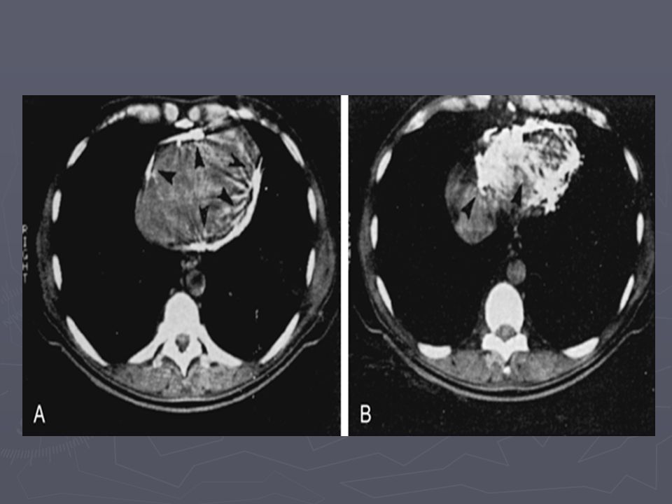
Miliary (disseminated) tuberculosis
Hematogenous spread of tubercle bacilli Recent infection or reactivation Yellowish granuloma up to 2mm

58
Clinical manifestation (miliaryTB)
History: fever, night sweat,anorexia, weakness,weight loss PH.E: Hepatomegaly,Splenomegaly, lymphadenopathy, choroidal tubercle, menangismus

59
Diagnosis miliary TB CXR :miliary reticonodular pattern
Sputum smear negative in 80% Anemia,leukopenia,leukocytosis DIC Elevated alk/ph, abnormal liver function test Negative PPD(50%) BAL and biopsy more positive
 BAL and biopsy more positive.")
64
Clinicopathologic patterns
Acute miliary tuberculosis(typical caseating granuloma) Cryptic miliary tuberculosis(more chronic, non specific clinical presentation) Non reactive miliary TB(acute septicemic) Risk factor: Age, alcohol abuse, malignancy, renal failure, CTD, diabetes, pregnancy, immunosuppresion
 Cryptic miliary tuberculosis(more chronic, non specific clinical presentation) Non reactive miliary TB(acute septicemic) Risk factor: Age, alcohol abuse, malignancy, renal failure, CTD, diabetes, pregnancy, immunosuppresion.")
66
HIV-associated TB Presentation varies with the stage
Late stage: primary pattern with miliary infiltrate and lymphadenopathy , no cavitation Higher incidence of extrapulmonary and pleural effusion Sputum smear frequently negative Lack of classic granuloma formation

68
Diagnosis Tuberculosis
High index suspicion Chest.x.ray AFB Microscopy: three sputum specimens, collected in the early morning Staining with ziehl neelsen or auramine-rhodamine

69
Diagnosis Culture: Egg based (lowenstein-jensen),
agar based (middelbrook); 4-8 week may be requird for growth Liquid media with radiometric growth detection (bactec-460) High pressure liquid chromatography of mycolic acid(2-3 week) Nucleic acid amplification
; 4-8 week may be requird for growth. Liquid media with radiometric growth detection (bactec-460) High pressure liquid chromatography of mycolic acid(2-3 week) Nucleic acid amplification.")
70
regional lymphadenitis,
BCG VACCINATION Derived from an attenuated strain of M.bovis Efficacy from 80% to nil Rarely complication: ulceration, regional lymphadenitis, osteomyelitis, dissemination

71
PPD Purified protein derivative PPD 5tu:0/1ml contain 0/0001mg
Mantoax method (interadermaly, volar surface) Wheal 6-10mm Reaction are read at 48 to72h(transverse diameter of induration in mm) A second skin test administered 1week or more after the first(two-step)
 Wheal 6-10mm. Reaction are read at 48 to72h(transverse diameter of induration in mm) A second skin test administered 1week or more after the first(two-step)")
72
Measuring TST Record induration (bump) only
Measure transverse (across arm) Record: Date given, date read, mm
 Record: Date given, date read, mm.")
75
TB Classification System (1)
Based on pathogenesis of TB Class Type Description No TB exposure Not infected No history of TB exposure Negative result to a TST or IGRA 1 TB exposure No evidence of infection History of TB exposure Negative result to a TST (given at least 8- 10 weeks after exposure) or IGRA 2 TB infection No TB disease Positive result to a TST or IGRA Negative smears and cultures (if done) No clinical or x-ray evidence of active TB disease Module 1 – Transmission and Pathogenesis of Tuberculosis
 or IGRA. 2. TB infection. No TB disease. Positive result to a TST or IGRA. Negative smears and cultures (if done) No clinical or x-ray evidence of active. TB disease. Module 1 – Transmission and Pathogenesis of Tuberculosis.")
76
TB Classification System (2)
Based on pathogenesis of TB Class Type Description 3 TB, clinically active Positive culture (if done) for M. tuberculosis Positive result to a TST or IGRA, and clinical, bacteriological, or x-ray evidence of TB disease 4 Previous TB disease (not clinically active) Medical history of TB disease Abnormal but stable x-ray findings Positive result to a TST or IGRA Negative smears and cultures (if done) No clinical or x-ray evidence of active TB disease 5 TB suspected Signs and symptoms of TB disease, but evaluation not complete Module 1 – Transmission and Pathogenesis of Tuberculosis
 for M. tuberculosis Positive result to a TST or IGRA, and clinical, bacteriological, or x-ray evidence of TB disease. 4. Previous TB disease (not clinically active) Medical history of TB disease. Abnormal but stable x-ray findings. Positive result to a TST or IGRA. Negative smears and cultures (if done) No clinical or x-ray evidence of active TB disease. 5. TB suspected. Signs and symptoms of TB disease, but evaluation not complete. Module 1 – Transmission and Pathogenesis of Tuberculosis.")
77
Short course treatment(6 month)
First line drugs: Rifampin, Isoniazid, Ethambutol, Pyrazinamid, streptomycin Short course treatment(6 month) 2month initial phase (bactericidal): rifampin, isoniazid, ethambutol, pyrazinamide 4month continuation (sterilizing): rifampin and isoniazid
 2month initial phase (bactericidal): rifampin, isoniazid, ethambutol, pyrazinamide. 4month continuation (sterilizing): rifampin and isoniazid.")
78
Treatment Pyridoxin (vit B6) should be given to: -ALCOHOLICS
-Malnurished -pregnant and lactating woman -chronic renal failure -diabetes -HIV infection

79
history of hepatic disease
Hepatitis Most common adverse reaction Baseline assessment of liver function in all adult patient monitoring monthly : Older patient alcoholics pregnancy HIV history of hepatic disease Treatment should be stopped: symptomatic hepatitis or three to five fold elevation LFT

80
First Line Drugs INH : RIF: GI-upset,Drug Raised LFT Interactions
Hepatitis Peripheral-neuropathy Drug interactions Mild effect on CNS drug induced lupus RIF: GI-upset,Drug Interactions Hepatitis Thrombocytopenia Rash Interstitial nephritis Flu like

81
PZA: Hepatitis, Rash GI upset Arthralgia Hyperuricemia rarerly gout EMB: Optic neuritis SM : Ototoxicity Nephrotoxicity

82
Adjunctive corticosteroid
Meningitis Pericarditis Acute life threatening pulmonary tuberculosis Low serum albumin Severe weight loss Adenitis in children

83
Monitoring response to treatment
Sputum culture monthly until become negative AFB smear conversion may follow culture conversion If culture not practical,AFB smear should be undertaken at 2,5and 6 months Culture/smear positive after 3month and smear positive after 5months are indicative treatment failure Serial c.x.ray not recommended ,except at the end of treatment

84
A cardinal rule in the latter situation
results of susceptibility testing are expected to become available within a few weeks changes in the regimen can be postponed until that time. If the patient's clinical condition is deteriorating, an earlier change in regimen may be indicated. A cardinal rule in the latter situation always add more than one drug at a time to a failing regimen: at least two and preferably three drugs that have never been used and to which the bacilli are likely to be susceptible The patient may continue to take isoniazid and rifampin along with these new agents pending the results of susceptibility tests.

85
Patients who experience a recurrence after apparently successful treatment (relapses) are less likely to harbor drug-resistant strains than are patients in whom treatment has failed. If the regimen administered initially does not contain rifampin, the probability of isoniazid resistance is high. begin the treatment of all patients who have relapsed with all four first-line drugs plus streptomycin, pending the results of susceptibility testing.

86
Drug-Resistant TB M. tuberculosis resistant to individual drugs spontaneous point mutations in the mycobacterial genome there is no cross-resistance among the commonly used drugs probability that a strain will be resistant to two drugs : product of the probabilities of resistance to each drug drug-resistant TB is invariably the result of Monotherapy —i.e., the failure of the health care provider to prescribe at least two drugs to which tubercle bacilli are susceptible the patient to take properly prescribed therapy

87
Drug-resistant TB may be either primary or acquired
Primary : develops in a strain infecting a patient who has not previously been treated Acquired : during treatment with an inappropriate regimen Multidrug-resistant tuberculosis (MDR-TB) defined as resistance to at least isoniazid and rifampin (also known as rifampicin in many countries),
 defined as resistance to at least isoniazid and rifampin (also known as rifampicin in many countries),")
88
Module 1 – Transmission and Pathogenesis of Tuberculosis
Drug-Resistant TB (3) Mono-resistant Resistant to any one TB treatment drug Poly-resistant Resistant to at least any 2 TB drugs (but not both isoniazid and rifampin) Multidrug resistant (MDR TB) Resistant to at least isoniazid and rifampin, the 2 best first-line TB treatment drugs Extensively drug resistant (XDR TB) Resistant to isoniazid and rifampin, PLUS resistant to any fluoroquinolone AND at least 1 of the 3 injectable second-line drugs (e.g., amikacin, kanamycin, or capreomycin) Module 1 – Transmission and Pathogenesis of Tuberculosis
 Mono-resistant. Resistant to any one TB treatment drug. Poly-resistant. Resistant to at least any 2 TB drugs (but not both isoniazid and rifampin) Multidrug resistant. (MDR TB) Resistant to at least isoniazid and rifampin, the 2 best first-line TB treatment drugs. Extensively drug resistant. (XDR TB) Resistant to isoniazid and rifampin, PLUS resistant to any fluoroquinolone AND at least 1 of the 3 injectable second-line drugs (e.g., amikacin, kanamycin, or capreomycin) Module 1 – Transmission and Pathogenesis of Tuberculosis.")
89
MDR strains that are resistant to : all fluoroquinolones and
XDR-TB MDR strains that are resistant to : all fluoroquinolones and at least one of three second-line injectable agents (amikacin, kanamycin, and capreomycin). Drug-resistant TB can be prevented by adherence : the inclusion of at least two bactericidal drugs to which the organism is susceptible the use of fixed-drug-combination products the verification that patients complete the prescribed course.
. Drug-resistant TB can be prevented by adherence : the inclusion of at least two bactericidal drugs to which the organism is susceptible. the use of fixed-drug-combination products. the verification that patients complete the prescribed course.")
Similar presentations


 is an infectious disease caused by bacteria whose scientific name is Mycobacterium tuberculosis. It was first isolated.>")


