Download presentation
Presentation is loading. Please wait.

1
Victor Sourjik ZMBH, University of Heidelberg
EMBO Practical course on Quantitative FRET, FRAP and FCS Live-cell FRET Victor Sourjik ZMBH, University of Heidelberg

2
Choose fluorescent labels
Measuring FRET in vivo Define the goal Choose fluorescent labels Choose your method Get data!

3
I. Goals of in vivo FRET measurements
Measuring molecular distances Detecting conformational changes Detecting interactions Localizing interactions Following interaction dynamics Reporting enzymatic activities and intracellular conditions

4
Measuring molecular distances using FRET
High efficiency FRET efficiency is very sensitive to the distance between fluorophores potential of FRET as a molecular ruler FRET efficiency for CFP/YFP FRET pair FRET Efficiency: E = R06/(R06+R6) 1/R6 No FRET at R > 11 nm (100 Å) GFP size ~ 5 nm (50 Å) R R0 R06 J*QD*n-4*2 Low efficiency
 1/R6. No FRET at R > 11 nm (100 Å) GFP size ~ 5 nm (50 Å) R. R0. R06 J*QD*n-4*2. Low efficiency.")
5
Measuring molecular distances using FRET
FRET efficiency is very sensitive to the distance between fluorophores potential of FRET as a molecular ruler Problems of in vivo FRET Fluorophores are usually large (fluorescent proteins) and coupled with flexible linkers Limited attachment sites for fluorophores Weak specific fluorescence (due low to moderate protein levels) High autofluorescence background Non-opimal ratio of donor to acceptor
 and coupled with flexible linkers. Limited attachment sites for fluorophores. Weak specific fluorescence (due low to moderate protein levels) High autofluorescence background. Non-opimal ratio of donor to acceptor.")
6
Measuring molecular distances using FRET
FRET efficiency is very sensitive to the distance between fluorophores potential of FRET as a molecular ruler Problems of in vivo FRET Fluorophores are usually large (fluorescent proteins) and coupled with flexible linkers Limited attachment sites for fluorophores Weak specific fluorescence (due low to moderate protein levels) High autofluorescence background Non-opimal ratio of donor to acceptor Possible (although not ideal) solution: Fix the cells and use fluorescently-labeled monoclonal antibodies
 and coupled with flexible linkers. Limited attachment sites for fluorophores. Weak specific fluorescence (due low to moderate protein levels) High autofluorescence background. Non-opimal ratio of donor to acceptor. Possible (although not ideal) solution: Fix the cells and use fluorescently-labeled monoclonal antibodies.")
7
Measuring molecular distances using FRET
FRET efficiency is very sensitive to the distance between fluorophores potential of FRET as a molecular ruler Problems of in vivo FRET Fluorophores are usually large (fluorescent proteins) and coupled with flexible linkers Limited attachment sites for fluorophores Weak specific fluorescence (due low to moderate protein levels) High autofluorescence background Non-opimal ratio of donor to acceptor Ideal solution: Labeling with small dyes
 and coupled with flexible linkers. Limited attachment sites for fluorophores. Weak specific fluorescence (due low to moderate protein levels) High autofluorescence background. Non-opimal ratio of donor to acceptor. Ideal solution: Labeling with small dyes.")
8
Detecting conformational changes using FRET
P High efficiency Low efficiency

9
Detecting conformational changes using FRET
Advantages Ratio of donor to acceptor is fixed P Problems Precision is frequently not high enough (general for measuring distances) Limited attachment sites for fluorophores
 Limited attachment sites for fluorophores.")
10
Detecting conformational changes using FRET
Advantages Ratio of donor to acceptor is fixed P Problems Precision is frequently not high enough (general for measuring distances) Limited attachment sites for fluorophores Most common current uses: Conformational changes in complexes Reporter of intracellular conditions
 Limited attachment sites for fluorophores. Most common current uses: Conformational changes in complexes. Reporter of intracellular conditions.")
11
Detecting conformational changes in complexes
Advantages Conformational changes are typically larger Problems Ratio of donor to acceptor is not fixed P P

12
Detecting conformational changes in complexes
Advantages Conformational changes are typically larger Problems Ratio of donor to acceptor is not fixed Possible solution: Use only one fluorophore (homo-FRET) P P
 P. P.")
13
FRET as reporter of intracellular conditions
Advantages Sensors are engineered to exhibit large conformational changes upon ligand binding or modification CaM Problems Only a limited number of sensors is available: Ca2+, cAMP, several kinases... Ca2+ CaM Based on conformational chenge, e.g. Cameleon (calcium sensor)
")
14
FRET as reporter of intracellular conditions
Advantages Sensors are engineered to exhibit large conformational changes upon ligand binding or modification Binding domain Phosphorylation domain Problems Only a limited number of sensors is available: Ca2+, cAMP, several kinases... P Based on intramolecular binding, e.g. kinase reporters

15
Detecting protein interactions using FRET
Interacting proteins (or, more exactly, proteins in one complex) Promises FRET as a generalized interaction- mapping technique Problems Strong spectral cross-talk between typical fluorophores (fluorescent proteins) Typically low FRET efficiency Limited attachment sites for fluorophores Weak specific fluorescence Non-opimal ratio of donor to acceptor Bulky fluorophores Detection of absolute strength of physiological interactions is non-trivial Non-interacting proteins
 Promises. FRET as a generalized interaction- mapping technique. Problems. Strong spectral cross-talk between typical fluorophores (fluorescent proteins) Typically low FRET efficiency. Limited attachment sites for fluorophores. Weak specific fluorescence. Non-opimal ratio of donor to acceptor. Bulky fluorophores. Detection of absolute strength of physiological interactions is non-trivial. Non-interacting proteins.")
16
Detecting protein interactions using FRET
+ Stimulus Possible solution: Detecting changes in protein interactions Relative concentrations of donor and acceptor do not change upon stimulation (i.e., internal control) Changes in FRET are more reliably detected than absolute values P - Stimulus
 Changes in FRET are more reliably detected than absolute values. P. - Stimulus.")
17
II. Fluorescent labels for in vivo FRET measurements
Fluorescent proteins In-vivo labeling with fluorescent dyes

18
Proteins vs dyes in fluorescence microscopy
Fluorescent proteins Can be genetically encoded (high specificity) Proteins are bulky (5 nm) Spectra are broad (strong cross-talk) Not very bright and photostable In-vivo labeling with fluorescent dyes Small size Bright and relatively photostable Narrow spectra and large spectral choice Specific in-vivo labeling is difficult
 Proteins are bulky (5 nm) Spectra are broad (strong cross-talk) Not very bright and photostable. In-vivo labeling with fluorescent dyes. Small size. Bright and relatively photostable. Narrow spectra and large spectral choice. Specific in-vivo labeling is difficult.")
19
Spectral requirements for FRET labels
CFP = cyan fluorescent protein (donor) YFP = yellow fluorescent protein (acceptor) Requirements for the FRET pair: excitation spectra of donor and acceptor are separated emission spectrum of donor overlaps with excitation spectrum of acceptor emission spectra of donor and acceptor are separated
 YFP = yellow fluorescent protein (acceptor) Requirements for the FRET pair: excitation spectra of donor and acceptor are separated. emission spectrum of donor overlaps with excitation spectrum of acceptor. emission spectra of donor and acceptor are separated.")
20
Fluorescent proteins for in vivo FRET measurements
Nathan C. Shaner, Paul A. Steinbach, & Roger Y. Tsien Nature Methods, Vol. 2: 905 – 909 Any two proteins with overlapping emission spectrum of donor and excitation spectrum of acceptor can be used a FRET pair (including the same protein as donor and acceptor)
")
21
Fluorescent proteins for in vivo FRET measurements
Caution: FRET efficiency with FPs as FRET pair is always far below 100%

22
Fluorescent dyes for in vivo FRET measurements
Fluorescent dyes with relatively specific binding to short peptide sequences (e.g., FlAsH or ReAsH) Miyawaki et al., supplement to Nature Cell Biol., 5 Fluorescent dyes specifically binding to protein tags (e.g., SNAP-tag or HaloTag) HaloTag, Promega Corporation
 Miyawaki et al., supplement to Nature Cell Biol., 5. Fluorescent dyes specifically binding to protein tags (e.g., SNAP-tag or HaloTag) HaloTag, Promega Corporation.")
23
Combining proteins and dyes for in vivo FRET measurements
Roger Y. Tsien’s web site

24
III. Methods to measure FRET in vivo
Spectral measurements Two-channel FRET (sensitized emission) One-channel FRET (acceptor photobleaching) One-channel FRET (donor photobleaching) Polarization imaging Life-time imaging
 One-channel FRET (acceptor photobleaching) One-channel FRET (donor photobleaching) Polarization imaging. Life-time imaging.")
25
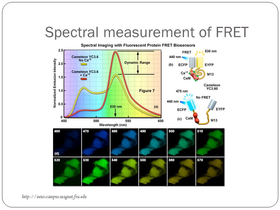
Spectral measurement of FRET
Advantages Complete spectral information Drawbacks Requires a specialized system (e.g., Zeiss LSM 710) Requires carefull image analysis
 Requires carefull image analysis.")
26
Spectral measurement of FRET
27
Spectral measurement of FRET
In a general case (so-called linear spectral unmixing): Acquire spectra at donor and acceptor excitation wavelength Acquire spectra for control samples with only donor and only acceptor Subtract donor and acceptor cross-talk (bleed-through) to get true FRET signal
: Acquire spectra at donor and acceptor excitation wavelength. Acquire spectra for control samples with only donor and only acceptor. Subtract donor and acceptor cross-talk (bleed-through) to get true FRET signal.")
28
Two-channel measurement of FRET
Advantages Can be performed on a simple wide-field microscope Drawbacks Limited spectral information Requires carefull image analysis

29
Two-channel measurement of FRET Sensitized emission
A B C Linear spectral unmixing Leica Microsystems

30
One-channel measurement of FRET Acceptor photobleaching
Procedure: Acquire signal of donor fluorescence Bleach acceptor Acquire signal of donor fluorescence again 510 nm

31
One-channel measurement of FRET Acceptor photobleaching
Advantages Is very simple and reliable Drawbacks One-time experiment 510 nm

32
One-channel measurement of FRET Acceptor photobleaching
Imaging Whole-field acquisition YFP CFP 510 nm Can be done either in imaging or whole-field acquisition mode

33
One-channel measurement of FRET Donor photobleaching
Donor (CFP) fluorescence + FRET - FRET Time (sec) Advantages Is comparatively simple Drawbacks One-time experiment Can be affected by other intracellular factors Procedure: Follow kinetics of donor bleaching
 fluorescence. + FRET. - FRET. Time (sec) Advantages. Is comparatively simple. Drawbacks. One-time experiment. Can be affected by other intracellular factors. Procedure: Follow kinetics of donor bleaching.")
34
Polarization (anisotropy) measurement of FRET
Weak (no) FRET = high anisotropy Strong FRET = low anisotropy Homo-FRET Advantages Allows measuring homo-FRET Is comparatively simple Drawbacks Requires specialized equipment Can be affected by other intracellular factors Procedure: Excite with polarized light Measure emission in two orthogonal directions of polarization
 FRET = high anisotropy. Strong FRET = low anisotropy. Homo-FRET. Advantages. Allows measuring homo-FRET. Is comparatively simple. Drawbacks. Requires specialized equipment. Can be affected by other intracellular factors. Procedure: Excite with polarized light. Measure emission in two orthogonal directions of polarization.")
35
Life-time measurement of FRET
Time (sec) ps fs ns Phizicky et al., Nature :208-15
 ps. fs. ns. Phizicky et al., Nature :")
36
Life-time measurement of FRET
Time (sec) Advantages Reports both FRET efficiency and fraction of interacting proteins Not sensitive to acceptor concentration Drawbacks Limited speed Limited spatial resolution Phizicky et al., Nature :208-15
 Advantages. Reports both FRET efficiency and fraction of interacting proteins. Not sensitive to acceptor concentration. Drawbacks. Limited speed. Limited spatial resolution. Phizicky et al., Nature :")
37
Our own work (just one slide!) FRET as a network mapping technique
Bacterial chemotaxis network A B

Similar presentations

 of molecular rotors maps.>")


, but how to study in cells? Do rafts really exist in cells? Are.>")


![Some structures Dansyl chloride 1,5-I-AEDANS Fluorescein isothiocyante ANS Ethidium bromide 5-[2-[(2-iodoacetyl)amino]ethylamino] naphthalene-1-sulfonic.](/15/4639132/big_thumb.jpg "Some structures Dansyl chloride 1,5-I-AEDANS Fluorescein isothiocyante ANS Ethidium bromide 5-[2-[(2-iodoacetyl)amino]ethylamino] naphthalene-1-sulfonic.>")


