Download presentation
Presentation is loading. Please wait.

1
Topic 12-I: Male Reproductive System
Animal Histology BIOL 241 Topic 12-I: Male Reproductive System Dr. Issa Al-Amri Department of Biological Sciences & Chemistry College of Arts & Sciences

2
Introduction Male Reproductive System consists of: testes, genital ducts, accessory genital glands (seminal vesicles, prostate gland, and bulbourethral glands), and penis. Function: produces spermatozoa (sperm), testosterone, and seminal fluid. Seminal fluid transports and nourishes sperm as they pass through excretory ducts. The penis delivers sperm to exterior and also serves as conduit for excretion of urine from the body.
, and penis. Function: produces spermatozoa (sperm), testosterone, and seminal fluid. Seminal fluid transports and nourishes sperm as they pass through excretory ducts. The penis delivers sperm to exterior and also serves as conduit for excretion of urine from the body.")
3
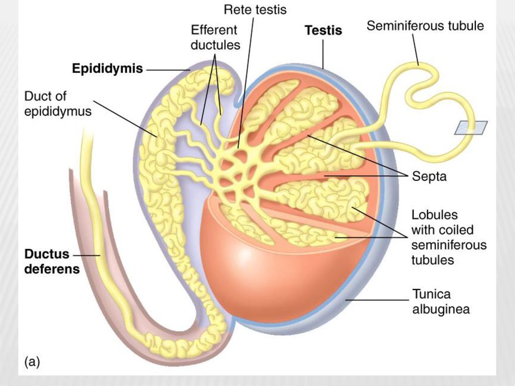
Introduction Testes Develop in abdominal cavity and later descend into scrotum, where they are suspended at ends of spermatic cords. They are the sites of spermatogenesis and production of male sex hormones, primarily testosterone.

4
Lobuli testis Lobuli testis:
Pyramidal intercommunicating compartments separated by incomplete septa. Each contains seminiferous tubules. Tubules embedded in meshwork of loose connective tissue containing blood and lymphatic vessels, nerves, and interstitial cells of Leydig.

7
Leydig cells Interstitial cells of Leydig:
Round to polygonal cells in interstitial regions between seminiferous tubules. Possess a large central nucleus, numerous mitochondria, well-developed Golgi complex, and many lipid droplets. Lipid droplets contain cholesterol esters, precursors of testosterone. Richly supplied with capillaries and lymphatic vessels. Function: endocrine cells produce and secrete testosterone. Secretion stimulated by luteinizing hormone (LH; interstitial cell–stimulating hormone) produced in pituitary gland. Leydig cells mature and begin to secrete during puberty.
 produced in pituitary gland. Leydig cells mature and begin to secrete during puberty.")
9
TEM of a Leydig cell in interstitial connective tissue of testis

10
TEM of a Leydig cell showing finely granulated nucleus (N),
mitochondria (M), and lipid droplets (LP) associated with SER.
, and lipid droplets (LP) associated with SER.")
11
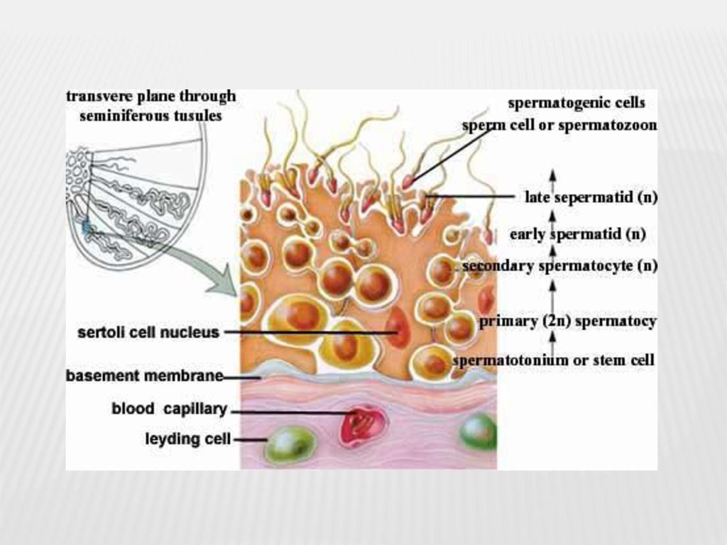
Seminiferous tubules Seminiferous tubules:
They are cm long, diameter of µm. Enveloped by fibrous connective tissue tunic (layers of fibroblasts and capillary beds). Form tortuous pathways through testicular lobules, then narrow into short, straight segments, tubuli recti, which connect with rete testis. Lined by thick epithelium (seminiferous or germinal epithelium). Epithelium consists of 4 – 8 cell layers and contains spermatogenic cells, from which germ cells develop (spermatogenesis), and Sertoli cells, which have several functions.
. Form tortuous pathways through testicular lobules, then narrow into short, straight segments, tubuli recti, which connect with rete testis. Lined by thick epithelium (seminiferous or germinal epithelium). Epithelium consists of 4 – 8 cell layers and contains spermatogenic cells, from which germ cells develop (spermatogenesis), and Sertoli cells, which have several functions.")
13
SEM of seminiferous tubule. The lumen is full of free spermatozoa

14
Figure 2: Testis, seminiferous tubules
-Tunica albuginea (TA), slender septa (Se) attach to it. -Seminiferous tubules (ST); highly convoluted. -Each lobule(Lo) packed with seminiferous tubules. -Connective tissue stroma (arrows) Figure 2: Testis, seminiferous tubules -Higher mag. of Figure 1. -Tunica vasculosa (TV) of tunica albuginea (TA) highly vascular region (arrows) -Blood vessels (BV) penetrate lobuli testis in connective tissue septa (Se). -Seminiferous tubules (ST) closely apposed to each other (arrowheads), Cellular stroma (St). Figure 3: Testis, seminiferous tubules -Two seminiferous tubules(ST): Myoid cells(MC), fibroblasts(F), fibromuscular connective tissue(CT). -Stratified seminiferous epithelium (SE) separated from tubular wall by basal membrane (arrowheads). -Spermatogonia (Sg) and Sertoli cells (SC) lie on basal membrane and in basal compartment(BC). -Primary spermatocytes (PS), secondary spermatocytes, spermatids( Sp), and spermatozoa(Sz) in adluminal compartment(AC). -Lumen(L) contains spermatozoa and cellular debris discarded during transformation of spermatids into spermatozoa. Figure 4: Testis, seminiferous tubules -Fbromuscular walls of two tubularcross-sections very close to each other (arrows). -Arterioles(A),venules(V) ,Sertoli cells (SC),nuclei and dense nucleoli(n). seminiferous epithelia (SE). -Dark spermatogonia A (Ad) dark, flattened nuclei; pale spermatogonia A (Ap) with flattened pale nuclei; and spermatogonia B (B) with round nuclei.
, slender septa (Se) attach to it. -Seminiferous tubules (ST); highly convoluted. -Each lobule(Lo) packed with seminiferous tubules. -Connective tissue stroma (arrows) Figure 2: Testis, seminiferous tubules. -Higher mag. of Figure 1. -Tunica vasculosa (TV) of tunica albuginea (TA) highly vascular region (arrows) -Blood vessels (BV) penetrate lobuli testis in connective tissue septa (Se). -Seminiferous tubules (ST) closely apposed to each other (arrowheads), Cellular stroma (St). Figure 3: Testis, seminiferous tubules. -Two seminiferous tubules(ST): Myoid cells(MC), fibroblasts(F), fibromuscular connective tissue(CT). -Stratified seminiferous epithelium (SE) separated from tubular wall by basal membrane (arrowheads). -Spermatogonia (Sg) and Sertoli cells (SC) lie on basal membrane and in basal compartment(BC). -Primary spermatocytes (PS), secondary spermatocytes, spermatids( Sp), and spermatozoa(Sz) in adluminal compartment(AC). -Lumen(L) contains spermatozoa and cellular debris discarded during transformation of spermatids into spermatozoa. Figure 4: Testis, seminiferous tubules. -Fbromuscular walls of two tubularcross-sections very close to each other (arrows). -Arterioles(A),venules(V) ,Sertoli cells (SC),nuclei and dense nucleoli(n). seminiferous epithelia (SE). -Dark spermatogonia A (Ad) dark, flattened nuclei; pale spermatogonia A (Ap) with flattened pale nuclei; and spermatogonia B (B) with round nuclei.")
15
Figure 1 : Interstitial cells, testis
Testis and Epididymis Figure 1 : Interstitial cells, testis -Stroma (St), surrounding seminiferous tubules (ST) have rich vascular supply (BV) and lymphatic drainage(LV). -Interstitial cells of Leydig (IC), produce testosterone: -Recognizable by round-to-oval nuclei (N) and presence of lipid (arrow) within their cytoplasm. Figure 2: Rete testis -Rete testis (RT), located in mediastinum testis (MT), made of labyrinthine, spaces lined by a simple cuboidal epithelium (Ep). -Dense collagenous connective tissue(CT) of mediastinum testis, -Seminiferous tubules (ST). Spermatozoa gain access to rete testis via short, straight tubuli recti (TR). Figure 3: Ductuli efferentes -First part of epididymis: ductuli efferentes( De). -Receives spermatozoa (Sz) from rete testis. -Lumina of ductuli lined by simple columnar epithelium (Ep) made of tall and short cells, responsible for uneven appearance of these tubules. -Thick fibroelastic connective tissue (CT) wall of ductuli has many smooth muscle cells (SM). Figure 4: Ductus epididymis -Ductus epididymis (DE) distinguished from ductuli efferentes by: -Nuclei (N) of pseudostratified epithelial lining (Ep) are two types, oval and round, whereas those of ductuli are round. -Lumen contains many spermatozoa (Sz) and epithelium sits on basal lamina. -Connective tissue wall of ductus epididymis differentiated easily from its circularly arranged smooth muscle coat (SM).
, surrounding seminiferous tubules (ST) have rich vascular supply (BV) and lymphatic drainage(LV). -Interstitial cells of Leydig (IC), produce testosterone: -Recognizable by round-to-oval nuclei (N) and presence of lipid (arrow) within their cytoplasm. Figure 2: Rete testis. -Rete testis (RT), located in mediastinum testis (MT), made of labyrinthine, spaces lined by a simple cuboidal epithelium (Ep). -Dense collagenous connective tissue(CT) of mediastinum testis, -Seminiferous tubules (ST). Spermatozoa gain access to rete testis via short, straight tubuli recti (TR). Figure 3: Ductuli efferentes. -First part of epididymis: ductuli efferentes( De). -Receives spermatozoa (Sz) from rete testis. -Lumina of ductuli lined by simple columnar epithelium (Ep) made of tall and short cells, responsible for uneven appearance of these tubules. -Thick fibroelastic connective tissue (CT) wall of ductuli has many smooth muscle cells (SM). Figure 4: Ductus epididymis. -Ductus epididymis (DE) distinguished from ductuli efferentes by: -Nuclei (N) of pseudostratified epithelial lining (Ep) are two types, oval and round, whereas those of ductuli are round. -Lumen contains many spermatozoa (Sz) and epithelium sits on basal lamina. -Connective tissue wall of ductus epididymis differentiated easily from its circularly arranged smooth muscle coat (SM).")
16
Sertoli cells Sertoli cells Structure:
Have pale, oval, nucleus (indentations) with large nucleolus. Well-developed SER, some RER, abundant mitochondria and lysosomes, and extensive Golgi complex. Receptors for follicle-stimulating hormone (FSH) present on plasma membranes. Form zonulae occludentes (tight junctions) with adjacent Sertoli cells near their bases, thus dividing lumen of seminiferous tubule into a basal and adluminal compartment. Tight junctions responsible for blood-testis barrier, which protects developing sperm cells from autoimmune reactions.
 with large nucleolus. Well-developed SER, some RER, abundant mitochondria and lysosomes, and extensive Golgi complex. Receptors for follicle-stimulating hormone (FSH) present on plasma membranes. Form zonulae occludentes (tight junctions) with adjacent Sertoli cells near their bases, thus dividing lumen of seminiferous tubule into a basal and adluminal compartment. Tight junctions responsible for blood-testis barrier, which protects developing sperm cells from autoimmune reactions.")
19
LM: Sertoli cells in seminiferous tubule

20
TEM: Sertoli cell (SR) attached to the basal lamina. nuclei (N)
appeared with numerous indentations. Interstitial tissue (IT).
.")
21
Sertoli cells Function:
Support, protect, and nourish spermatogenic cells. Phagocytose excess cytoplasm discarded by maturing spermatids. Secrete fructose-rich fluid into lumen that nourishes and facilitates transport of spermatozoa through seminiferous tubules to genital ducts. Synthesize androgen-binding protein (ABP) under influence of FSH. ABP maintain necessary concentration of testosterone in seminiferous tubule so that spermatogenesis can progress. Secrete inhibin; hormone inhibits synthesis and release of FSH by anterior pituitary. Establish blood-testis barrier. Synthesize and release antimüllerian hormone, which determines maleness.
 under influence of FSH. ABP maintain necessary concentration of testosterone in seminiferous tubule so that spermatogenesis can progress. Secrete inhibin; hormone inhibits synthesis and release of FSH by anterior pituitary. Establish blood-testis barrier. Synthesize and release antimüllerian hormone, which determines maleness.")
22
Spermatogenesis Spermatogenesis:
Process of spermatozoon formation divided into three phases: Spermatocytogenesis: differentiation of spermatogonia into primary spermatocytes. Meiosis—reduction division to reduce diploid chromosomal complement of primary spermatocytes to form haploid spermatids. Spermiogenesis—transformation of spermatids into spermatozoa. Spermatogenesis does not occur simultaneously in all seminiferous tubules, but in wavelike sequences of maturation (cycles of seminiferous epithelium). During spermatogenesis: daughter cells remain connected to each other via intercellular bridges (responsible for synchronous development of germ cells along any one seminiferous tubule).
. During spermatogenesis: daughter cells remain connected to each other via intercellular bridges (responsible for synchronous development of germ cells along any one seminiferous tubule).")
23
Spermatogenesis

24
Spermatogenesis Spermatogenic cells:
Spermatogonia : diploid germ cells adjacent to basal lamina of seminiferous epithelium. At puberty, testosterone influences them to enter cell cycle. Pale type A spermatogonia: contain pale-staining nucleus, spherical mitochondria, small Golgi complex, abundant, free ribosomes. Mitotically give rise either to more cells of same type (maintain supply) or to type B spermatogonia. Dark type A spermatogonia: mitotically inactive (reserve) cells with dark nuclei; have potential to resume mitosis and produce pale type A cells. Type B spermatogonia: undergo mitosis and give rise to primary spermatocytes.
 or to type B spermatogonia. Dark type A spermatogonia: mitotically inactive (reserve) cells with dark nuclei; have potential to resume mitosis and produce pale type A cells. Type B spermatogonia: undergo mitosis and give rise to primary spermatocytes.")
25
Spermatogenesis Spermatocytes: Spermatids:
Primary spermatocytes large diploid cells, undergo first meiotic division (reductional division) to form secondary spermatocytes. Secondary spermatocytes haploid cells, undergo second meiotic division (equatorial division) to form spermatids. Spermatids: Spermatids small haploid cells. Located near lumen of seminiferous tubule. Nuclei display regions of condensed chromatin. Contain pair of centrioles, mitochondria, free ribosomes, SER, and well-developed Golgi complex.
 to form secondary spermatocytes. Secondary spermatocytes haploid cells, undergo second meiotic division (equatorial division) to form spermatids. Spermatids: Spermatids small haploid cells. Located near lumen of seminiferous tubule. Nuclei display regions of condensed chromatin. Contain pair of centrioles, mitochondria, free ribosomes, SER, and well-developed Golgi complex.")
26
Spermatogenesis processes in seminiferous tubules

27
Spermatogenesis processes in seminiferous tubules

28
Spermiogenesis Spermiogenesis:
Process of cytodifferentiation: spermatids shed much of their cytoplasm and transform into spermatozoa (released inlumen of seminiferous tubule). Four phases of Spermiogenesis: Golgi phase: Formation of acrosomal granule, enclosed within acrosomal vesicle, which becomes attached to anterior end of nuclear envelope of spermatid. In this phase, centrioles migrate away from nucleus to form flagellar axoneme. Centrioles then migrate back toward nucleus to assist in forming connecting piece associated with the tail.
. Four phases of Spermiogenesis: Golgi phase: Formation of acrosomal granule, enclosed within acrosomal vesicle, which becomes attached to anterior end of nuclear envelope of spermatid. In this phase, centrioles migrate away from nucleus to form flagellar axoneme. Centrioles then migrate back toward nucleus to assist in forming connecting piece associated with the tail.")
29
Diagram of spermiogenesis processes

31
Spermiogenesis Cap phase:
Expansion of acrosomal vesicle over much of nucleus, forming acrosomal cap. c. Acrosomal phase: Nucleus: becomes condensed, flattened, and located in head region. Mitochondria: aggregate around proximal portion of flagellum, which develops into middle piece of the tail. Spermatid elongation: process aided by a temporary cylinder of microtubules called manchette. Acrosomal phase: ends as spermatid is oriented with its acrosome pointing toward base of seminiferous tubule.

32
Flagellum oriented towards lumen (L) . Sertoli cells (SR),
TEM of midpiece (MD), principal piece (PP) and endpiece (ED) of flagellum. Flagellum oriented towards lumen (L) . Sertoli cells (SR), nucleus (N), mitochondria (M), lumen (L).
, principal piece (PP) and endpiece (ED) of flagellum. Flagellum oriented towards lumen (L) . Sertoli cells (SR), nucleus (N), mitochondria (M), lumen (L).")
33
Spermiogenesis Maturation phase characterized by the following:
Loss of excess cytoplasm and intercellular bridges connecting spermatids into asyncytium. Discarded cytoplasm phagocytized by Sertoli cells. Maturation phase ends when nonmotile spermatozoa are released (tail first) into lumen of seminiferous tubule. Spermatozoa remain immotile until they leave epididymis. They become capacitated (capable of fertilizing) in female reproductive system.
 into lumen of seminiferous tubule. Spermatozoa remain immotile until they leave epididymis. They become capacitated (capable of fertilizing) in female reproductive system.")
34
Spermiogenesis Spermatozoon: Head of spermatozoon:
Spermatozoon head is flattened and contains dense, homogeneous nucleus with 23 chromosomes: (22 plus Y chromosome or 22 plus X chro-mosome). Also contain acrosome, which contains hydrolytic enzymes (e.g., acid phosphatase, neuraminidase, hyaluronidase, and proteases) assist sperm in penetrating corona radiata and zona pellucida of oocyte. Release of these enzymes termed acrosomal reaction.
. Also contain acrosome, which contains hydrolytic enzymes (e.g., acid phosphatase, neuraminidase, hyaluronidase, and proteases) assist sperm in penetrating corona radiata and zona pellucida of oocyte. Release of these enzymes termed acrosomal reaction.")
35
Spermiogenesis Tail of the spermatozoon:
Includes neck, middle piece, principal piece, and end piece. Neck: includes centrioles and connecting piece; attached to nine outer dense fibers of remainder of tail. Middle piece: extends from neck to annulus, contains axoneme, nine outer dense fibers, and spirally arranged sheath of mitochondria. Principal piece: extends from annulus to he end piece; contains axoneme with its surrounding dense fibers, which in turn encircled by fibrous sheath (has circumferential ribs). End piece: consists of axoneme and surrounding plasma membrane.
. End piece: consists of axoneme and surrounding plasma membrane.")
37
Regulation of spermatogenesis
Critical testicular temperature (35oC for spermatogenesis): Testicular arteries arise from aorta accompany testes into scrotum providing each testis with a vascular supply. As convoluted arteries approach testes, they are surrounded by pampiniform plexus of veins that dissipates heat of arterial blood resulting in its temperature being reduced to 35oC. Hormonal interactions: a. Certain neurons in hypothalamus produce luteinizing hormone-releasing hormone (LHRH), also known as gonadotropin-releasing hormone (GnRH). LHRH initiates release of LH and FSH from adenohypophysis (anterior pituitary).
: Testicular arteries arise from aorta accompany testes into scrotum providing each testis with a vascular supply. As convoluted arteries approach testes, they are surrounded by pampiniform plexus of veins that dissipates heat of arterial blood resulting in its temperature being reduced to 35oC. Hormonal interactions: a. Certain neurons in hypothalamus produce luteinizing hormone-releasing hormone (LHRH), also known as gonadotropin-releasing hormone (GnRH). LHRH initiates release of LH and FSH from adenohypophysis (anterior pituitary).")
38
Genital ducts 1. Ductus epididymis:
Together with ductuli efferentes, constitutes epididymis. Surrounded by smooth muscle layer; undergo peristaltic contractions, conveying sperm toward ductus deferens. Lined by pseudostratified columnar epithelium, which supported by basal lamina and contains two cell types: Basal cells: round; serve as precursors of principal cells. Principal cells: columnar and contain nonmotile stereocilia on luminal surfaces. Have large Golgi complex, RER, lysosomes, and many apical pinocytotic and coated vesicles (fluid resorption). Secrete glycerophosphocholine; inhibits capacitation. Thus, capacitation occurs only after sperm enters female genital tract
. Secrete glycerophosphocholine; inhibits capacitation. Thus, capacitation occurs only after sperm enters female genital tract.")
40
Genital ducts 2. Ductus (vas) deferens:
Has thick muscular wall with inner and outer layers of longitudinal smooth muscle, separated by middle circular layer. Contain narrow, irregular lumen lined by pseudostratified columnar epithelium. 3. Ejaculatory duct: Straight continuation of ductus deferens; receives duct of seminal vesicle. Lacks muscular wall. Enters prostate gland and terminates in a slit on colliculus seminalis located in prostatic urethra.

42
Epididymis, Ductus Deferens, Simenal Vesicles
Figure 1 : Ductus epididymis -Pseudostratified stereociliated columnar epithelium (Ep): -Two types of cells: short basal cells( BC), recognizable by their round nuclei, and tall columnar principal cells (PC), whose oval nuclei display one or more nucleoli (n). -Smooth muscle (SM) cells, surrounded by connective tissue(CT) elements. -In box: round nuclei of basal cells(BC) and oval nuclei of the principal cells (PC). -Clumped stereocilia (arrows) extend into spermatozoa (Sz) filled lumen. Figure 2: Ductus deferens -Muscular: layers of smooth muscle: outer longitudinal (OL), middle circular (MC), inner longitudinal (IL). -Fbroelastic lamina propria (LP), receive vascular supply (BV) from vessels (arrow). -Pseudostratified columnar epithelium (Ep), spermatozoa-filled lumen(L -Box: Pseudostratified columnar epithelium (Ep) displays presence of stereocilia (Sc). Figure 3: Seminal vesicle -Highly folded mucous membrane (MM) made of pseudostratified epithelium (Ep) with thin connective tissue core(CT). -Folded membrane anastomoses with itself, partitioning off small spaces (asterisks) that, although continuous with central lumen, appear to be discrete regions. Figure 4: Seminal vesicle -Higher magnification of Fig. 3. -Tall columnar cells (CC): basally located, round nuclei(N). -Cytoplasm with secretory granules (arrows). -Short basal cells (BC), lumen (L), capillaries (C) in connective tissue, spermatozoa (Sz).
: -Two types of cells: short basal cells( BC), recognizable by their round nuclei, and tall columnar principal cells (PC), whose oval nuclei display one or more nucleoli (n). -Smooth muscle (SM) cells, surrounded by connective tissue(CT) elements. -In box: round nuclei of basal cells(BC) and oval nuclei of the principal cells (PC). -Clumped stereocilia (arrows) extend into spermatozoa (Sz) filled lumen. Figure 2: Ductus deferens. -Muscular: layers of smooth muscle: outer longitudinal (OL), middle circular (MC), inner longitudinal (IL). -Fbroelastic lamina propria (LP), receive vascular supply (BV) from vessels (arrow). -Pseudostratified columnar epithelium (Ep), spermatozoa-filled lumen(L. -Box: Pseudostratified columnar epithelium (Ep) displays presence of stereocilia (Sc). Figure 3: Seminal vesicle. -Highly folded mucous membrane (MM) made of pseudostratified epithelium (Ep) with thin connective tissue core(CT). -Folded membrane anastomoses with itself, partitioning off small spaces (asterisks) that, although continuous with central lumen, appear to be discrete regions. Figure 4: Seminal vesicle. -Higher magnification of Fig. 3. -Tall columnar cells (CC): basally located, round nuclei(N). -Cytoplasm with secretory granules (arrows). -Short basal cells (BC), lumen (L), capillaries (C) in connective tissue, spermatozoa (Sz).")
Similar presentations


 produce androgen-testosterone ---gernital ducts: store.>")


 PROF. DR. FAUZIAH OTHMAN DEPT OF HUMAN ANATOMY.>")
