Download presentation
Presentation is loading. Please wait.

1
Dr Amit Gupta Associate Professor Dept of Surgery
Spleen Dr Amit Gupta Associate Professor Dept of Surgery

2
Word "spleen" in English is "ill temper

3
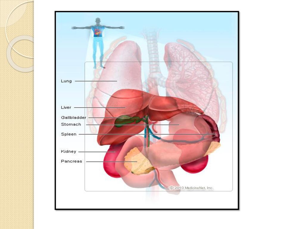
Anatomy Largest reticuloendothelial organ in the body
Intra-abdominal wedge shaped organ. Left hypochondrium & epigastrium. Soft , highly vascular. Variable size & weight Average 12.5 X 7.5 X 2.5 in size gm in weight.

5
It derives most of its blood from the splenic artery
Small amount from short gastric vessels Venous drainage: splenic vein Total splenic inflow of blood is approximately 250 to 300 mL/min

6
Physiology Filtration Host defence Storage Hematopoiesis

7
Congenital Anomalies Complete absence is rare
associated with other congenital abnormalities such as situs inversus and cardiac malformations. Hypoplasia: more common finding Accessory spleens (spleniculi) are common Generally situated in the gastrosplenic ligament or the tail of the pancreas,omentum or mesenteries of the small or large intestine. In splenectomy if an accessory spleen is overlooked, the benefit of removal of the definitive spleen can be lost
 are common. Generally situated in the gastrosplenic ligament or the tail of the pancreas,omentum or mesenteries of the small or large intestine. In splenectomy if an accessory spleen is overlooked, the benefit of removal of the definitive spleen can be lost.")
8
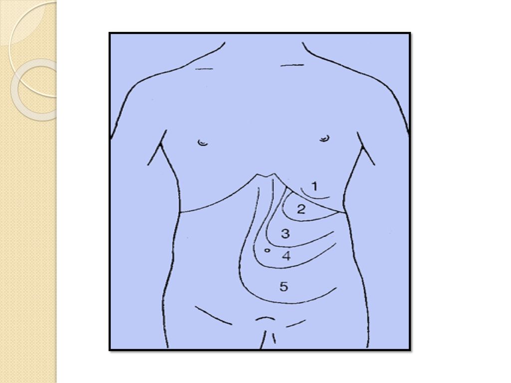
Splenomegaly Means enlargement of spleen. Normal spleen not palpable.
has to enlarge 2 time to be detectable. Enlarges from left hypochondrium to right illiac fossa.

11
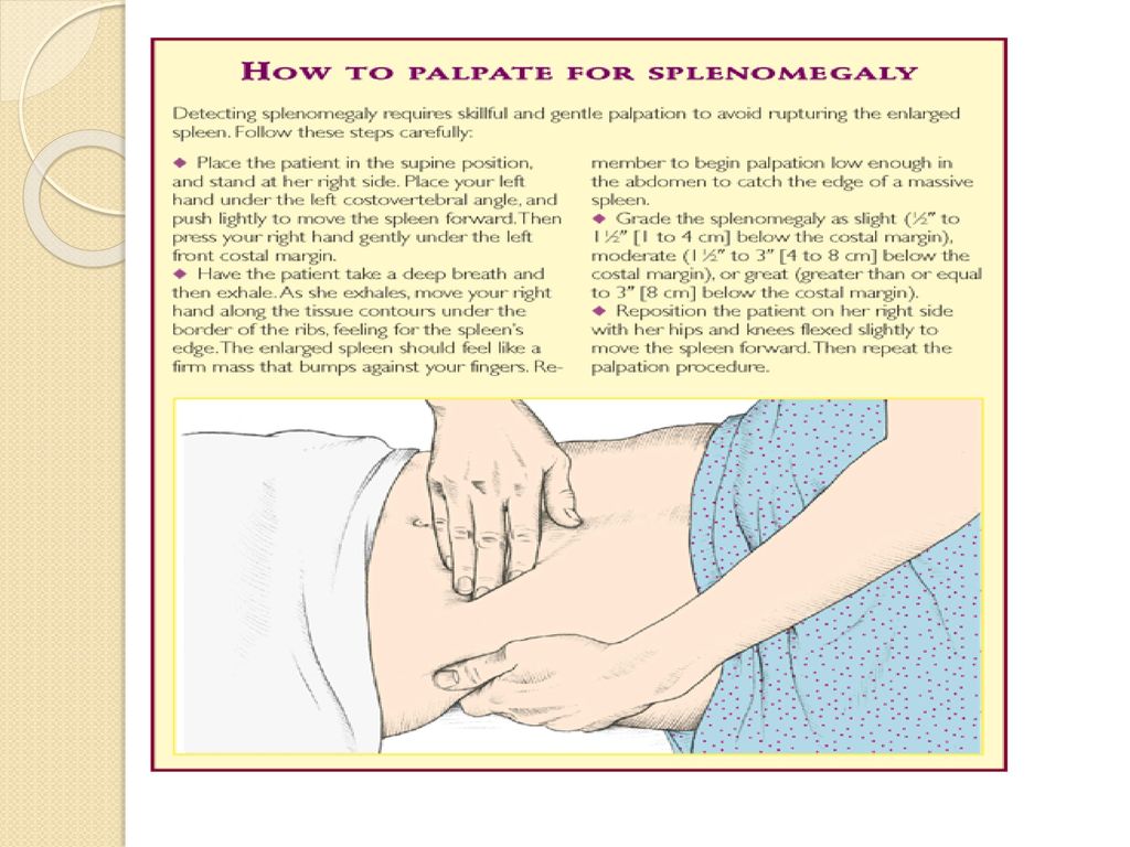
Examination

12
Examination

13
Examination

14
Classification Of Splenomegaly
Alotaibi G et al. classification splenomegaly as: Moderate : 11–20 cm Severe : >20 cm Another classification acc. to extent below coastal margin: Mild : <5 cm Moderate : 5-8 cm Severe : >8 cm

15
Splenomegaly Grading (Hacket’s Grading)
")
16
Pathology Basically splenomegaly is due to: Increased function
Abnormal blood flow Infiltration

17
Increased Function Removal of defective RBCs Spherocytosis Thalassemia
Hemoglobinopathies Nutritional anemias Early sickle cell anemia

18
Increased Function Immune hyperplasia
Response to infection (viral, bacterial, fungal,parasitic) mononucleosis, AIDS, viral hepatitis subacute bacterial endocarditis, bacterial septicemia splenic abscess, typhoid fever brucellosis, leptospirosis, tuberculosis histoplasmosis malaria, leishmaniasis, trypanosomiasis
 mononucleosis, AIDS, viral hepatitis. subacute bacterial endocarditis, bacterial septicemia. splenic abscess, typhoid fever. brucellosis, leptospirosis, tuberculosis. histoplasmosis. malaria, leishmaniasis, trypanosomiasis.")
19
Increased Function Immune hyperplasia Disordered immunoregulation
Rheumatoid arthritis Systemic lupus erythematosus Serum sickness Autoimmune hemolytic anemia Sarcoidosis

20
Increased Function Extramedullary hematopoiesis Myelofibrosis
marrow infiltration by tumors, leukemias marrow damage by radiation, toxins

21
Abnormal Blood Flow Organ Failure Vascular cirrhosis
hepatic vein obstruction portal vein obstruction Budd–Chiari syndrome splenic vein obstruction

22
Infiltration Metabolic diseases Gauchers disease Niemann–pick disease
Hurler syndrome Mucopolysaccharidoses Amyloidosis

23
Infiltration Benign and malignant “infiltrations”
leukemias (acute, chronic, lymphoid, and myeloid) lymphomas (Hodgkins and non-Hodgkins) myeloproliferative disease metastatic tumors (commonly melanoma) histiocytosis X hemangioma, lymphangioma splenic cysts hamartomas
 lymphomas (Hodgkins and non-Hodgkins) myeloproliferative disease. metastatic tumors (commonly melanoma) histiocytosis X. hemangioma, lymphangioma. splenic cysts. hamartomas.")
24
Mild Splenomegaly Malaria Typhoid Disseminated TB Viral hepatitis
Septicemia Thalessemia minor HIV

25
Moderate Splenomegaly
Cirrhosis Lymphomas Leukaemia Infectious mononeucleosis Hemolytic anemia Splenic abcess Amylodosis hemochromatosis

26
Severe Splenomegaly Chronic malaria Kala azar CML Portal hypertension
Thalessemia major Infiltrative & metabolic disorders

27
Management Depends on cause
Various investigations as per clinical features & epidemiology are employed Basic investigations done are: CBC USG CECT scan

28
Treatment Can be medical or surgical
Medical management involves treatment of cause if possible Surgical treatment is splenectomy

29
Indications Of Splenectomy
Trauma : splenic rupture (MC) ITP Hemolytic anemias CLL, Lymphomas Primary Myelofibrosis Tropical splenomegaly
 ITP. Hemolytic anemias. CLL, Lymphomas. Primary Myelofibrosis. Tropical splenomegaly.")
30
Preoperative Considerations
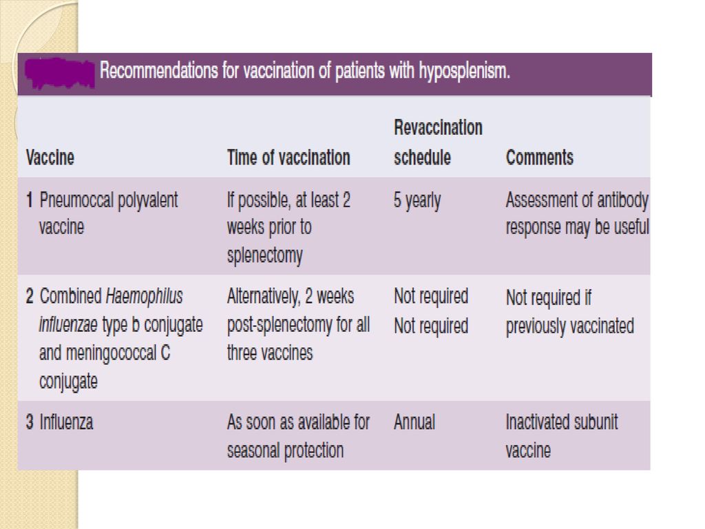
Splenic Artery Embolization Vaccination Deep Venous Thrombosis Prophylaxis

31
Splenectomy Techniques
Open Splenectomy Technique Laparoscopic Splenectomy Partial Splenectomy

32
Complications Pulmonary Hemorrhagic Infectious Pancreatic
Left lower lobe atelectasis, pleural effusion, pneumonia Hemorrhagic Infectious Subphrenic abscess. Wound infection Pancreatic Pancreatitis, pseudocyst, pancreatic fistula Thromboembolic

33
Overwhelming Post splenectomy Infection (OPSI)
loss of the ability to filter and phagocytose bacteria loss of a significant source of antibody production MC source of infection:Streptococcus pneumoniae Others: H.influenzae type B, meningococcus, group A streptococci , Babesia microti

Similar presentations


>")


 Injuries are often associated.>")


