Download presentation
Presentation is loading. Please wait.

1
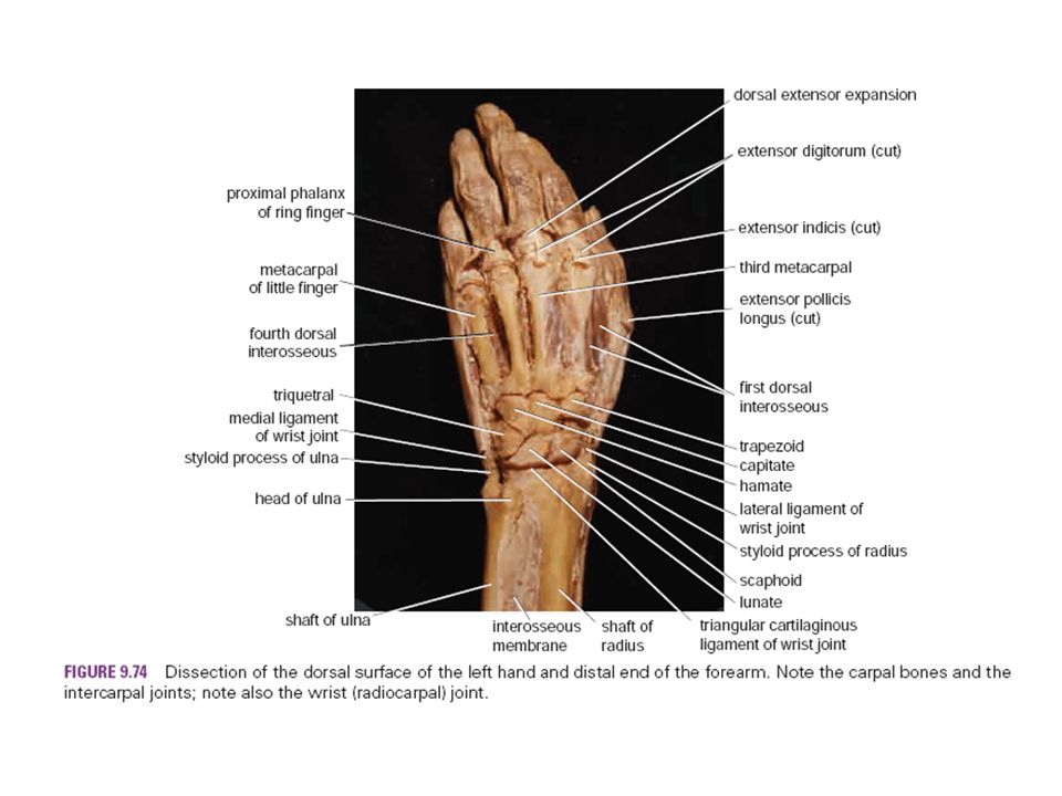
Lecture 26-Dorsum of the hand, retinaculum and wrist joint

2
– Enlist bones forming the wrist joint. – Describe the blood and nerve supply of the wrist joint. – Discuss the movements of the wrist joint. – Discuss the inferior radioulnar joint. – Enumerate different joints of the hand. – Define and explain anatomy of retinaculum. – Enlist blood supply, nerve supply and lymphatics of the dorsum of the hand. – Identify clinical application.

3
References: - Clinical anatomy by regions 9 th edition (Pages 394 – 412). - Gray’s anatomy for student, 2ed edition (Pages 751 – 774).
..")
4
Wrist joint-articulating bones The wrist joint is an articulation between the distal end of the radius along with an articular disc above, and the scaphoid, lunate and triquetral bones below.

7
Contd Nerve Supply: – Anterior interosseous nerve and the deep branch of radial nerve. Blood Supply: – Anterior interosseous artery (branch of ulnar artery) – Anterior and posterior carpal branches radial and ulnar arteries. – Palmar and dorsal metacarpal arteries of deep palmar arch.
 – Anterior and posterior carpal branches radial and ulnar arteries. – Palmar and dorsal metacarpal arteries of deep palmar arch..")
8
Movements of the wrist joint Flexion: – Flexor carpi radialis, flexor carpi ulnaris and palmaris longus. Extension: – Ext. carpi radialis longus, extensor carpi radialis brevis, extensor carpi ulnaris Abduction: – flexor carpi radialis, extensor carpi radialis and brevis. Adduction: flexor and extensor carpi ulnaris

9
Inferior radioulnar joint An articulation between rounded head of ulna and ulnar notch on radius. Type of joint: Synovial, pivot joint. Nerve Supply: Anterior interosseous nerve, deep branch of radial nerve. Movements: – Pronation: by pronator teres and pronator quadratus muscles – Supination: by biceps brachii and supinator muscles

11
Different joints of hand Intercarpal joints: allow gliding movements to occur which increase the range of extension and, more particularly, flexion permitted at the wrist joint. Carpometacarpal and intermetacarpal joints Metacarpophalyngeal joints Interphalyngeal joints

12
Retinacula Thickening of the deep fascia on anterior and posterior aspect of lower part of the forearm to protect the long tendons: – Flexor: Medial attachment to pisiform and hook of hamate, lateral to tubercle of scaphoid and trapezium bone---Carpal tunnel. – Extensor: Medial attachment to pisiform and hook of hamate, and lateral to distal end of radius--- Extensor compartments.

16
Dorsum of the hand Nerve supply: Sensory nerve supply to the skin is derived from: – Superficial branch of radial nerve supplies the lateral two thirds of the dorsum of the hand. It divides into several dorsal digital nerves that supply the thumb, the index and middle fingers, and the lateral side of the ring finger. – Post cut branch of ulnar nerve. supplies the medial third of the dorsum of the hand. It divides into several dorsal digital nerves that supply the medial side of the ring and the sides of the little fingers. Blood Supply: Branches of the radial artery.

18
Radial artery on dorsum of the hand The radial artery winds around the lateral margin of the wrist joint, beneath the tendons of the abductor pollicis longus and extensor pollicis brevis, and lies on the lateral ligament of the joint. On reaching the dorsum of the hand, the artery descends beneath the tendon of the extensor pollicis longus to reach the interval between the two heads of the first dorsal interosseous muscle; here, the artery turns forward to enter the palm of the hand. Branches of the radial artery on the dorsum of the hand take part in the anastomosis around the wrist joint. Dorsal digital arteries pass to the thumb and index finger.

21
Lymphatic drainage Superficial lymph vessels from: – Thumb, lateral fingers and lateral areas of hand, and forearm follow the cephalic vein → infra- clavicular nodes. – Medial fingers, medial areas of hand, and the forearm follow the basilic vein to cubital fossa → then to → axilla (lateral group).
..")
24
Clinical applications Carpal tunnel syndrome: the carpal tunnel, formed by the concave anterior surface of the carpal bones and closed by the flexor retinaculum, is tightly packed with the long flexor tendons of the fingers, with their surrounding synovial sheaths, and the median nerve. Clinically, the syndrome consists of a burning pain or “pins and needles” along the distribution of the median nerve to the lateral three and a half fingers and weakness of the thenar muscles. It is produced by compression of the median nerve within the tunnel.

25
Trigger finger: there is a palpable and even audible snapping when a patient is asked to flex and extend the fingers. It is caused by the presence of a localized swelling of one of the long flexor tendons Mallet finger: Avulsion of the insertion of one of the extensor tendons into the distal phalanges can occur if the distal phalanx is forcibly flexed when the extensor tendon is taut. The last 20° of active extension is lost, resulting in a condition known as mallet finger. Wrist joint injuries:

Similar presentations