Download presentation
Presentation is loading. Please wait.

1
Protein overexpression and induction in E. coli Determination of the protein b/w in the pellet and soluble state Protein purification using Ni-chelated resin Examination of protein purification SDS-PAGE Coomassie staining Western blotting

2
SDS-PAGE (polyacrylamide gel electrophoresis): denatured gel Polyacrylamide gel:-SDS: native gel +SDS:denatured gel reducing reagent: beta-mercaptoethanol, DTT (dithiothreitol) SDS-PAGE separates protein samples by the mobility of each macromolecule depending on the linear length of proteins’ primary structure and its mass-to-charge ratio.
: denatured gel Polyacrylamide gel:-SDS: native gel +SDS:denatured gel reducing reagent: beta-mercaptoethanol, DTT (dithiothreitol) SDS-PAGE separates protein samples by the mobility of each macromolecule depending on the linear length of proteins’ primary structure and its mass-to-charge ratio.")
3
2.475 ml 1.575 ml 62.5 ul 5 ul (Ammonium persulfate) Tetra-methyl-ethylene-di-amine
 Tetra-methyl-ethylene-di-amine")
4
Protein loading (sample) buffer 4X stock for protein sample buffer 2.0 ml1M Tris-HCl pH 6.8 0.8 gSDS 4.0 ml100% glycerol 0.4 ml14.7 M β-mercaptoethanol 1.0 ml0.5 M EDTA 8 mgbromophenol Blue SDS contained in the sample buffer makes proteins negatively charged proportionally to their length. 2-mercaptoethanol/DTT breaks disulphide bonds.

5
Staining solution 0.1% Coomassie Brilliant Blue R-250 50% methanol 10% glacial acetic acid D.W Destaining solution 40% methanol 10% glacial acetic acid D.W Staining an SDS-PAGE gel with Coomassie blue 1. Stain a gel in staining solution for 20 min with gentle agitation. 2. Destain a gel in destaining solution. Replenish the solution several times until background of the gel is fully destained. 3. Check the expressed protein in a destained gel 4. Store the destained gel in D.W

6
Coomassie blue staining

7
http://www.youtube.com/watch?v=b- 1dXzU4iOw How to detect protein in an SDS-PAGE?

8
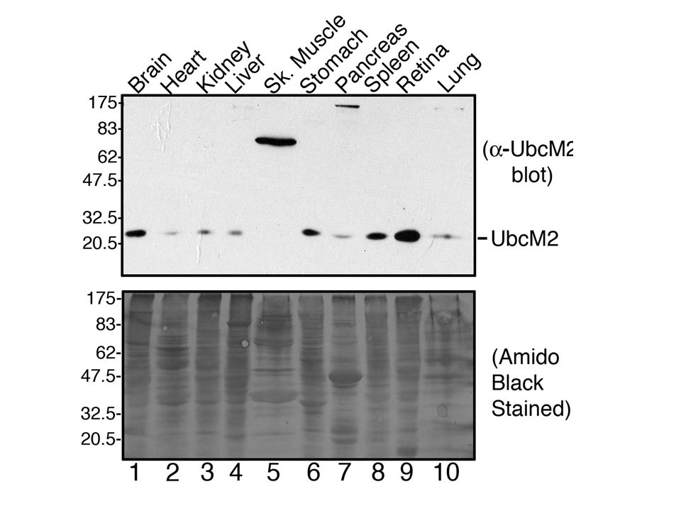
Western Blotting

9
1.Running a SDS-PAGE gel 2.Protein transfer from SDS-PAGE gel to PVDF (Polyvinylidene fluoride)/nitrocellulose membrane 3.Western blotting a.Blocking b.Incubation with primary antibody c.Washing d.Incubation with secondary antibody e.Washing f.Chemiluminescence Flow chart https://www.youtube.com/watch?v=VgAuZ6dBOfs
/nitrocellulose membrane 3.Western blotting a.Blocking b.Incubation with primary antibody c.Washing d.Incubation with secondary antibody e.Washing f.Chemiluminescence Flow chart v=VgAuZ6dBOfs")
10
Transfer buffer (blotting buffer) 48 mM Tris base 39 mM glycie 20% Methanol Fill up D.W up to 1 litre Blot onto PVDF membrane (Transfer) 1.cut PVDF membrane to size = 8 x 6cm 2.place a blotting cassette black side down in a large box of cold blotting buffer 3.immerse 2 blotting pads and a piece of Whatman paper (x2) 4.wet this in methanol & then equilibrate in blotting buffer 5.assemble the blotting sandwich (smooth out any air bubbles at each stage) Black side of cassette blotting pad a piece of Whatman paper resolving gel (stacker has been removed) PVDF membrane 1 piece of Whatman paper blotting pad white side of the cassette 6.place in blotting module in running tank 7.if only one gel is to be blotted then fill the other space with a second cassette containing just two blotting pads 8.insert a cooling block containing frozen water 9.fill tank with transfer buffer & place in an ice bucket 10.run at 100V for 1 hour
 48 mM Tris base 39 mM glycie 20% Methanol Fill up D.W up to 1 litre Blot onto PVDF membrane (Transfer) 1.cut PVDF membrane to size = 8 x 6cm 2.place a blotting cassette black side down in a large box of cold blotting buffer 3.immerse 2 blotting pads and a piece of Whatman paper (x2) 4.wet this in methanol & then equilibrate in blotting buffer 5.assemble the blotting sandwich (smooth out any air bubbles at each stage) Black side of cassette blotting pad a piece of Whatman paper resolving gel (stacker has been removed) PVDF membrane 1 piece of Whatman paper blotting pad white side of the cassette 6.place in blotting module in running tank 7.if only one gel is to be blotted then fill the other space with a second cassette containing just two blotting pads 8.insert a cooling block containing frozen water 9.fill tank with transfer buffer & place in an ice bucket 10.run at 100V for 1 hour")
11
1. Blocking Place blot in 5% Skim milk in TBS-T for at least 1 hour rinse blot in TBS-T, then wash in fresh TBS while preparing the primary antibodies 2. Primary antibody prep: Dilute primary antibody by a factor that fits in a 15 ml conical tube with 5% skim milk in TBS-T (dilution factor is depends on type of antibody & manufacturer recommendation ) Incubate primary with shaking in a clean container for at least 1 hour or o/n 3. Wash the membrane with TBS-T 5 min in TBS-T x 2 10 min in TBS-T x 2 15 min in TBS-T x 1 4. Secondary antibody (conjugate with Horseradish Peroxidase (HRP) attached, in fridge) 1/2000 (it is changeable) in 5% skim milk in TBS-T -use 10 ml per blot and incubate with shaking for at least 1 hour 5. Wash the membrane with TBS-T, then: 5 min in TBS-T x 2 10 min in TBS-T x 2 15 min in TBS-T x 2
 Incubate primary with shaking in a clean container for at least 1 hour or o/n 3. Wash the membrane with TBS-T 5 min in TBS-T x 2 10 min in TBS-T x 2 15 min in TBS-T x 1 4. Secondary antibody (conjugate with Horseradish Peroxidase (HRP) attached, in fridge) 1/2000 (it is changeable) in 5% skim milk in TBS-T -use 10 ml per blot and incubate with shaking for at least 1 hour 5. Wash the membrane with TBS-T, then: 5 min in TBS-T x 2 10 min in TBS-T x 2 15 min in TBS-T x 2.")
12
http://www.piercenet.com/method/chemilumi nescent-western-blotting

13
Detect the signal by chemiluminescence 1.Place some OHP film in a light resistant cassette 2.Using forceps grasp a edge of the membrane, remove excess liquid, and place on OHP film 3.mix 0.5 ml of each of HRP reagents 1 and 2 together per blot (once these are mixed then the light emitting reaction starts) 4.working quickly, pipette one ml onto each blot ensuring the entire surface is covered 5.leave for 1 min 6.place a second piece of film on top and smooth out any air bubbles 7.In dark room expose blot to X-ray film and develop - exposure times of 1 min and 5 min are good starting point (more exposure time is available, if the signal is weak) Development of the X-ray film After exposure X-ray film soaking in developer solution – water – fixer – water (you can see the band on film at this point)
 4.working quickly, pipette one ml onto each blot ensuring the entire surface is covered 5.leave for 1 min 6.place a second piece of film on top and smooth out any air bubbles 7.In dark room expose blot to X-ray film and develop - exposure times of 1 min and 5 min are good starting point (more exposure time is available, if the signal is weak) Development of the X-ray film After exposure X-ray film soaking in developer solution – water – fixer – water (you can see the band on film at this point)")
14
3-aminophthalate Horse radish peroxidase (HRP) subustrates: Luminol Light TMB, DAB, ABTS Coloured products Alkaline phosphatase (AP) substrates:NBT/BCIP Luminol Oxidation
 subustrates: Luminol Light TMB, DAB, ABTS Coloured products Alkaline phosphatase (AP) substrates:NBT/BCIP Luminol Oxidation")
Similar presentations


![Lab#6 Western Blotting BCH 462[practical].](/18/6070629/big_thumb.jpg "Lab#6 Western Blotting BCH 462[practical].>")