Download presentation
Presentation is loading. Please wait.

1
Nervous System Part 3: Neurons & Nerve Impulses

2
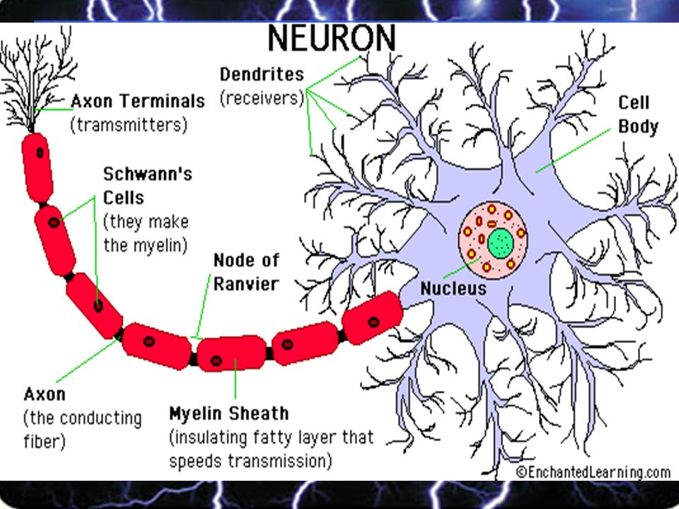
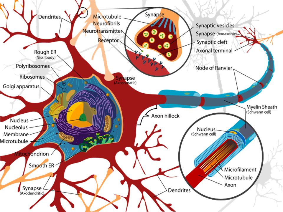
Neuron Structure A neuron is a nerve cellA neuron is a nerve cell The nucleus of a neuron and most of its organelles are located in the cell bodyThe nucleus of a neuron and most of its organelles are located in the cell body Dendrites are membrane-covered extensions that extend from the cell body in different directionsDendrites are membrane-covered extensions that extend from the cell body in different directions –They receive information form other neurons or other cells and carry the info toward the cell body An axon is a long, membrane-bound projectionAn axon is a long, membrane-bound projection –It transmits info away from the cell body via action potentials

3
Neuron Structure Continued Neurons may have a single axon or branching axonsNeurons may have a single axon or branching axons The end of an axon is called the axon terminal, which may contact and communicate with a muscle cell, a gland cell or another neuronThe end of an axon is called the axon terminal, which may contact and communicate with a muscle cell, a gland cell or another neuron Most axons are covered with a lipid layer called the myelin sheathMost axons are covered with a lipid layer called the myelin sheath The myelin sheath speeds up transmission of action potentialsThe myelin sheath speeds up transmission of action potentials

4
Neuron Structure Continued Schwann cells, which are found in neurons not of the brain or spinal cord, surround the axon and produce myelinSchwann cells, which are found in neurons not of the brain or spinal cord, surround the axon and produce myelin In the CNS, myelin is produced by a type of neuroglia called an oligodendrocyteIn the CNS, myelin is produced by a type of neuroglia called an oligodendrocyte Gaps in the myelin sheath along the length of the axon are called nodes of RanvierGaps in the myelin sheath along the length of the axon are called nodes of Ranvier On top of the myelin sheath is the neurilemma (neurilemmal sheath), but it is not present in the brain or spinal cord.On top of the myelin sheath is the neurilemma (neurilemmal sheath), but it is not present in the brain or spinal cord.
, but it is not present in the brain or spinal cord.On top of the myelin sheath is the neurilemma (neurilemmal sheath), but it is not present in the brain or spinal cord.")
6
Neuron Classification Neurons are classified in two ways: structural differences and functional differencesNeurons are classified in two ways: structural differences and functional differences There are 3 structural classifications: multipolar, bipolar and unipolarThere are 3 structural classifications: multipolar, bipolar and unipolar There are also 3 functional classifications: sensory, interneuron and motorThere are also 3 functional classifications: sensory, interneuron and motor How they are connected is found on p.368 in table 10.1 of your bookHow they are connected is found on p.368 in table 10.1 of your book

7
Neuron Classification

8
Neuron Structural Classification 1.Multipolar neurons: –have many processes arising from their cell bodies –Only one process is an axon & the rest are dendrites –Found mostly in the brain and spinal cord –Picture on p.367

9
Neuron Structural Classification 2.Bipolar neurons: –The cell bodies have only two processes, one on each end –One process is an axon and the other is a dendrite –They are found in specialized parts of the eyes, nose and ears

10
Neuron Structural Classification 3.Unipolar neurons: –Have a single process extending from their cell bodies –A short distance from the cell body, this process divides into two branches, which function as a single axon –One branch (peripheral process) is associated with the dendrites near a peripheral body part –The other branch (central process) enter the brain or spinal cord –The cell bodies of some unipolar neurons aggregate in specialized masses of nerve tissue called ganglia, which are located outside of the CNS
 is associated with the dendrites near a peripheral body part –The other branch (central process) enter the brain or spinal cord –The cell bodies of some unipolar neurons aggregate in specialized masses of nerve tissue called ganglia, which are located outside of the CNS")
11
Neuron Functional Classification 1.Sensory Neurons: –Also known as afferent neurons –Conduct impulses from peripheral body parts to the brain or spinal cord –At their distal ends, the dendrites or specialized structures act as sensory receptors –Most are unipolar, but some are bipolar

12
Neuron Functional Classification 2.Interneurons: –Also known as association or internuncial neurons –They lie within the brain or spinal cord –They are multipolar and form links with other neurons –They relay information from one part of the CNS to another part –They direct incoming sensory information to appropriate regions for processing

13
Neuron Functional Classification 3.Motor Neurons: –Also known as efferent neurons –Are multipolar and conduct impulses out of the brain or spinal cord to effectors –Motor neurons of skeletal muscles are under voluntary control –Motor neurons of cardiac and smooth muscles are involuntarily controlled.

14
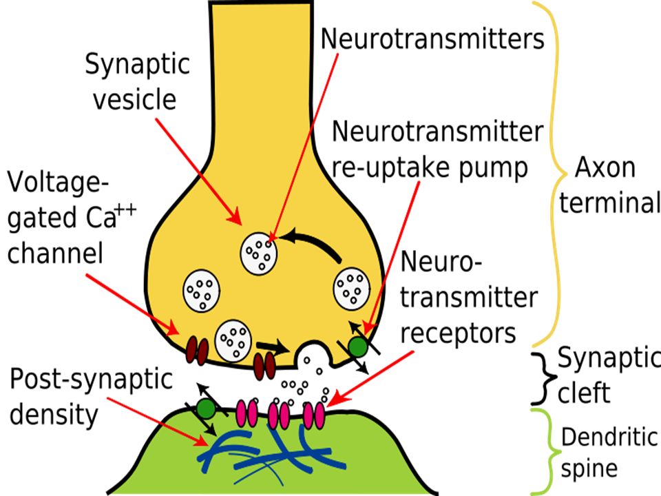
Neuron Communication Neurons communicate with other neurons and other cells at special junctions called synapsesNeurons communicate with other neurons and other cells at special junctions called synapses Neurons usually do not touch each other or other cellsNeurons usually do not touch each other or other cells A small gap, called a synaptic cleft, is present between the axon terminal and the receiving cellA small gap, called a synaptic cleft, is present between the axon terminal and the receiving cell Electrical activity in the neuron usually causes the release of chemicals called neurotransmitters into the synaptic cleftElectrical activity in the neuron usually causes the release of chemicals called neurotransmitters into the synaptic cleft

16
Synaptic Terminology At a synapse, the transmitting neuron is called a presynaptic neuronAt a synapse, the transmitting neuron is called a presynaptic neuron The receiving cell is called a postsynaptic cellThe receiving cell is called a postsynaptic cell

18
Nerve Impulses All cells, including neurons, have an electrical charge inside the cell that is different from the electrical charge outside the cellAll cells, including neurons, have an electrical charge inside the cell that is different from the electrical charge outside the cell This difference in electrical charge across a membrane is called a membrane potentialThis difference in electrical charge across a membrane is called a membrane potential Membrane potentials are produced by the movement of ions across a cellular membraneMembrane potentials are produced by the movement of ions across a cellular membrane

19
Resting Potential A neuron is at rest when it is not receiving or sending a signalA neuron is at rest when it is not receiving or sending a signal In most neurons, the resting potential isIn most neurons, the resting potential is -70 millivolts -70 millivolts

20
Action Potential When a dendrite or cell body is stimulated, the permeability of the neuron’s membrane changes suddenlyWhen a dendrite or cell body is stimulated, the permeability of the neuron’s membrane changes suddenly This action begins an action potentialThis action begins an action potential

21
At Membrane Level At the resting potential, sodium channels are closed and some potassium channels are openAt the resting potential, sodium channels are closed and some potassium channels are open During an action potential, sodium channels open, allowing sodium ions to move into the axonDuring an action potential, sodium channels open, allowing sodium ions to move into the axon

22
Nerve Impulse Animation!!!!Animation!!!!Animation!!!! The essential steps are outlined in the animationThe essential steps are outlined in the animation

Similar presentations


 1.Microglial cells –Scattered throughout CNS –Support neurons.>")


 from one part of the body to another. ◦ Major regions.>")


