Download presentation
Presentation is loading. Please wait.

1
Hypertrophic Pyloric Stenosis (HPS) Jenelle Beadle 2/11/16
 Jenelle Beadle 2/11/16")
2
Hyperplasia & hypertrophy of pyloric muscle x2 layers of muscle – longitudinal and circular visualized as one hypoechoic layer on ultrasound

3
Pathogenesis is not understood Immunohistochemical Genetic Infectious Hyperacidity

4
Pathogenesis is not understood Immunohistochemical Genetic Infectious Hyperacidity Associations Turner Syndrome Tracheo-esophageal fistula Esophageal atresia Trisomy 18 Renal anomalies

5
Pathogenesis is not understood Immunohistochemical Genetic Infectious Hyperacidity Associations Turner Syndrome Tracheo-esophageal fistula Esophageal atresia Trisomy 18 Renal anomalies Incidence: 2-5 per 1,000 per year caucasian male first born family history

6
Upper abdominal distension Olive-shaped palpable RUQ mass Noticeable left-to-right peristaltic waves Projectile vomiting followed by hunger (eager feeding) Signs of dehydration & malnutrition sunken anterior fontanel & eyes decreased elasticity of the skin failure to thrive
 Signs of dehydration & malnutrition sunken anterior fontanel & eyes decreased elasticity of the skin failure to thrive")
7
Presents at 3-6wks gradual onset of worsening vomiting very rare in infants > 12wks Treatment is surgical pylormyomotomy successful with few complications not emergent, so infant’s hydration/electrolyte imbalance is corrected prior to surgery

8
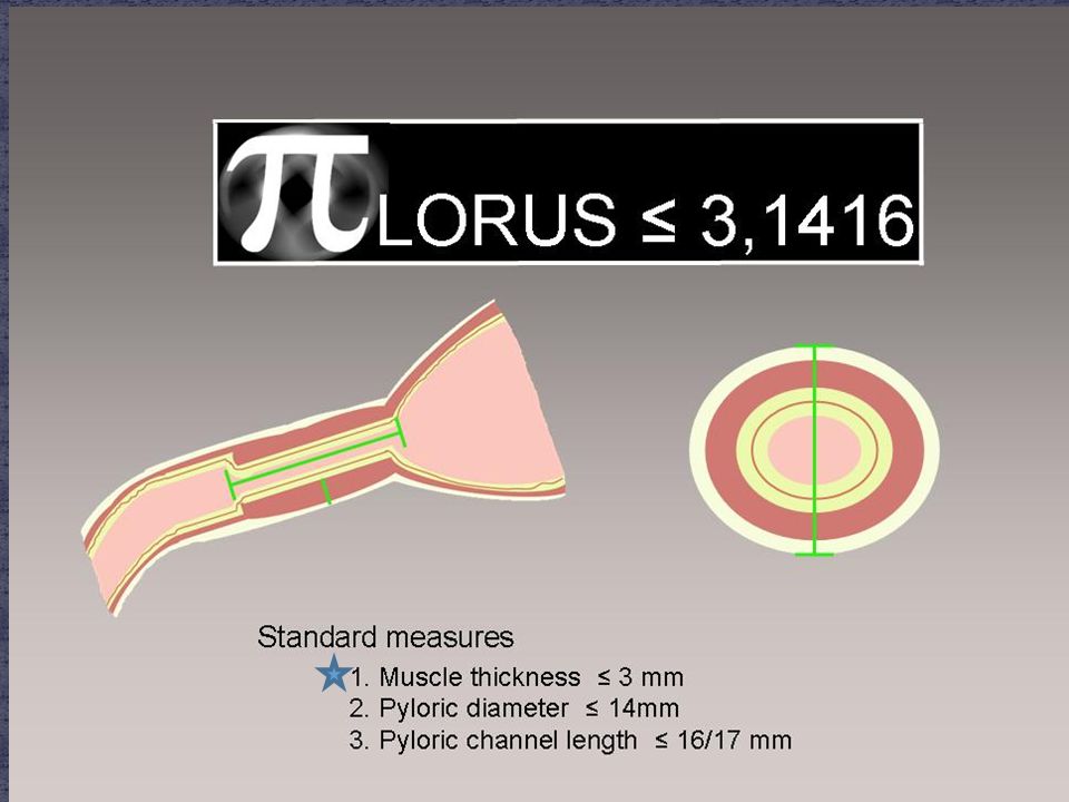
Fasting Longitudinal and transverse Measurements muscle layer thickeness normal <3mm length normal <15mm Post-prandial Longitudinal fluid passing through the pylorus images and cines

10
Pylorus Layers

11
Serosa – echogenic very thin

12
Serosa – echogenic Muscularis – hypoechoic thickest layer composed of x2 layers of muscle longitudinal circular hypertrophic > 3mm

13
Hypertrophic Normal

14
Serosa – echogenic Muscularis – hypoechoic Submucosa – echogenic Muscularis Mucosae – hypoechoic These x2 layers are often indistinguishable from the adjacent muscle ?problems with measurement

15
Serosa – echogenic Muscularis – hypoechoic Submucosa – echogenic Muscularis Mucosae – hypoechoic Mucosa – echogenic (usually) with IPS, it may protrude into antrum of the stomach “nipple sign”
 with IPS, it may protrude into antrum of the stomach nipple sign")
16
Inner mucosa appearance is varied Relatively thin usually IPS positive Relatively thick usually IPS negative

17
Inner mucosa appearance is varied Relatively thin usually IPS positive Relatively thick usually IPS negative

18
Inner mucosa appearance is varied Single echogenic Most common x2 echogenic layers with a hypoechoic layer in between

19
Inner mucosa appearance is varied Single echogenic Most common x2 echogenic layers with a hypoechoic layer in between

20
Serosa – echogenic Muscularis – hypoechoic Submucosa – echogenic Muscularis Mucosae – hypoechoic Mucosa – echogenic (sometimes with a hypoechoic center layer) Lumen – potential space normal: contents moving through IPS: empty (mucosa walls coapted)
 Lumen – potential space normal: contents moving through IPS: empty (mucosa walls coapted)")
21
Bad Measurement Including all layers Technically Good Measurement Muscle layer only

22
But what about this one? A more typical appearance? Submucosa and muscularis mucosae can not be distinguished from the overlying muscle, so they are included in the measurement Will this result in inaccuracies?

23
Submucosa and muscularis mucosae are clearly distinguishable from muscle

24
Submucosa and muscularis mucosae are not clearly visualized included in muscle measurement

25
I don’t know. more digging through the literature is necessary The most reproducible measurement is to include submucosa and muscularis mucosae layers in the muscle measurement, as shown below, but this would be unconventional. Does it matter? it is always important to be accurate, but it could make a difference in borderline measurements as seen in premature infants or in early findings of pyloric stenosis.

26
Typical indications vomiting failure to thrive Patient Preparation Fasting for 4 hours Parent should be prepared to feed the infant during the course of the examination Warm blankets & warm gel Sterile gel should be used during the neonatal period Pacifier 9L Transducer

27
Post-prandial Milk Water

28
Survey in a transverse plane to find the longitundinal pylorus use the liver and/or GB as a window slide up and a angle inferiorly

29
Position the infant in a RPO position a rolled blanket may be helpful this will direct gas bubbles away from the area of interest and fluid towards the pylorus

30
Distended stomach can result in posterior displacement of the pylorus do not overfeed the infant for the post- prandial portion of the exam pylorus similar in appearance to a cervix positioning the infant in a LLD may improve visualization

31
Easier to identify than a normal pylorus Abnormal Measurements muscle layer >3mm length >15mm

32
Nipple Sign Thickening of the mucosa protruding through the antrum

Similar presentations


 versus partial wrap (Thal/Toupet) H. Ahmed , U. Rolle, H. Till Department.>")


 Department of Pediatrics>")


