Download presentation
Presentation is loading. Please wait.

1
MEDICAL USES FOR RADIATION Andres Perez P.1 Level 4 MT10

2
RADIATION FOR MEDICAL USE In medicine it is required to utilize X-rays in order for a doctor to properly diagnose a disease, such as Cancer, but also, it can be used for radiotherapy in order to try and cure a disease. Radiation is very helpful in the Medical Field, it has MANY benefits that allow it to be a most commonly used treatment & diagnostic, especially when it comes to Cancer. But while it may have many benefits, it also has quite a few risks that come along with utilizing it. If used improperly and unprofessionally, it is MOST likely that the disease would only become even worse and the consequences could be very fatal.

3
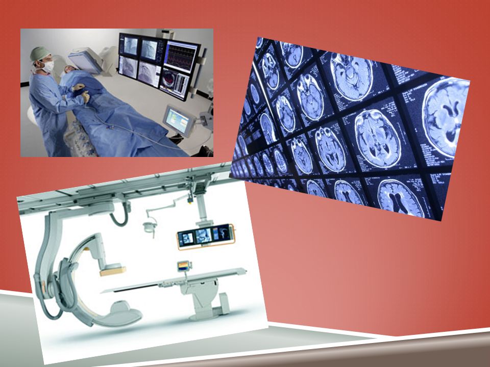
DIAGNOSIS WITH RADIATION Diagnostic Radiology: CT Scan: Use of X-rays to examine patients X-rays penetrate flesh, & bone on photographic film In some cases the images are captured & processed electronically. Radiation from a machine passes through patient Parts of Body Commonly Examined: Chest Limbs Teeth Doses are fairly low For Example: About 0.1 mSv from chest exam. The use has increased considerably in recent years. Make up approximately 5% of all procedures (in developed countries) A fan-shaped beam of X-rays is rotated around the patient and registered on the opposite side by a row of detectors. An image of a slice or section through the patient is then reconstructed by a computer and expresses superior diagnostic info. Doses can be an order of magnitude or more higher than those from conventional X-ray exams.
 A fan-shaped beam of X-rays is rotated around the patient and registered on the opposite side by a row of detectors. An image of a slice or section through the patient is then reconstructed by a computer and expresses superior diagnostic info. Doses can be an order of magnitude or more higher than those from conventional X-ray exams..")
4
INTERVENTIONAL RADIOLOGY Diagnostic procedure that gives the highest doses. If it is not controlled carefully, it can lead to similarly high doses to surgeons. The physician uses a series of X-rays to “see” into the patient in real time while performing a procedure inside the patient. Allows a procedure on an internal organ to be done without the complicated surgery that might have been needed to get to the organ. Procedures can give patient doses in the range 10 -100mSv Doses from procedures have been high enough to cause “deterministic effects” in patients & surgeons alike.

6
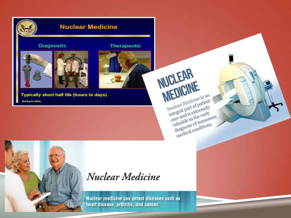
NUCLEAR MEDICINE The patient is given a radionuclide in a carrying substance, which is preferentially taken up by the tissue or organ under study. Administration may be by injection, ingestion, or inhalation The radionuclide emits gamma rays When used to treat instead of diagnosis, much greater activities are given to the patient and much higher doses are given to the target tissues or organs. Treatment of an overactive thyroid gland-hyperthyroidsim-is probably the most common therapeutic procedure(the radionuclide used is iodine-131) Many make us of the radionuclide techetium-99m Has a half-life of 6hours Gives off gamma rays with an energy of 0.14MeV Can be conveniently prepared in the hospital Readily labels a variety of carrying substances Although they may have short half- lives, taking to account of the fact that activity still remains in the body of the patient that received the radionuclide for a while after the procedure, is advised.
 Many make us of the radionuclide techetium-99m Has a half-life of 6hours Gives off gamma rays with an energy of 0.14MeV Can be conveniently prepared in the hospital Readily labels a variety of carrying substances Although they may have short half- lives, taking to account of the fact that activity still remains in the body of the patient that received the radionuclide for a while after the procedure, is advised..")
8
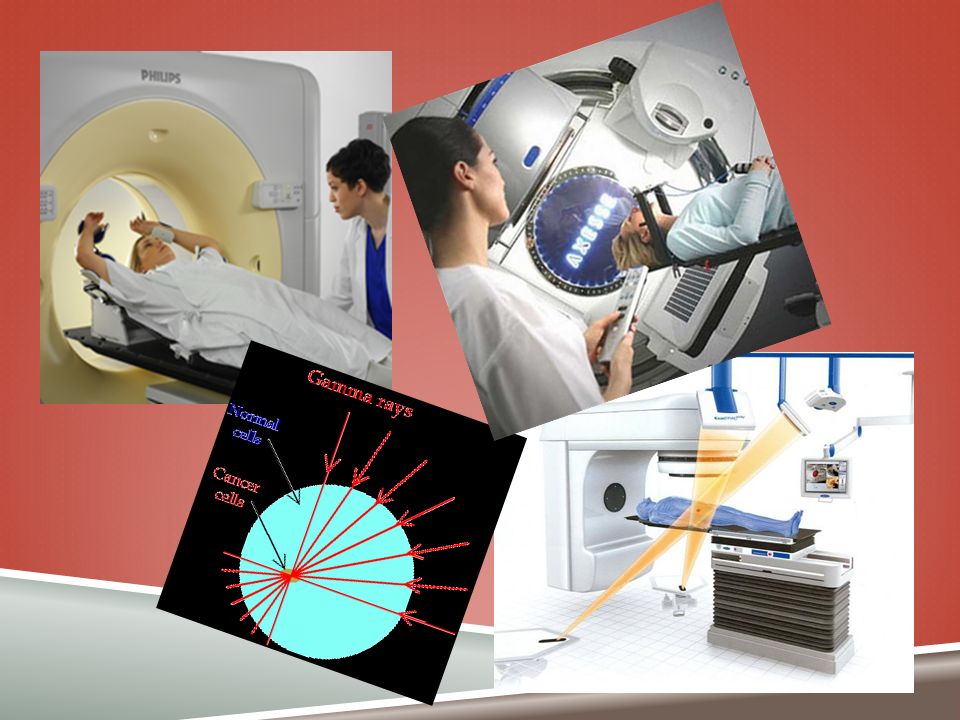
RADIOTHERAPY Technique used to cure Cancers or at least alleviate the most distressing symptoms, by killing cancerous cells. A beam of high energy X-rays, gamma rays or electrons is directed towards the diseased tissue so it can receive a high dose while sparing the surrounding healthy tissue. If tumor is deep in the body, beam is pointed at it from several directions to reduce the incidental damage. Brachytherapy: Another form of treatment, in which a radiation source is placed in/on the body for a short period (is used for some cancers.) Since radiotherapy doses are strong, this treatment is only used when the outlook for a cure or relief is good and when other methods for treatment would be less effective. Although it can cure the original cancer, it may possibly cause cancer in other tissues or “adverse hereditary effects in subsequent generations.” Most people who receive this therapy are: Past the age to have children Too old for delayed cancers to occur
 Since radiotherapy doses are strong, this treatment is only used when the outlook for a cure or relief is good and when other methods for treatment would be less effective. Although it can cure the original cancer, it may possibly cause cancer in other tissues or adverse hereditary effects in subsequent generations. Most people who receive this therapy are: Past the age to have children Too old for delayed cancers to occur.")
9
RADIOTHERAPY Its aim is to maximize the effectiveness of treatment while minimizing the opposing side effects. Tumors need absorbed doses of “tens of gray” to kill the cancer cells successfully. Prescribed doses to tissues are typically in the range 20- 60 Gy normally given in fractions over a period of several weeks. Considerable care is required to deliver accurate doses. Too low or too high doses may lead to incomplete treatment or unacceptable side-effects. Consequences may be grave. A miscalibrated radiotherapy beam in Costa Rica in 1996 resulted in more than 100 patients receiving higher doses then intended; leading to death or serious injury.

11
CAUTIONS WITH RADIATION Guidance Levels for Medical Exposure Methods of Minimizing Doses Its very important to avoid unnecessary exposures and keep the essential exposures as low as possible. Decision to prescribe an X- ray exam is a matter of medical judgment made in the best interests of the patient. The dose to the patient should be the lowest possible compatible with accurate diagnosis. Use of good equipment Well-maintained, properly adjusted, & skillfully operated Having a programmer of quality in the X-ray Department. Young people do not have many X-rays and the likelihood of needing an examination increases with age. This implies a lower probability, in general, of any consequential cancers being expressed.

13
WORKS CITED http://www.iaea.org/Publications/Booklets/RadPeopleEnv/pdf/chapter_8. pdf http://www.iaea.org/Publications/Booklets/RadPeopleEnv/pdf/chapter_8. pdf

Similar presentations


>")


 CT scanning or (CAT scanning) is using X-rays to create a 3D image of the inside of an object. CT stands for computed tomography.>")

>")


 Topic:>")

