Download presentation
Presentation is loading. Please wait.

1
Radiation Exposure from X-ray and CT Examinations Evan Lum

2
What is an X-ray An x-ray is a form of electromagnetic radiation which falls in between gamma rays and ultra violet rays on the electromagnetic spectrum. They have a wavelength of anywhere between 0.01 nm and 10 nm. Corresponds to an energy of 0.1 to about 123 keV (kiloelectron volts)
.")
3
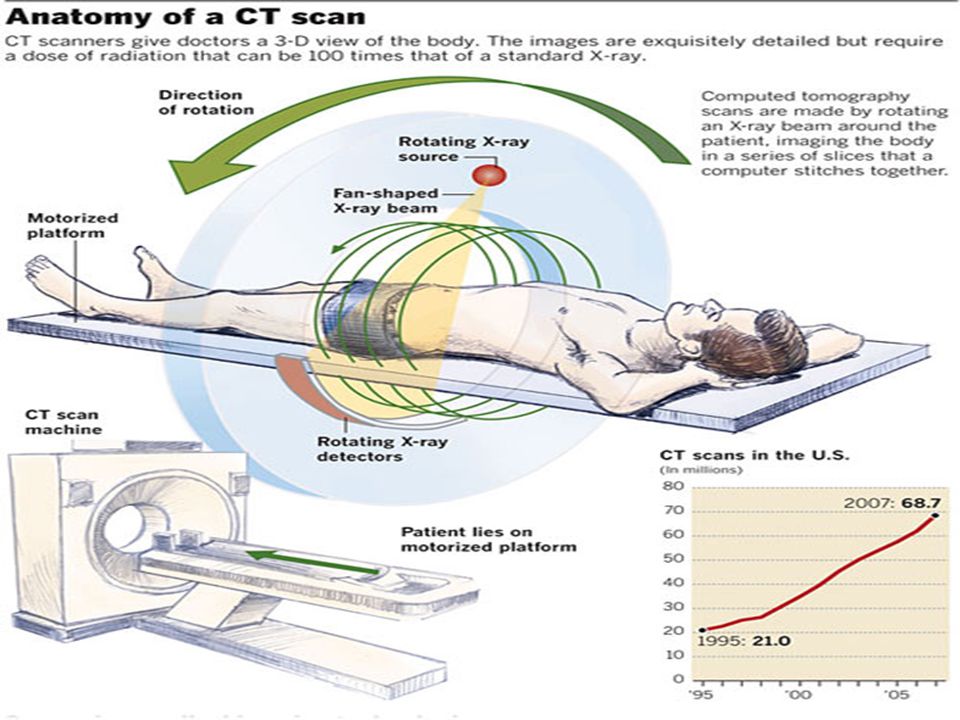
What is a CT scan? CT stands for computed tomography which is a medical imaging method employing tomography (imaging in sections) created by computer processing. Using a series of x-ray images about a single axis of rotation, CT produces volume data such as 3D images of internal organs which can be manipulated through a process called “windowing”.
 created by computer processing. Using a series of x-ray images about a single axis of rotation, CT produces volume data such as 3D images of internal organs which can be manipulated through a process called windowing ..")
4
Medical Uses for X-ray Plain x-rays are mostly used to detect pathology in the skeletal system. Sometimes used for soft tissue: – Chest x-ray to identify lung diseases like pneumonia or lung cancer. – Abdominal x-ray to detect intestinal obstruction, free air or free fluid.

5
Medical Uses for CT Scan Head – detect infarction, tumors, hemorrhage, bone trauma. Lungs – shows internal aspects of the lungs which plain x-ray can not. Cardiac – excellent imaging of coronary arteries Abdominal/Pelvic – sensitive method for diagnoses of abdominal diseases. Extremities – can image complex fractures, especially around joints. Also ligament injuries and dislocations can be seen.

7
Radiation Exposure The Sievert is the SI unit for radiation dose which is equivalent to one joule per kilogram. The average American is exposed to about 3 mSv per year from naturally occurring radioactive materials and cosmic radiation from outer space. This amount of radiation exposure over such a long period of time is basically harmless to us.

8
Radiation from X-rays Extremities – 0.001 mSv per x-ray (not harmful) Spine – 1.5 mSv per x-ray (about 6 months worth of natural exposure) GI tract – 6-8 mSv per x-ray (2-3 years worth of natural exposure) Each of these are considered not to be relatively harmful
 Spine – 1.5 mSv per x-ray (about 6 months worth of natural exposure) GI tract – 6-8 mSv per x-ray (2-3 years worth of natural exposure) Each of these are considered not to be relatively harmful")
9
Head – 2 mSv per CT scan (under 1 year) Spine – 6 mSv per CT scan (2 years) Chest – 7 mSv per CT scan (over 2 years) Abdomen/Pelvis – 15 mSv per CT scan (5 years) Abdomen/Pelvis without contrast material – 30 mSv per CT scan (10 years) Coronary Computed Tomography Angiography (CTA) – 16 mSv (5 years) Radiation from CT Scan
 Spine – 6 mSv per CT scan (2 years) Chest – 7 mSv per CT scan (over 2 years) Abdomen/Pelvis – 15 mSv per CT scan (5 years) Abdomen/Pelvis without contrast material – 30 mSv per CT scan (10 years) Coronary Computed Tomography Angiography (CTA) – 16 mSv (5 years) Radiation from CT Scan")
10
Why is this dangerous? Although radiation can be used to treat cancer, it can also cause it. The delivery of energy waves through our body can alter atoms’ molecular structure causing cells in our body to mutate and become cancerous.

11
Higher risk procedures can leave patients with up to a 1 in 500 chance of getting cancer. The united states gives over 60 million CT scans per year. It has been estimated recently that CT scanning has been the cause of about 30,000 cases of cancer yearly. Statistics

12
What Can We Do? Due to their major contribution to the medical field, the benefits of x-ray imaging and CT scanning usually outweigh the risks from radiation exposure. Something we can do to avoid this is avoiding unnecessary procedures. This decision should only be made by a professional not a patient!

13
Sources http://www.usatoday.com/news/health/2009-12-15- radiation15_st_N.htm http://www.usatoday.com/news/health/2009-12-15- radiation15_st_N.htm http://en.wikipedia.org/wiki/X- ray_computed_tomography http://en.wikipedia.org/wiki/X- ray_computed_tomography http://www.radiologyinfo.org/en/pdf/sfty_xray.pdf http://www.worldculturepictorial.com/blog/content/ct -scan-study-shows-increased-radiation-exposure- cancer-risks-tests-often-unnecessary http://www.worldculturepictorial.com/blog/content/ct -scan-study-shows-increased-radiation-exposure- cancer-risks-tests-often-unnecessary http://www.hanskellner.com/2005/06/27/left- shoulder-xray/ http://www.hanskellner.com/2005/06/27/left- shoulder-xray/ “The Physics of Medical Imaging”, by S. Webb, New York: Adam Hilger, 1990.

14
THE END

Similar presentations


 Ionizing Radiation.>")

 theory was developed 1972: The CT scan was invented by Godfrey.>")


 CT scanning or (CAT scanning) is using X-rays to create a 3D image of the inside of an object. CT stands for computed tomography.>")


>")


 Topic:>")


