Download presentation
Presentation is loading. Please wait.

2
Muhammad Sohaib Shahid (Lecturer & Course Co-ordinator BS-MIT) University Institute of Radiological Sciences & Medical Imaging Technology (UIRSMIT)
 University Institute of Radiological Sciences & Medical Imaging Technology (UIRSMIT)")
4
The vertebral column is the central bony pillar of the body. It supports the skull, pectoral girdle, upper limbs, and thoracic cage and, by way of the pelvic girdle, transmits body weight to the lower limbs. Within its cavity lie the spinal cord, the roots of the spinal nerves, and the covering meninges, to which the vertebral column gives great protection. THE VERTEBRAL COLUMN

5
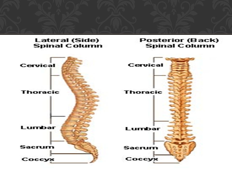
The vertebral column is composed of 33 vertebrae 7 cervical, 12 thoracic, 5 lumbar, 5 sacral (fused to form the sacrum), and 4 coccygeal (the lower 3 are commonly fused). Because it is segmented and made up of vertebrae, joints, and pads of fibrocartilage called intervertebral discs, it is a flexible structure. The intervertebral discs form about one fourth the length of the column. COMPOSITION OF THE VERTEBRAL COLUMN

7
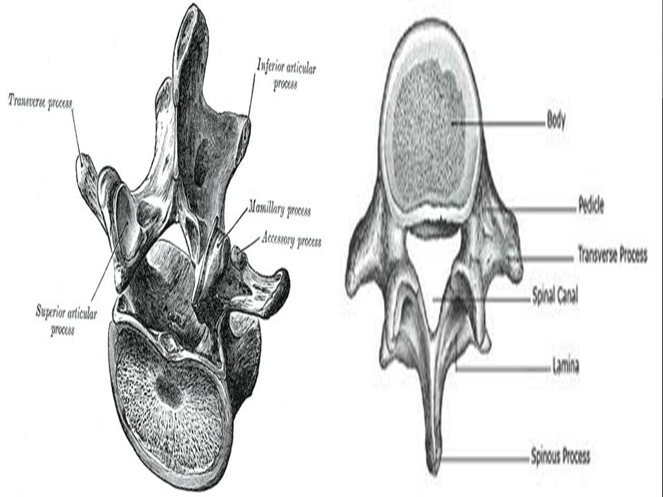
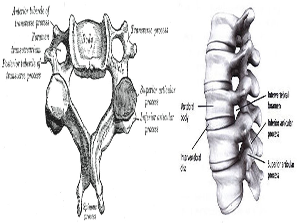
A typical vertebra consists of a rounded body anteriorly and a vertebral arch posteriorly. These enclose a space called the vertebral foramen, through which run the spinal cord and its coverings. The vertebral arch consists of a pair of cylindrical pedicles, which form the sides of the arch, and a pair of flattened laminae, which complete the arch posteriorly. The vertebral arch gives rise to seven processes: one spinous, two transverse, and four articular. The spinous process, or spine, is directed posteriorly from the junction of the two laminae. The transverse processes are directed laterally from the junction of the laminae and the pedicles. Both the spinous and transverse processes serve as levers and receive attachments of muscles and ligaments. GENERAL CHARACTERISTICS OF A VERTEBRA

9
The articular processes are vertically arranged and consist of two superior and two inferior processes. They arise from the junction of the laminae and the pedicles, and their articular surfaces are covered with hyaline cartilage. The two superior articular processes of one vertebral arch articulate with the two inferior articular processes of the arch above, forming two synovial joints. The pedicles are notched on their upper and lower borders, forming the superior and inferior vertebral notches. On each side, the superior notch of one vertebra and the inferior notch of an adjacent vertebra together form an intervertebral foramen These foramina, in an articulated skeleton, serve to transmit the spinal nerves and blood vessels. The anterior and posterior nerve roots of a spinal nerve unite within these foramina with their coverings of Dura to form the segmental spinal nerves.

12
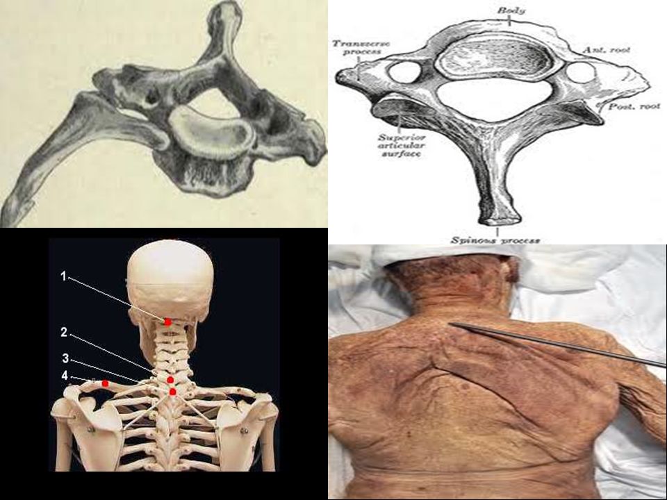
A typical cervical vertebra has the following characteristics: The transverse processes possess a foramen transversarium for the passage of the vertebral artery and veins (note that the vertebral artery passes through the transverse processes C1 to 6 and not through C7). The spines are small and bifid. The body is small and broad from side to side. The vertebral foramen is large and triangular. The superior articular processes have facets that face backward and upward; the inferior processes have facets that face downward and forward. CHARACTERISTICS OF A TYPICAL CERVICAL VERTEBRA

13
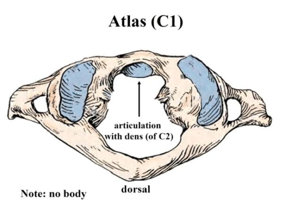
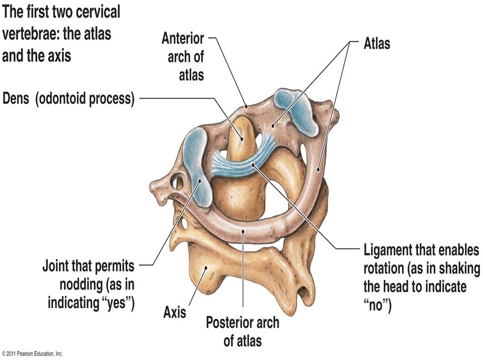
The first, second, and seventh cervical vertebrae are atypical. The first cervical vertebra, or atlas, does not possess a body or a spinous process. It has an anterior and posterior arch. It has a lateral mass on each side with articular surfaces on its upper surface for articulation with the occipital condyles (atlanto-occipital joints) and articular surfaces on its lower surface for articulation with the axis (atlantoaxial joints). The second cervical vertebra, or axis, has a peg like odontoid process that projects from the superior surface of the body (representing the body of the atlas that has fused with the body of the axis). The seventh cervical vertebra, or vertebra prominens, is so named because it has the longest spinous process, and the process is not bifid. The transverse process is large, but the foramen transversarium is small and transmits the vertebral vein or veins. CHARACTERISTICS OF THE ATYPICAL CERVICAL VERTEBRAE
 and articular surfaces on its lower surface for articulation with the axis (atlantoaxial joints). The second cervical vertebra, or axis, has a peg like odontoid process that projects from the superior surface of the body (representing the body of the atlas that has fused with the body of the axis). The seventh cervical vertebra, or vertebra prominens, is so named because it has the longest spinous process, and the process is not bifid. The transverse process is large, but the foramen transversarium is small and transmits the vertebral vein or veins. CHARACTERISTICS OF THE ATYPICAL CERVICAL VERTEBRAE.")
17
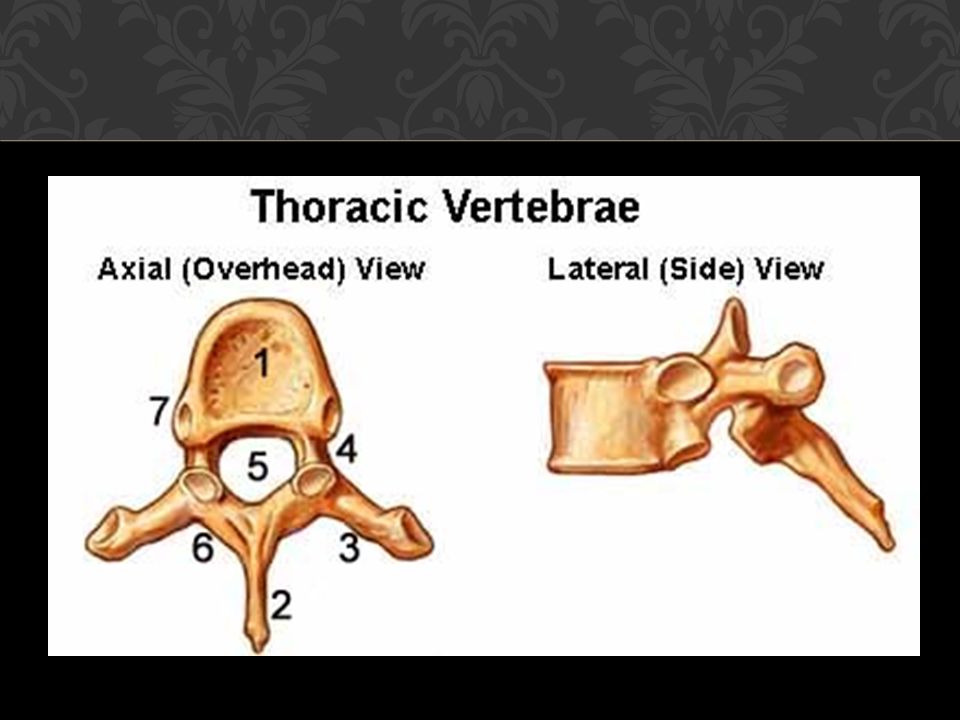
A typical thoracic vertebra has the following characteristics : The body is medium size and heart shaped. The vertebral foramen is small and circular. The spines are long and inclined downward. Costal facets are present on the sides of the bodies for articulation with the heads of the ribs. Costal facets are present on the transverse processes for articulation with the tubercles of the ribs (T11 and 12 have no facets on the transverse processes). The superior articular processes bear facets that face backward and laterally, whereas the facets on the inferior articular processes face forward and medially. The inferior articular processes of the 12th vertebra face laterally, as do those of the lumbar vertebrae. CHARACTERISTICS OF A TYPICAL THORACIC VERTEBRA
. The superior articular processes bear facets that face backward and laterally, whereas the facets on the inferior articular processes face forward and medially. The inferior articular processes of the 12th vertebra face laterally, as do those of the lumbar vertebrae. CHARACTERISTICS OF A TYPICAL THORACIC VERTEBRA.")
19
A typical lumbar vertebra has the following characteristics : The body is large and kidney shaped. The pedicles are strong and directed backward. The laminae are thick. The vertebral foramina are triangular. The transverse processes are long and slender. The spinous processes are short, flat, and quadrangular and project backward. The articular surfaces of the superior articular processes face medially, and those of the inferior articular processes face laterally. Note that the lumbar vertebrae have no facets for articulation with ribs and no foramina in the transverse processes. CHARACTERISTICS OF A TYPICAL LUMBAR VERTEBRA

21
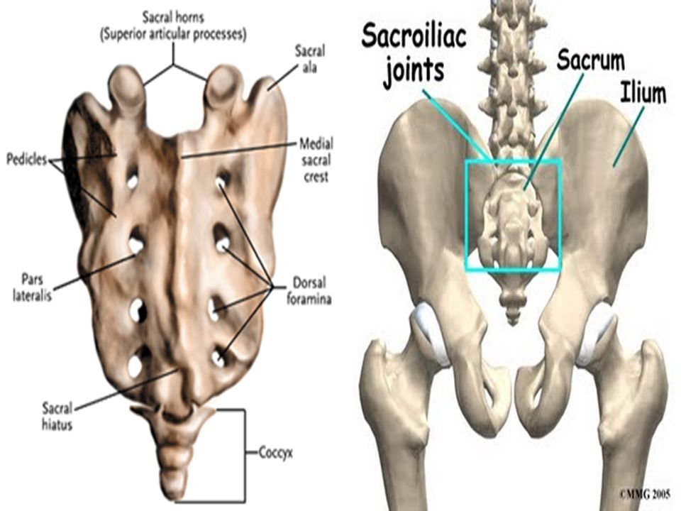
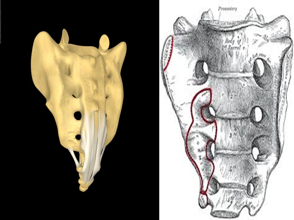
The sacrum consists of five rudimentary vertebrae fused together to form a wedge-shaped bone, which is concave anteriorly. The upper border, or base, of the bone articulates with the fifth lumbar vertebra. The narrow inferior border articulates with the coccyx. Laterally, the sacrum articulates with the two iliac bones to form the sacroiliac joints. The anterior and upper margin of the first sacral vertebra bulges forward as the posterior margin of the pelvic inlet and is known as the sacral promontory. The sacral promontory in the female is of considerable obstetric importance and is used when measuring the size of the pelvis. The vertebral foramina are present and form the sacral canal. The laminae of the fifth sacral vertebra, and sometimes those of the fourth also, fail to meet in the midline, forming the sacral hiatus. The sacral canal contains the anterior and posterior roots of the sacral and coccygeal spinal nerves, the filum terminale, and fibrofatty material. It also contains the lower part of the subarachnoid space down as far as the lower border of the second sacral vertebra. The anterior and posterior surfaces of the sacrum each have four foramina on each side for the passage of the anterior and posterior rami of the upper four sacral nerves. SACRUM

24
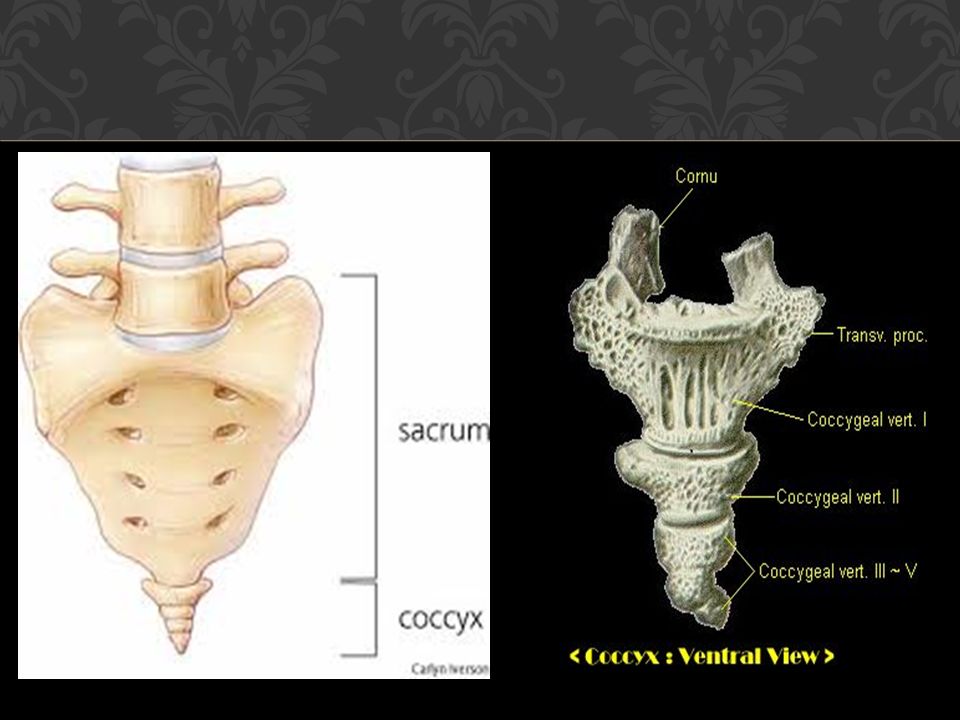
The coccyx consists of four vertebrae fused together to form a single, small triangular bone that articulates at its base with the lower end of the sacrum. The first coccygeal vertebra is usually not fused or is incompletely fused with the second vertebra. Knowledge of the preceding basic anatomy of the vertebral column is important when interpreting radiographs and when noting the precise sites of bony pathologic features relative to soft tissue injury. COCCYX

26
The number of cervical vertebrae is constant, but the seventh cervical vertebra may possess a cervical rib. The thoracic vertebrae may be increased in number by the addition of the first lumbar vertebra, which may have a rib. The fifth lumbar vertebra may be incorporated into the sacrum; this is usually incomplete and may be limited to one side. The first sacral vertebra may remain partially or completely separate from the sacrum and resemble a sixth lumbar vertebra. A large extent of the posterior wall of the sacral canal may be absent because the laminae and spines fail to develop. The coccyx, which usually consists of four fused vertebrae, may have three or five vertebrae. The first coccygeal vertebra may be separate. In this condition, the free vertebra usually projects downward and anteriorly from the apex of the sacrum. IMPORTANT VARIATIONS IN THE VERTEBRAE

27
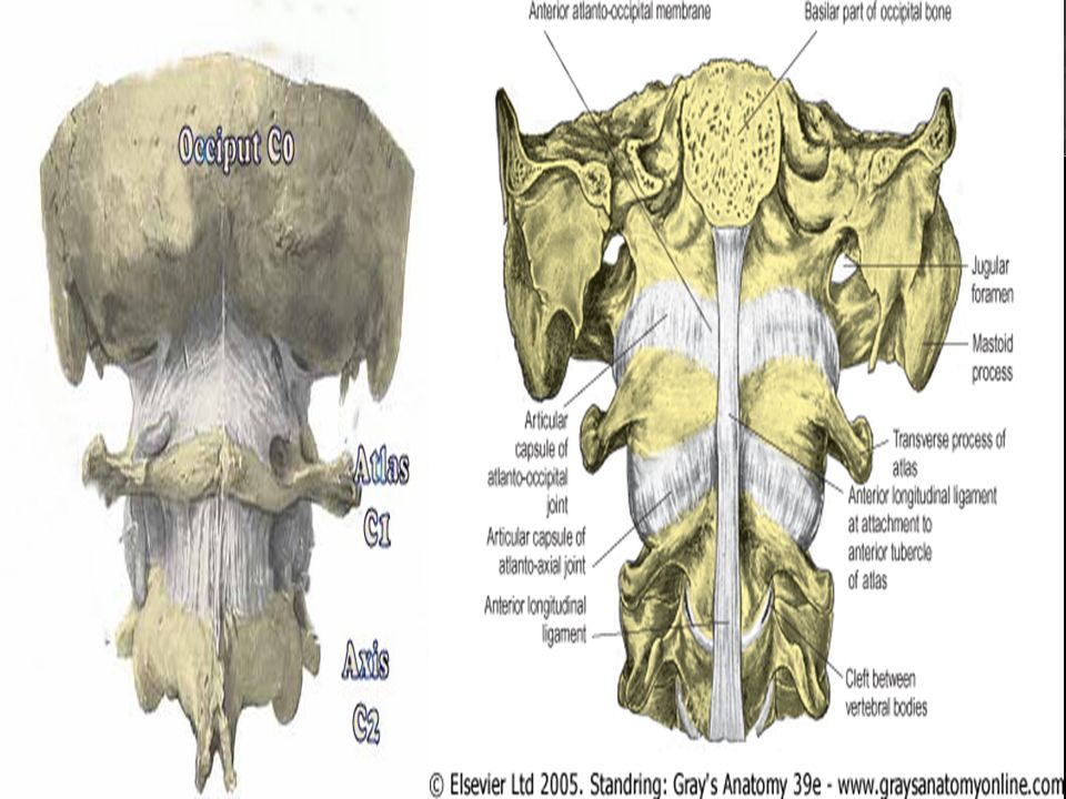
Atlanto-Occipital Joints The atlanto-occipital joints are synovial joints that are formed between the occipital condyles, which are found on either side of the foramen magnum above and the facets on the superior surfaces of the lateral masses of the atlas below. They are enclosed by a capsule. Ligaments Anterior atlanto-occipital membrane: This is a continuation of the anterior longitudinal ligament, which runs as a band down the anterior surface of the vertebral column. The membrane connects the anterior arch of the atlas to the anterior margin of the foramen magnum. Posterior atlanto-occipital membrane: This membrane is similar to the ligamentum flavum and connects the posterior arch of the atlas to the posterior margin of the foramen magnum. Movements Flexion, extension, and lateral flexion. No rotation is possible. JOINTS OF THE VERTEBRAL COLUMN

30
The atlantoaxial joints are three synovial joints: one is between the odontoid process and the anterior arch of the atlas, and the other two are between the lateral masses of the bones. The joints are enclosed by capsules. Ligaments Apical ligament: This median-placed structure connects the apex of the odontoid process to the anterior margin of the foramen magnum. Alar ligaments: These lie one on each side of the apical ligament and connect the odontoid process to the medial sides of the occipital condyles. Cruciate ligament: This ligament consists of a transverse part and a vertical part. The transverse part is attached on each side to the inner aspect of the lateral mass of the atlas and binds the odontoid process to the anterior arch of the atlas. The vertical part runs from the posterior surface of the body of the axis to the anterior margin of the foramen magnum. Membrana tectoria: This is an upward continuation of the posterior longitudinal ligament. It is attached above to the occipital bone just within the foramen magnum. It covers the posterior surface of the odontoid process and the apical, alar, and cruciate ligaments. Movements There can be extensive rotation of the atlas and thus of the head on the axis ATLANTOAXIAL JOINTS

31
Joints of the Vertebral Column Below the Axis With the exception of the first two cervical vertebrae, the remainder of the mobile vertebrae articulate with each other by means of cartilaginous joints between their bodies and by synovial joints between their articular processes. Joints Between Two Vertebral Bodies The upper and lower surfaces of the bodies of adjacent vertebrae are covered by thin plates of hyaline cartilage. Sandwiched between the plates of hyaline cartilage is an intervertebral disc of fibrocartilage. The collagen fibers of the disc strongly unite the bodies of the two vertebrae. In the lower cervical region, small synovial joints are present at the sides of the intervertebral disc between the upper and lower surfaces of the bodies of the vertebrae.

32
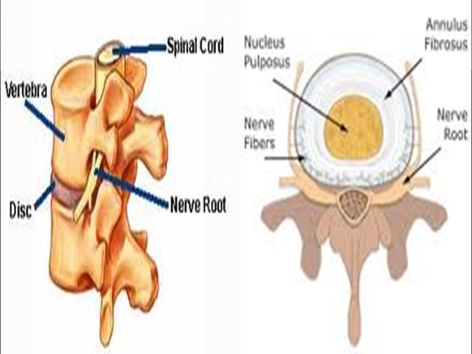
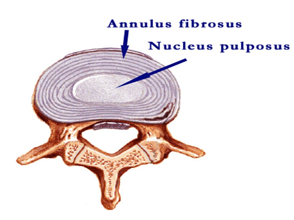
The intervertebral discs are responsible for one fourth of the length of the vertebral column. They are thickest in the cervical and lumbar regions, where the movements of the vertebral column are greatest. They may be regarded as semi-elastic discs, which lie between the rigid bodies of adjacent vertebrae. Their physical characteristics permit them to serve as shock absorbers when the load on the vertebral column is suddenly increased, as when one is jumping from a height. Their elasticity allows the rigid vertebrae to move one on the other. Unfortunately, their resilience is gradually lost with advancing age. Each disc consists of : The annulus fibrosus The nucleus pulposus The annulus fibrosus is composed of fibrocartilage, in which the collagen fibers are arranged in concentric layers or sheets. The collagen bundles pass obliquely between adjacent vertebral bodies, and their inclination is reversed in alternate sheets. The more peripheral fibers are strongly attached to the anterior and posterior longitudinal ligaments of the vertebral column. INTERVERTEBRAL DISCS

36
The nucleus pulposus in children and adolescents is an ovoid mass of gelatinous material containing a large amount of water, a small number of collagen fibers, and a few cartilage cells. It is normally under pressure and situated slightly nearer to the posterior than to the anterior margin of the disc. NOTE: The upper and lower surfaces of the bodies of adjacent vertebrae that abut onto the disc are covered with thin plates of hyaline cartilage. No discs are found between the first two cervical vertebrae or in the sacrum or coccyx.

37
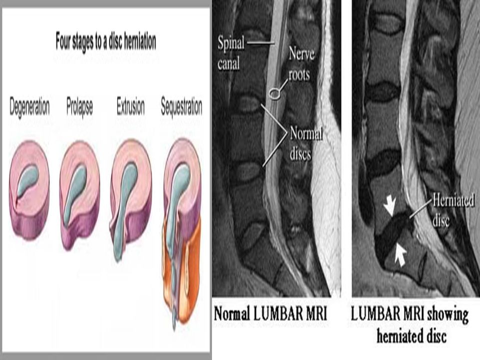
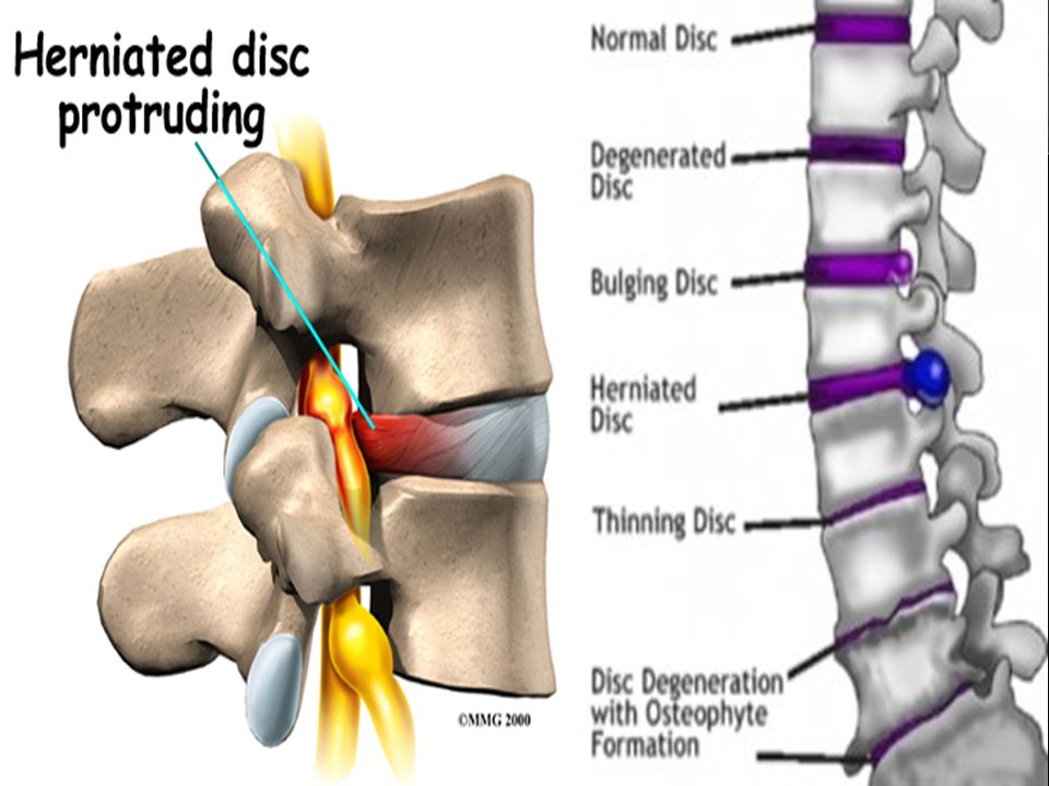
The semifluid nature of the nucleus pulposus allows it to change shape and permits one vertebra to rock forward or backward on another, as in flexion and extension of the vertebral column. A sudden increase in the compression load on the vertebral column causes the semifluid nucleus pulposus to become flattened. The outward thrust of the nucleus is accommodated by the resilience of the surrounding annulus fibrosus. Sometimes, the outward thrust is too great for the annulus fibrosus and it ruptures, allowing the nucleus pulposus to herniate and protrude into the vertebral canal, where it may press on the spinal nerve roots, the spinal nerve, or even the spinal cord. With advancing age, the water content of the nucleus pulposus diminishes and is replaced by fibrocartilage. The collagen fibers of the annulus degenerate and, as a result, the annulus cannot always contain the nucleus pulposus under stress. In old age the discs are thin and less elastic, and it is no longer possible to distinguish the nucleus from the annulus. FUNCTION OF THE INTERVERTEBRAL DISCS

38
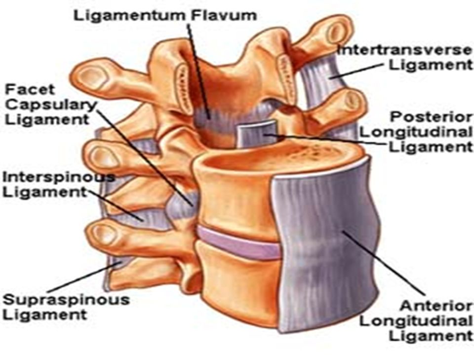
The anterior and posterior longitudinal ligaments run as continuous bands down the anterior and posterior surfaces of the vertebral column from the skull to the sacrum. The anterior ligament is wide and is strongly attached to the front and sides of the vertebral bodies and to the intervertebral discs. The posterior ligament is weak and narrow and is attached to the posterior borders of the discs. These ligaments hold the vertebrae firmly together but at the same time permit a small amount of movement to take place between them. LIGAMENTS

39
The joints between the vertebral bodies are innervated by the small meningeal branches of each spinal nerve. The nerve arises from the spinal nerve as it exits from the intervertebral foramen. It then re-enters the vertebral canal through the intervertebral foramen and supplies the meninges, ligaments, and intervertebral discs. The joints between the articular processes are innervated by branches from the posterior rami of the spinal nerves. It should be noted that the joints of any particular level receive nerve fibers from two adjacent spinal nerves. NERVE SUPPLY OF VERTEBRAL JOINTS

40
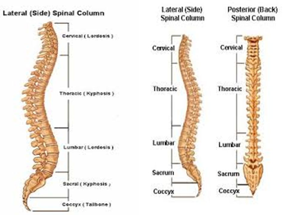
Curves in the Sagittal Plane In the fetus, the vertebral column has one continuous anterior concavity. As development proceeds, the lumbosacral angle appears. After birth, when the child becomes able to raise his or her head and keep it poised on the vertebral column, the cervical part of the vertebral column becomes concave posteriorly. Toward the end of the first year, when the child begins to stand upright, the lumbar part of the vertebral column becomes concave posteriorly. The development of these secondary curves is largely caused by modification in the shape of the intervertebral discs.In the adult in the standing position, the vertebral column therefore exhibits in the sagittal plane the following regional curves: cervical, posterior concavity thoracic, posterior convexity lumbar, posterior concavity sacral, posterior convexity CURVES OF THE VERTEBRAL COLUMN

42
During the later months of pregnancy, with the increase in size and weight of the fetus, women tend to increase the posterior lumbar concavity in an attempt to preserve their center of gravity. In old age, the intervertebral discs atrophy, resulting in a loss of height and a gradual return of the vertebral column to a continuous anterior concavity. Curves in the Coronal Plane In late childhood, it is common to find the development of minor lateral curves in the thoracic region of the vertebral column. This is normal and is usually caused by the predominant use of one of the upper limbs. For example, right- handed persons will often have a slight right-sided thoracic convexity. Slight compensatory curves are always present above and below such a curvature.

43
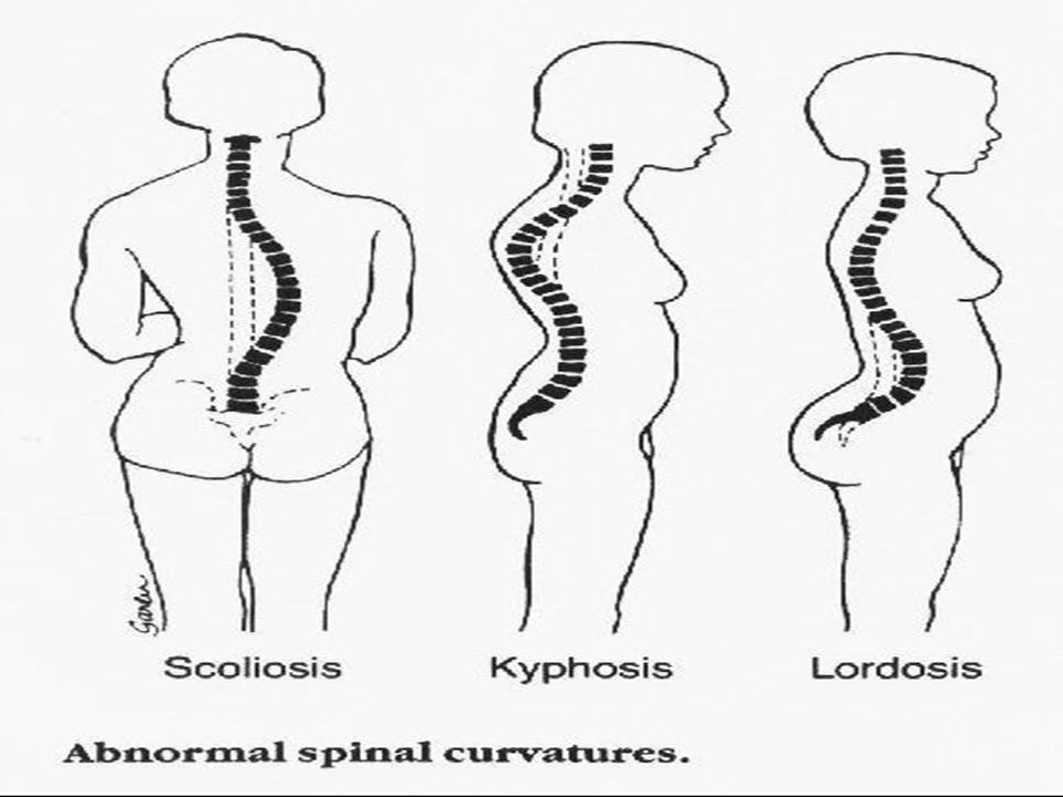
Kyphosis is an exaggeration in the sagittal curvature present in the thoracic part of the vertebral column. It can be caused by muscular weakness or by structural changes in the vertebral bodies or by intervertebral discs. In sickly adolescents, for example, where the muscle tone is poor, long hours of study or work over a low desk can lead to a gently curved kyphosis of the upper thoracic region. The person is said to be around- shouldered. Crush fractures or tuberculous destruction of the vertebral bodies leads to acute angular kyphosis of the vertebral column. In the aged, osteoporosis (abnormal rarefaction of bone) and/or degeneration of the intervertebral discs leads to senile kyphosis, involving the cervical, thoracic, and lumbar regions of the column. KYPHOSIS
 and/or degeneration of the intervertebral discs leads to senile kyphosis, involving the cervical, thoracic, and lumbar regions of the column. KYPHOSIS.")
45
Lordosis is an exaggeration in the sagittal curvature present in the lumbar region. Lordosis may be caused by an increase in the weight of the abdominal contents, as with the gravid uterus or a large ovarian tumor, or it may be caused by disease of the vertebral column such as spondylolisthesis. The possibility that it is a postural compensation for a kyphosis in the thoracic region or a disease of the hip joint (congenital dislocation) must not be overlooked. Scoliosis is a lateral deviation of the vertebral column. This is most commonly found in the thoracic region and may be caused by muscular or vertebral defects. Paralysis of muscles caused by poliomyelitis can cause severe scoliosis. The presence of a congenital hemi vertebra can cause scoliosis. Often scoliosis is compensatory and may be caused by a short leg or hip disease.
 must not be overlooked. Scoliosis is a lateral deviation of the vertebral column. This is most commonly found in the thoracic region and may be caused by muscular or vertebral defects. Paralysis of muscles caused by poliomyelitis can cause severe scoliosis. The presence of a congenital hemi vertebra can cause scoliosis. Often scoliosis is compensatory and may be caused by a short leg or hip disease..")
46
The vertebral column consists of several separate vertebrae accurately positioned one on the other and separated by intervertebral discs. The vertebrae are held in position relative to one another by strong ligaments that severely limit the degree of movement possible between adjacent vertebrae. Nevertheless, the summation of all these movements gives the vertebral column as a whole a remarkable degree of mobility. The following movements are possible: flexion, extension, lateral flexion, rotation, and circumduction Flexion is a forward movement, and extension is a backward movement. Both are extensive in the cervical and lumbar regions but restricted in the thoracic region Lateral flexion is the bending of the body to one or the other side. It is extensive in the cervical and lumbar regions but restricted in the thoracic region. Rotation is a twisting of the vertebral column. This is least extensive in the lumbar region. Circumduction is a combination of all these movements. MOVEMENTS OF THE VERTEBRAL COLUMN

47
The muscles of the back may be divided into three groups: The superficial muscles connected with the shoulder girdle. The intermediate muscles involved with movements of the thoracic cage. The deep muscles or postvertebral muscles belonging to the vertebral column MUSCLES OF THE BACK

48
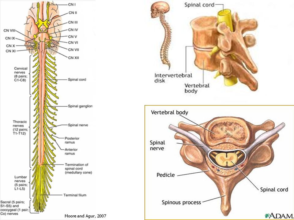
The spinal cord is a cylindrical, grayish white structure that begins above at the foramen magnum, where it is continuous with the medulla oblongata of the brain. It terminates below in the adult at the level of the lower border of the first lumbar vertebra. In the young child, it is relatively longer and ends at the upper border of the third lumbar vertebra. The spinal cord in the cervical region, where it gives origin to the brachial plexus, and in the lower thoracic and lumbar regions, where it gives origin to the lumbosacral plexus, has fusiform enlargements called cervical and lumbar enlargements. Inferiorly, the spinal cord tapers off into the conus medullaris, from the apex of which a prolongation of the pia mater, the filum terminale, descends to be attached to the back of the coccyx. The cord possesses in the midline anteriorly a deep longitudinal fissure, the anterior median fissure, and on the posterior surface a shallow furrow, the posterior median sulcus. SPINAL CORD

51
Along the whole length of the spinal cord are attached 31 pairs of spinal nerves by the anterior, or motor, roots and the posterior, or sensory, roots. Each root is attached to the cord by a series of rootlets, which extend the whole length of the corresponding segment of the cord. Each posterior nerve root possesses a posterior root ganglion, the cells of which give rise to peripheral and central nerve fibers. The spinal nerve roots pass laterally from each spinal cord segment to the level of their respective intervertebral foramina, where they unite to form a spinal nerve. Here, the motor and sensory fibers become mixed so that a spinal nerve is made up of a mixture of motor and sensory fibers. Because of the disproportionate growth in length of the vertebral column during development compared to that of the spinal cord, the length of the roots increases progressively from above downward. ROOTS OF THE SPINAL NERVES

52
In the upper cervical region the spinal nerve roots are short and run almost horizontally, but the roots of the lumbar and sacral nerves below the level of the termination of the cord (lower border of the first lumbar vertebra in the adult) form a vertical leash of nerves around the filum terminale. The lower nerve roots together are called the cauda equina. After emergence from the intervertebral foramen, each spinal nerve immediately divides into a large anterior ramus and a smaller posterior ramus, which contain both motor and sensory fibers.

53
The spinal cord receives its arterial supply from three small, longitudinally running arteries: The two posterior spinal arteries and one anterior spinal artery. The posterior spinal arteries, which arise either directly or indirectly from the vertebral arteries, run down the side of the spinal cord, close to the attachments of the posterior spinal nerve roots. The anterior spinal arteries, which arise from the vertebral arteries, unite to form a single artery, which runs down within the anterior median fissure. The posterior and anterior spinal arteries are reinforced by radicular arteries, which enter the vertebral canal through the intervertebral foramina. The veins of the spinal cord drain into the internal vertebral venous plexus. BLOOD SUPPLY OF THE SPINAL CORD

54
Vertebrae Spinal Segment CervicalAdd 1 Upper thoracicAdd 2 Lower thoracic (T7 to 9)Add 3 Tenth thoracicL1 and 2 cord segments Eleventh thoracicL3 and 4 cord segments Twelfth thoracicL5 cord segment First lumbarSacral and coccygeal cord segments RELATIONSHIPS OF SPINAL CORD SEGMENTS TO VERTEBRAL NUMBERS
Add 3 Tenth thoracicL1 and 2 cord segments Eleventh thoracicL3 and 4 cord segments Twelfth thoracicL5 cord segment First lumbarSacral and coccygeal cord segments RELATIONSHIPS OF SPINAL CORD SEGMENTS TO VERTEBRAL NUMBERS")
55
Dura Mater The Dura mater is the most external membrane and is a dense, strong, fibrous sheet that encloses the spinal cord and cauda equina. It is continuous above through the foramen magnum with the meningeal layer of Dura covering the brain. Inferiorly, it ends on the filum terminale at the level of the lower border of the second sacral vertebra. The Dural sheath lies loosely in the vertebral canal and is separated from the walls of the canal by the extradural space (epidural space). This contains loose areolar tissue and the internal vertebral venous plexus. The Dura mater extends along each nerve root and becomes continuous with connective tissue surrounding each spinal nerve (epineurium) at the intervertebral foramen. The inner surface of the Dura mater is separated from the arachnoid mater by the potential subdural space. MENINGES OF THE SPINAL CORD
. This contains loose areolar tissue and the internal vertebral venous plexus. The Dura mater extends along each nerve root and becomes continuous with connective tissue surrounding each spinal nerve (epineurium) at the intervertebral foramen. The inner surface of the Dura mater is separated from the arachnoid mater by the potential subdural space. MENINGES OF THE SPINAL CORD.")
56
The arachnoid mater is a delicate impermeable membrane covering the spinal cord and lying between the pia mater internally and the Dura mater externally. It is separated from the Dura by the subdural space that contains a thin film of tissue fluid. The arachnoid is separated from the pia mater by a wide space, the subarachnoid space, which is filled with cerebrospinal fluid. The arachnoid is continuous above through the foramen magnum with the arachnoid covering the brain. Inferiorly, it ends on the filum terminale at the level of the lower border of the second sacral vertebra. Between the levels of the conus medullaris and the lower end of the subarachnoid space lie the nerve roots of the cauda equina bathed in cerebrospinal fluid. The arachnoid mater is continued along the spinal nerve roots, forming small lateral extensions of the subarachnoid space. ARACHNOID MATER

57
The pia mater is a vascular membrane that closely covers the spinal cord. It is continuous above through the foramen magnum with the pia covering the brain; below it fuses with the filum terminale. The pia mater is thickened on either side between the nerve roots to form the ligamentum denticulate which passes laterally to be attached to the Dura. It is by this means that the spinal cord is suspended in the middle of the Dural sheath. The pia mater extends along each nerve root and becomes continuous with the connective tissue surrounding each spinal nerve. PIA MATER

58
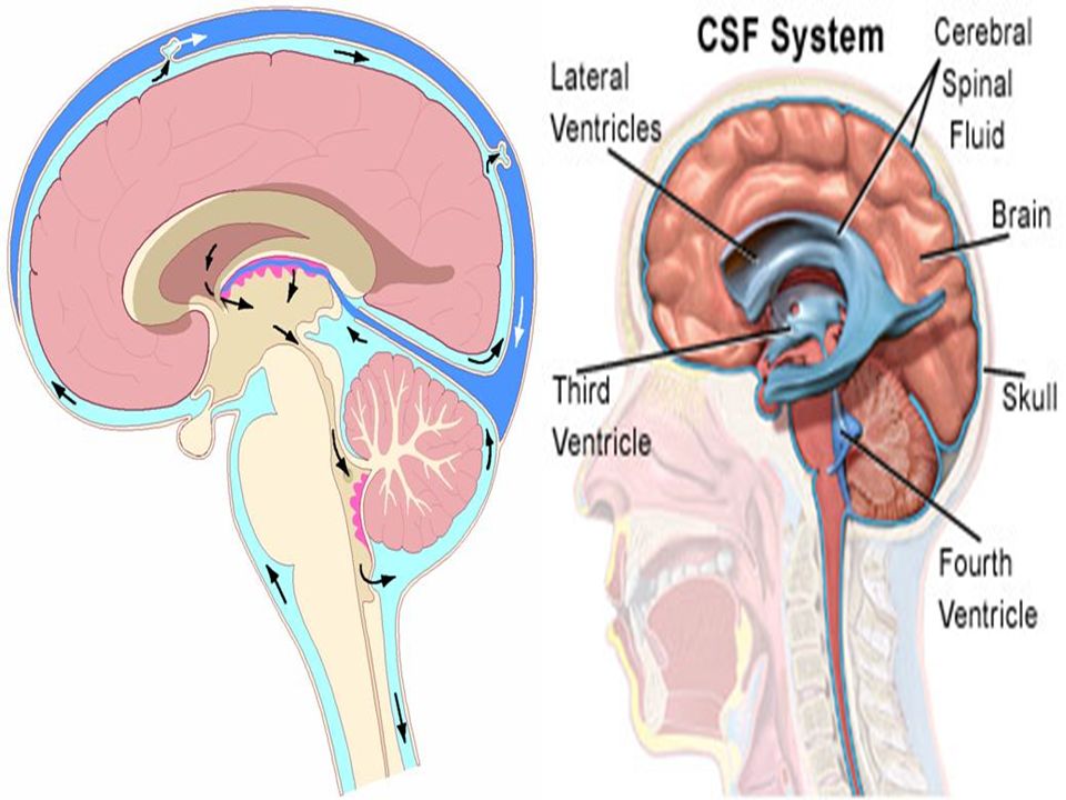
The cerebrospinal fluid is a clear, colorless fluid formed mainly by the choroid plexuses, within the lateral, third, and fourth ventricles of the brain. The fluid circulates through the ventricular system and enters the subarachnoid space through the three foramina in the roof of the fourth ventricle. It circulates both upward over the surface of the cerebral hemispheres and downward around the spinal cord. The spinal part of the subarachnoid space extends down as far as the lower border of the second sacral vertebra, where the arachnoid fuses with the filum terminale. Eventually, the fluid enters the bloodstream by passing through the arachnoid villi into the Dural venous sinuses, in particular the superior sagittal venous sinus. In addition to removing waste products associated with neuronal activity, the cerebrospinal fluid provides a fluid medium that surrounds the spinal cord. This fluid, together with the bony and ligamentous walls of the vertebral canal, effectively protects the spinal cord from trauma. CEREBROSPINAL FLUID

Similar presentations